

 PDF
PDF ePub
ePub Citation
 Print
Print


Abstract
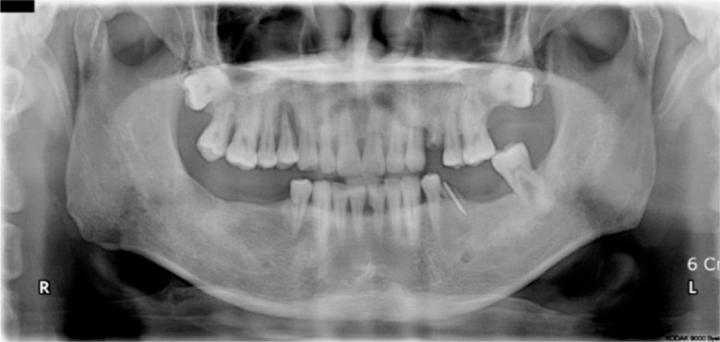
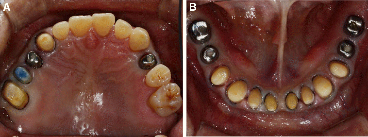
Loss of posterior support may cause extrusion of antagonistic teeth and excessive wear of remaining teeth which often leads to the destruction of the occlusal plane. In such cases, it is critical to verify the need to increase vertical dimension of occlusion (VDO). Should you increase VDO, provisionalization is crucial in evaluating function and esthetics. Double scanning technique is a useful method when fabricating definitive restoration that mimic provisional restoration. In this case, a patient with apparently no loss of VDO and insufficient interocclusal space for dental materials due to loss of posterior support and extrusion of antagonistic teeth was rehabilitated using double scanning technique. (J Korean Acad Prosthodont 2017;55:205-11)
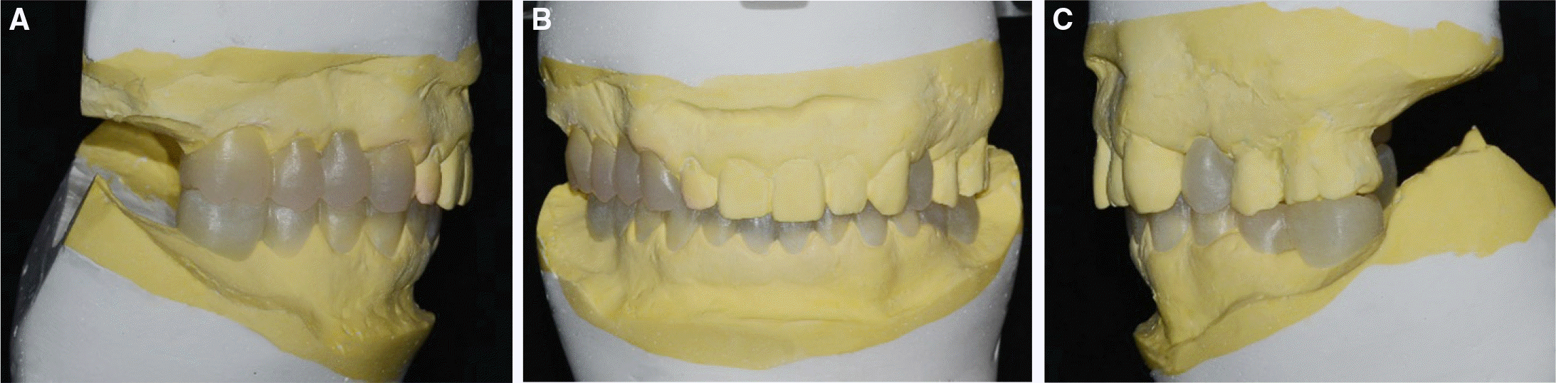
Go to : 
REFERENCES
1.Carr AB., Brown DT. McCracken's removable partial prosthodontics. 12th ed.St. Louis: USA; Elsvier;2011. p. 3–5.
2.Turner KA., Missirlian DM. Restoration of the extremely worn dentition. J Prosthet Dent. 1984. 52:467–74.

3.Rosenstiel SF., Land MF., Fujimoto J. Contemporary fixed prosthodontics. 4th ed.St. Louis: USA; Elsevier;2008. p. 87–8.
4.The Korean Academy of Stomatognathic Function & Occlusion. Textbook of human jaw function & occlusion. Yenang, Seoul,. 2014. 130.
5.Yang DH., Yang HS., Park SW., Lim HP., Yun KD., Vang MS. Full mouth implant rehabilitation with double scanning of provisional restoration. J Korean Acad Prosthodont. 2014. 52:252–7.
6.Seo JM. Full mouth rehabilitation in a patient with severely worn dentition. J Dent Rehabil Appl Sci. 2010. 26:463–76.
7.Triwatana P., Nagaviroj N., Tulapornchai C. Clinical performance and failures of zirconia-based fixed partial dentures: a review literature. J Adv Prosthodont. 2012. 4:76–83.
8.Kim KS., Lim YJ., Kim MJ., Kwon HB., Yang JH., Lee JB., Yim SH. Variation in the total lengths of abutment/implant assemblies generated with a function of applied tightening torque in external and internal implant-abutment connection. Clin Oral Implants Res. 2011. 22:834–9.
Go to :
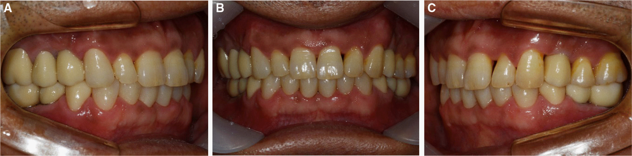
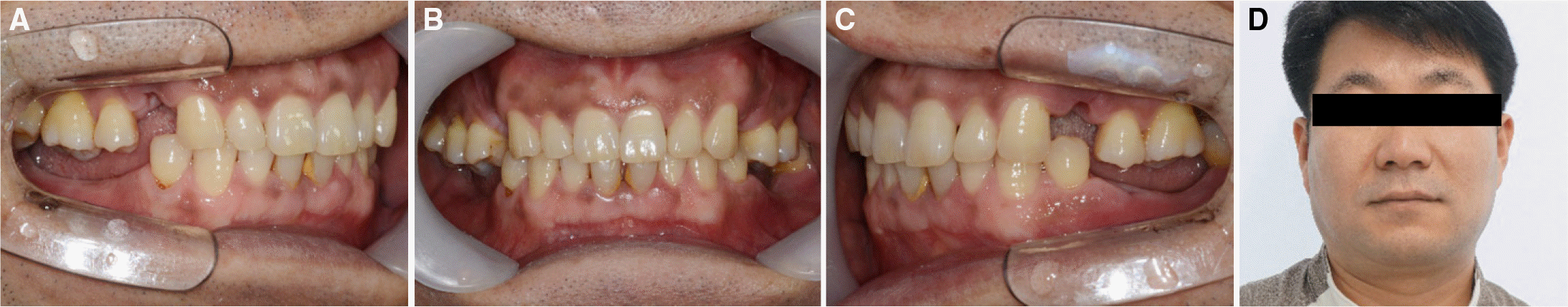 | Fig. 2.Initial photographs. (A) Right view, (B) Frontal view, (C) Left view, (D) Extraoral photograph. |
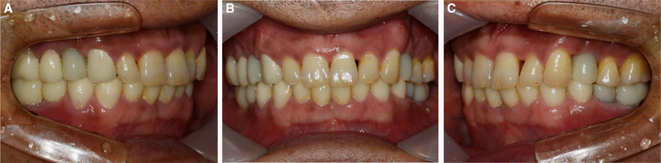
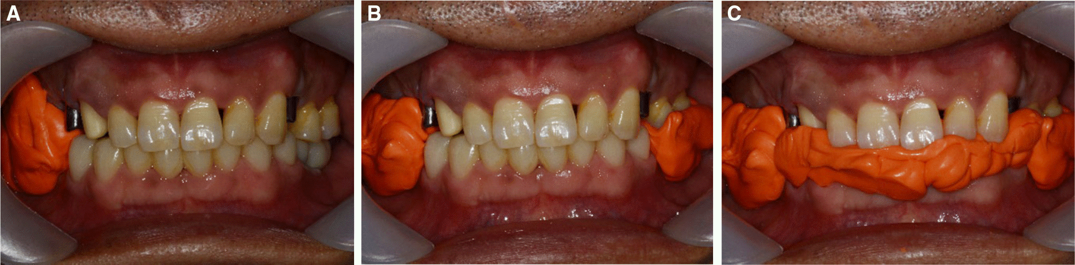 | Fig. 6.Interocclusal registration. (A) Right posterior provisional restoration removed, (B) Left posterior provisional restoration removed, (C) Anterior provisional restoration removed. |
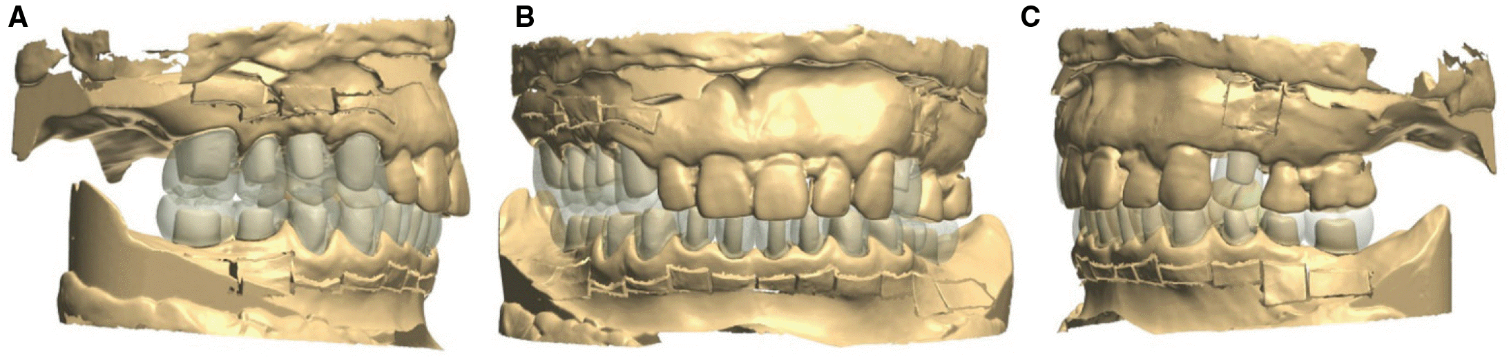 | Fig. 7.Superimposition of diagnostic wax up and working model. (A) Right view, (B) Frontal view, (C) Left view. |
 XML Download