Abstract
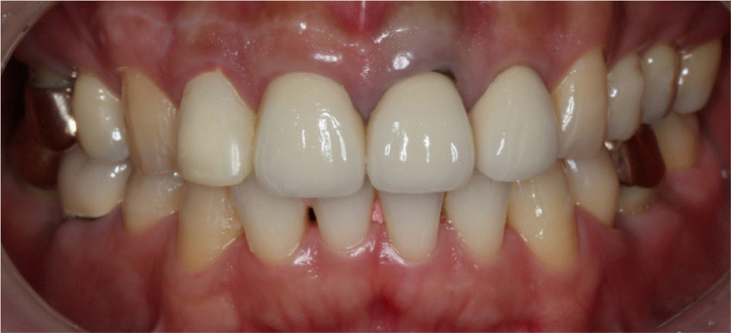
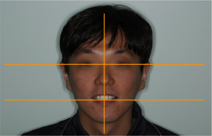
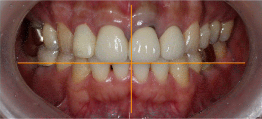
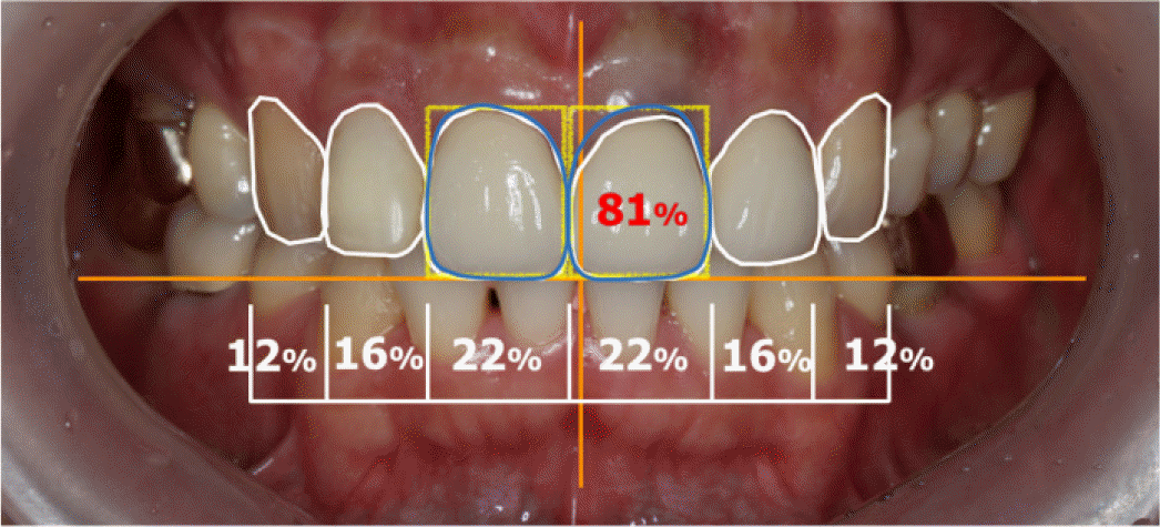
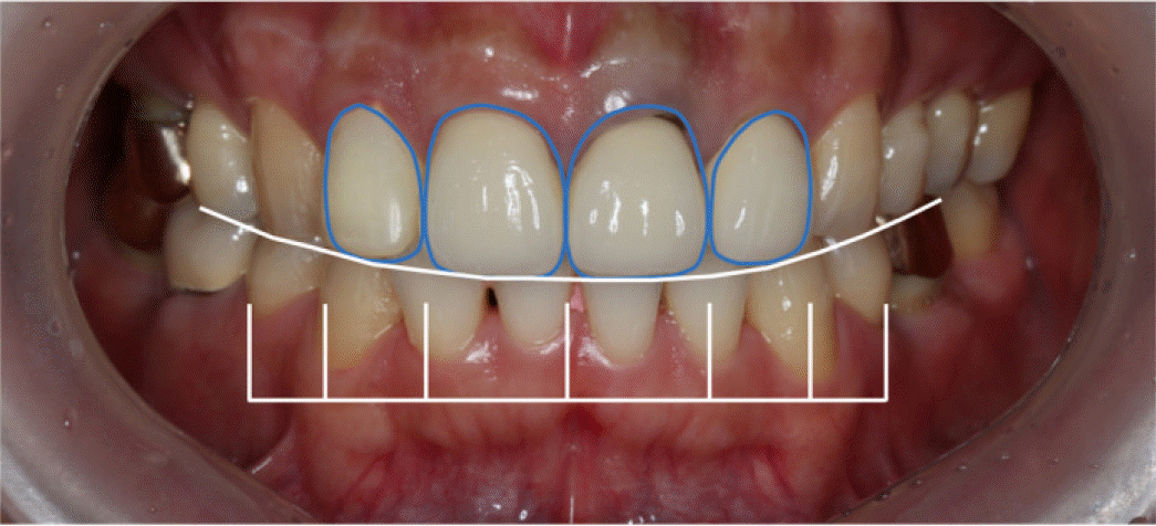
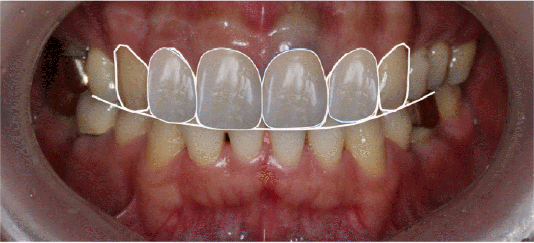
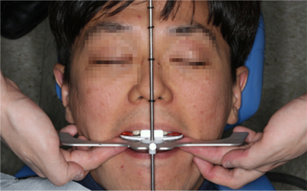
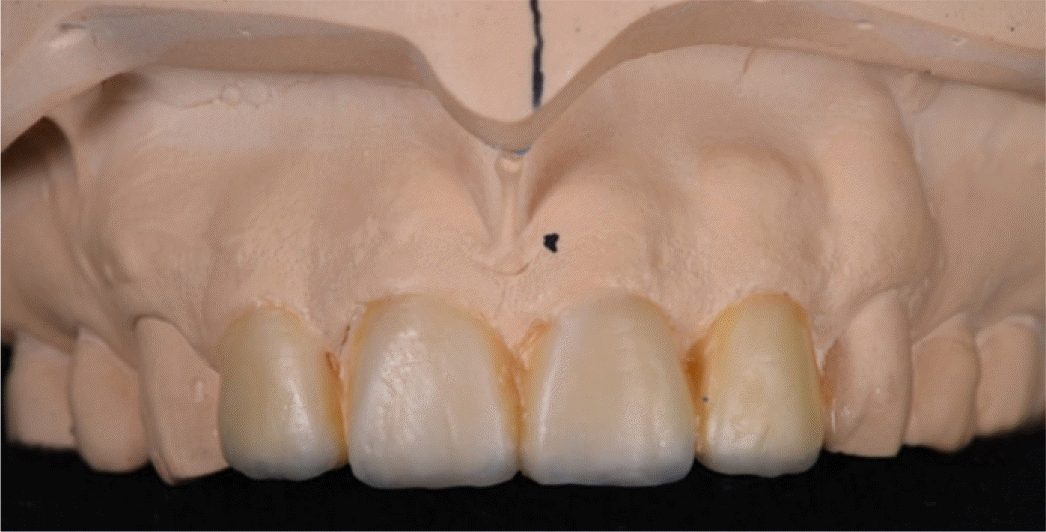
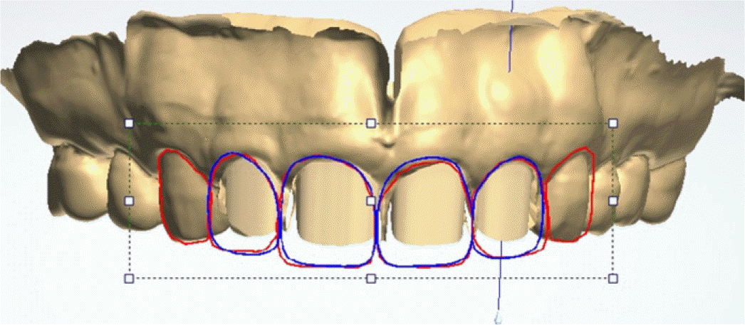
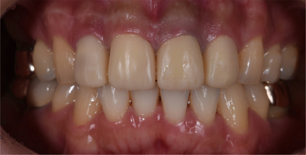
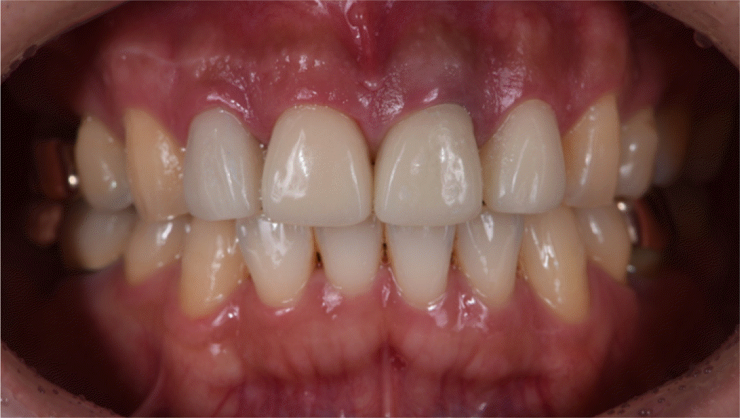
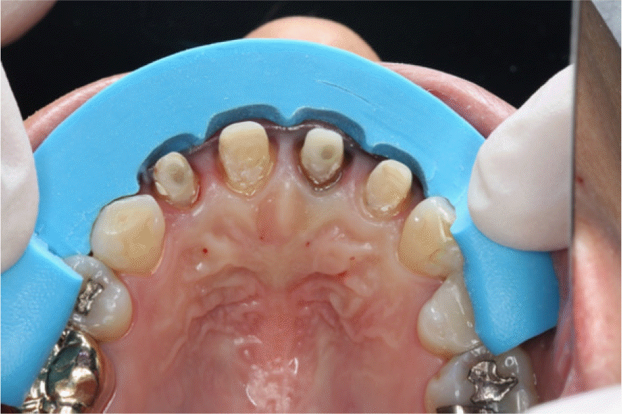
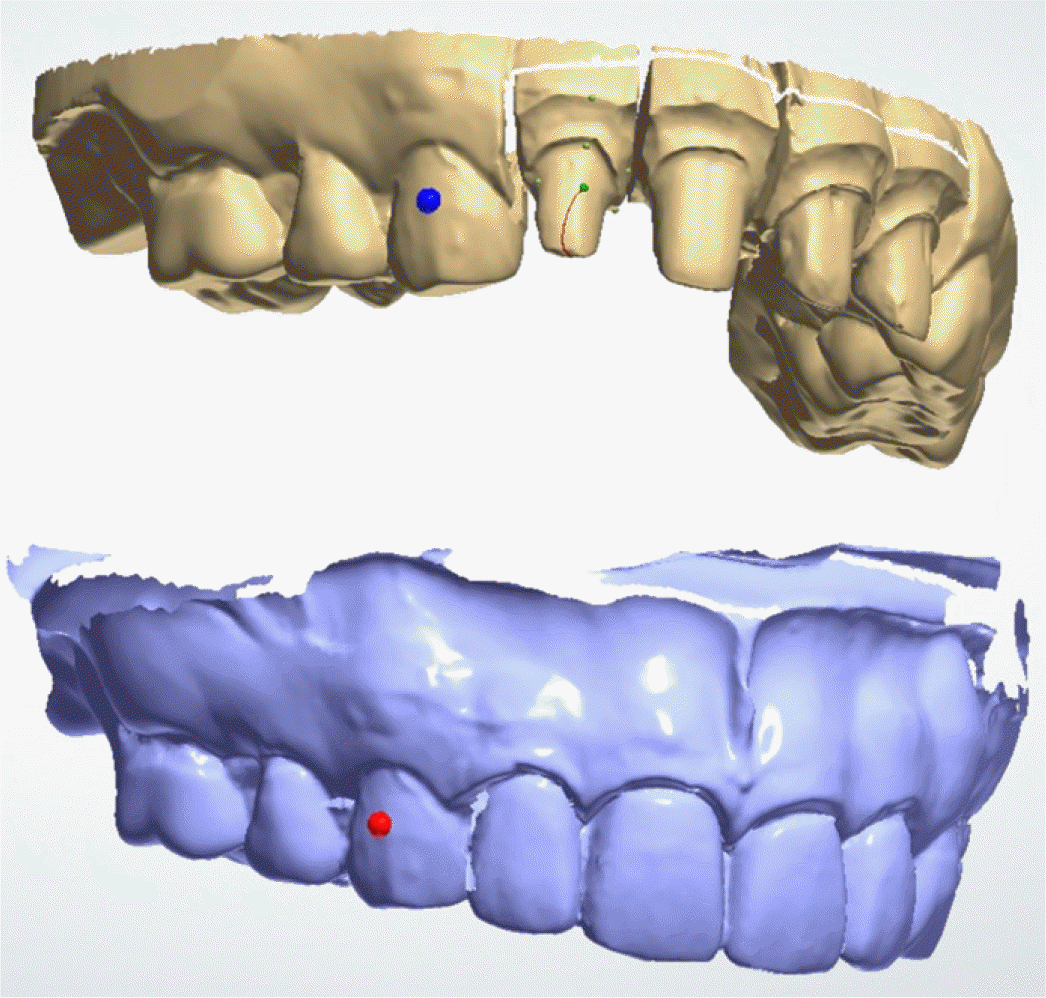
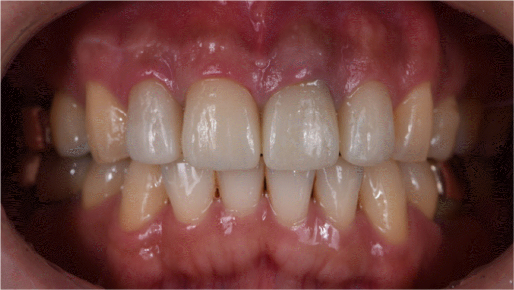
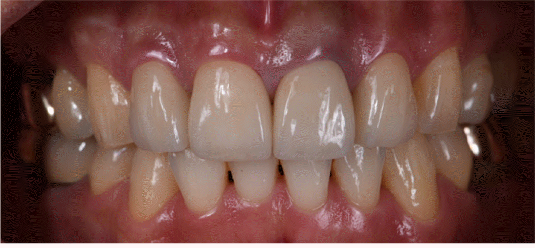
When restoring their anterior dentition, patients become more demanding on esthetics compared to posterior region during treatment planning phase. Digital Smile Design (DSD) procedure is performed in presentation software and digital photographs. This can widen diagnostic visualization and aid in transferring information between clinician, patient, and technician. This case presented is that of patient with dissatisfaction of his anterior old restoration. Retreatment procedures were carried out in two different manners: (1) using DSD protocol for diagnosis, smile simulation, communication and fabricating interim and definitive prosthesis by totally digitized workflow. (2) Using diagnostic wax-up for smile design and fabricating restorations by conventional workflow. Comparing two methods, DSD was easier to communicate between the dental team than the diagnostic wax-up method. But the final result obtained failed to meet total esthetic factors. Therefore, to obtain predictable esthetic results, more advanced design tool would be needed, including consideration of various esthetic factors besides successful communications. (J Korean Acad Prosthodont 2017;55:164-70)
Go to : 
REFERENCES
1.Paolucci B., Calamita M., Coachman C., Gürel G., Shayder A., Hallawell P. Visagism: The art of dental composition. Quintessence Dent Tech. 2012. 187–200.
2.Mclaren EA., Culp L. Smile analysis: The Photoshop smile design technique. Part I. J Cosmet Dent. 2013. 29:94–108.
3.Coachman C., Van Dooren E., GUrel G., Landsberg CJ., Calamita MA., Bichacho N. Smile design: From digital treatment planning to clinical reality. In: Cohen M (ed). Interdisciplinary treatment planning. Vol 2: Comprehensive case studies. Chicago: Quintessence;2012. p. 119–74.
4.Chu SJ. Range and mean distribution frequency of individual tooth width of the maxillary anterior dentition. Pract Proced Aesthet Dent. 2007. 19:209–15.
5.Magne P., Gallucci GO., Belser UC. Anatomic crown width/length ratios of unworn and worn maxillary teeth in white subjects. J Prosthet Dent. 2003. 89:453–61.

6.Murthy BV., Ramani N. Evaluation of natural smile: Golden proportion, RED or Golden percentage. J Conserv Dent. 2008. 11:16–21.

7.Zandinejad A., Lin WS., Atarodi M., Abdel-Azim T., Metz MJ., Morton D. Digital workflow for virtually designing and milling ceramic lithium disilicate veneers: a clinical report. Oper Dent. 2015. 40:241–6.

8.Lux LH., Thompson GA., Waliszewski KJ., Ziebert GJ. Comparison of the Kois Dento-Facial Analyzer System with an earbow for mounting a maxillary cast. J Prosthet Dent. 2015. 114:432–9.

9.Imburgia M. Patient and team communication in the iPad era - a practical appraisal. Int J Esthet Dent. 2014. 9:26–39.
10.Murray H., Locker D., Mock D., Tenenbaum H. Patient satisfaction with a consultation at a cranio-facial pain unit. Community Dent Health. 1997. 14:69–73.
Go to : 


 PDF
PDF ePub
ePub Citation
 Print
Print


 XML Download