

 PDF
PDF ePub
ePub Citation
 Print
Print


Abstract
Purpose
This research was conducted to compare the marginal and internal fit of zirconia prostheses fabricated with the model scan method and the intraoral scan method.
Materials and methods
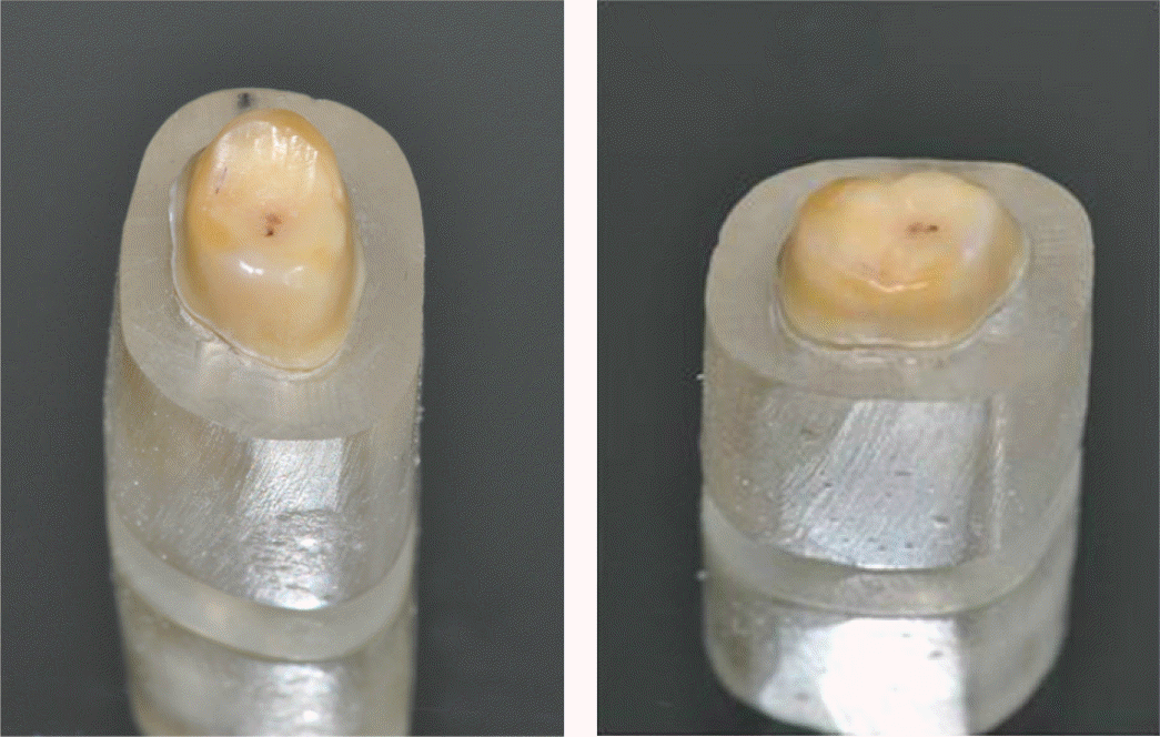
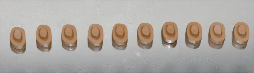
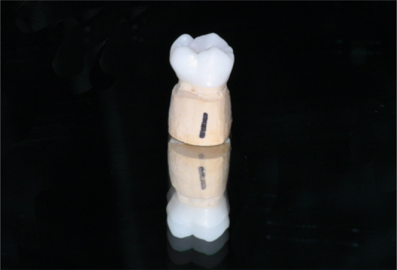
In this study, 20 extracted human mandibular first molar was used in the preparation of abutment tooth for the fabrication of zirconia prostheses. In the first group, the model scan method was applied on 10 prepared teeth. In the other group, the intraoral scan method was used on other 10 prepared teeth. Datum of both groups were transmitted to the software system. Afterwards, 20 zirconia prostheses were fabricated using the Ceramill system. Weight technique was used to evaluate the internal gap of the zirconia prostheses. In the Replica technique, marginal gap of the zirconia prostheses were analyzed by optical microscopy. Statistical analysis was based on one-way ANOVA.
Go to : 
REFERENCES
1.Conrad HJ., Seong WJ., Pesun IJ. Current ceramic materials and systems with clinical recommendations: a systematic review. J Prosthet Dent. 2007. 98:389–404.

2.Martl′nez-Rus F., Sua′rez MJ., Rivera B., Pradl′es G. Evaluation of the absolute marginal discrepancy of zirconia-based ceramic copings. J Prosthet Dent. 2011. 105:108–14.
3.Patzelt SB., Lamprinos C., Stampf S., Att W. The time efficiency of intraoral scanners: an in vitro comparative study. J Am Dent Assoc. 2014. 145:542–51.
4.Cho YB., Chung CH., Kim HJ. Marginal and internal fit of copings made by CAD/CAM using different scanning methods. J Dent Rehab App Sci. 2013. 29:366–76.
5.Jacobs MS., Windeler AS. An investigation of dental luting cement solubility as a function of the marginal gap. J Prosthet Dent. 1991. 65:436–42.
6.Felton DA., Kanoy BE., Bayne SC., Wirthman GP. Effect of in vivo crown margin discrepancies on periodontal health. J Prosthet Dent. 1991. 65:357–64.
7.Holden JE., Goldstein GR., Hittelman EL., Clark EA. Comparison of the marginal fit of pressable ceramic to metal ceramic restorations. J Prosthodont. 2009. 18:645–8.
8.Quintas AF., Oliveira F., Bottino MA. Vertical marginal discrepancy of ceramic copings with different ceramic materials, finish lines, and luting agents: an in vitro evaluation. J Prosthet Dent. 2004. 92:250–7.
9.Tan PL., Gratton DG., Diaz-Arnold AM., Holmes DC. An in vitro comparison of vertical marginal gaps of CAD/CAM titanium and conventional cast restorations. J Prosthodont. 2008. 17:378–83.
10.Pera P., Gilodi S., Bassi F., Carossa S. In vitro marginal adaptation of alumina porcelain ceramic crowns. J Prosthet Dent. 1994. 72:585–90.
11.Kohorst P., Brinkmann H., Dittmer MP., Borchers L., Stiesch M. Influence of the veneering process on the marginal fit of zirconia fixed dental prostheses. J Oral Rehabil. 2010. 37:283–91.
12.Stappert CF., Dai M., Chitmongkolsuk S., Gerds T., Strub JR. Marginal adaptation of three-unit fixed partial dentures constructed from pressed ceramic systems. Br Dent J. 2004. 196:766–70.
13.Wolfart S., Wegner SM., Al-Halabi A., Kern M. Clinical evaluation of marginal fit of a new experimental all-ceramic system before and after cementation. Int J Prosthodont. 2003. 16:587–92.
14.Balkaya MC., Cinar A., Pamuk S. Influence of firing cycles on the margin distortion of 3 all-ceramic crown systems. J Prosthet Dent. 2005. 93:346–55.
15.Fonseca JC., Henriques GE., Sobrinho LC., de Go′es MF. Stress-relieving and porcelain firing cycle influence on marginal fit of commercially pure titanium and titanium-aluminum-vanadium copings. Dent Mater. 2003. 19:686–91.
16.Wettstein F., Sailer I., Roos M., Hämmerle CH. Clinical study of the internal gaps of zirconia and metal frameworks for fixed partial dentures. Eur J Oral Sci. 2008. 116:272–9.
17.Gonzalo E., Sua′rez MJ., Serrano B., Lozano JF. A comparison of the marginal vertical discrepancies of zirconium and metal ceramic posterior fixed dental prostheses before and after cementation. J Prosthet Dent. 2009. 102:378–84.
18.Valderrama S., Van Roekel N., Andersson M., Goodacre CJ., Munoz CA. A comparison of the marginal and internal adaptation of titanium and gold-platinum-palladium metal ceramic crowns. Int J Prosthodont. 1995. 8:29–37.
19.Baig MR., Tan KB., Nicholls JI. Evaluation of the marginal fit of a zirconia ceramic computer-aided machined (CAM) crown system. J Prosthet Dent. 2010. 104:216–27.
20.Han HS., Yang HS., Lim HP., Park YJ. Marginal accuracy and internal fit of machine-milled and cast titanium crowns. J Prosthet Dent. 2011. 106:191–7.
21.Reich S., Wichmann M., Nkenke E., Proeschel P. Clinical fit of all-ceramic three-unit fixed partial dentures, generated with three different CAD/CAM systems. Eur J Oral Sci. 2005. 113:174–9.
22.Jesu′s Sua′rez M., Lozano JF., Paz Salido M., Martl′nez F. Marginal fit of titanium metal-ceramic crowns. Int J Prosthodont. 2005. 18:390–1.
23.Castillo de Oyagüe R., Sa′nchez-Jorge MI., Sa′nchez Turrio′n A., Monticelli F., Toledano M., Osorio R. Influence of CAM vs. CAD/CAM scanning methods and finish line of tooth preparation in the vertical misfit of zirconia bridge structures. Am J Dent. 2009. 22:79–83.
24.Holmes JR., Bayne SC., Holland GA., Sulik WD. Considerations in measurement of marginal fit. J Prosthet Dent. 1989. 62:405–8.
25.Flügge TV., Schlager S., Nelson K., Nahles S., Metzger MC. Precision of intraoral digital dental impressions with iTero and extraoral digitization with the iTero and a model scanner. Am J Orthod Dentofacial Orthop. 2013. 144:471–8.
26.Vojdani M., Torabi K., Farjood E., Khaledi A. Comparison the marginal and internal fit of metal copings cast from wax patterns fabricated by CAD/CAM and conventional wax up techniques. J Dent (Shiraz). 2013. 14:118–29.
27.McLean JW., von Fraunhofer JA. The estimation of cement film thickness by an in vivo technique. Br Dent J. 1971. 131:107–11.
28.Fransson B., Oilo G., Gjeitanger R. The fit of metal-ceramic crowns, a clinical study. Dent Mater. 1985. 1:197–9.
29.Moldovan O., Rudolph H., Quaas S., Bornemann G., Luthardt RG. Internal and external fit of CAM-made zirconia bridge frameworks-a pilot study. Dtsch Zahnärztl Z. 2006. 61:38–42.
30.Sakrana AA. In vitro evaluation of the marginal and internal discrepancies of different esthetic restorations. J Appl Oral Sci. 2013. 21:575–80.
31.Sorensen JA. A standardized method for determination of crown margin fidelity. J Prosthet Dent. 1990. 64:18–24.
32.Molin M., Karlsson S. The fit of gold inlays and three ceramic inlay systems. A clinical and in vitro study. Acta Odontol Scand. 1993. 51:201–6.
33.Kokubo Y., Nagayama Y., Tsumita M., Ohkubo C., Fukushima S., Vult von Steyern P. Clinical marginal and internal gaps of In-Ceram crowns fabricated using the GN-I system. J Oral Rehabil. 2005. 32:753–8.
34.Colpani JT., Borba M., Della Bona A. Evaluation of marginal and internal fit of ceramic crown copings. Dent Mater. 2013. 29:174–80.
Go to :
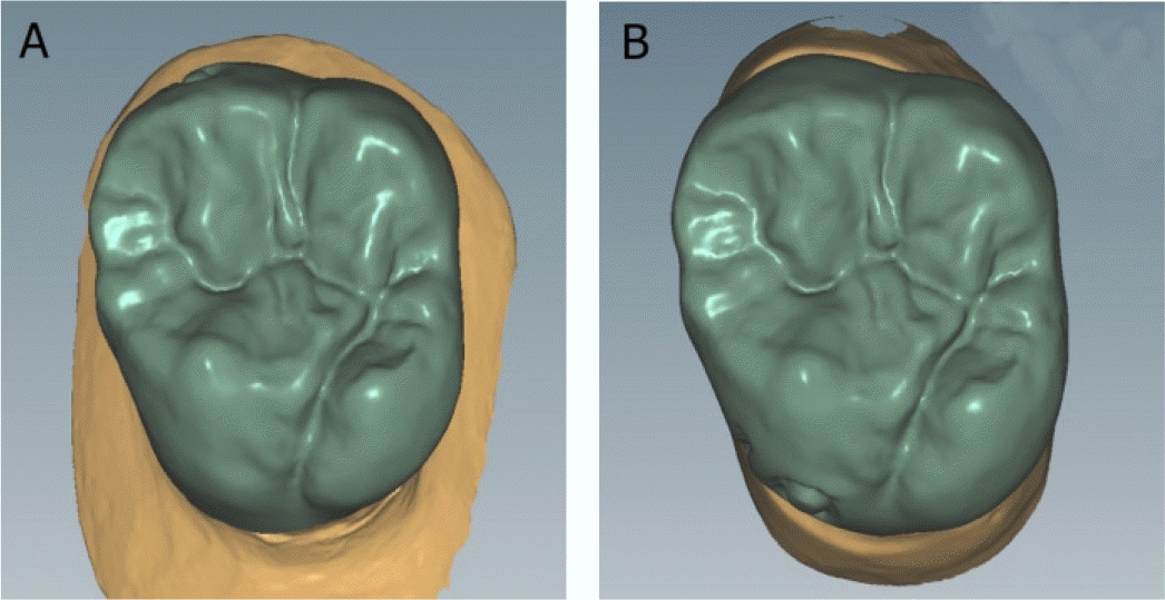 | Fig. 4.Design of the zirconia prostheses for each group. (A) Intraoral scan, (B) model scan. |
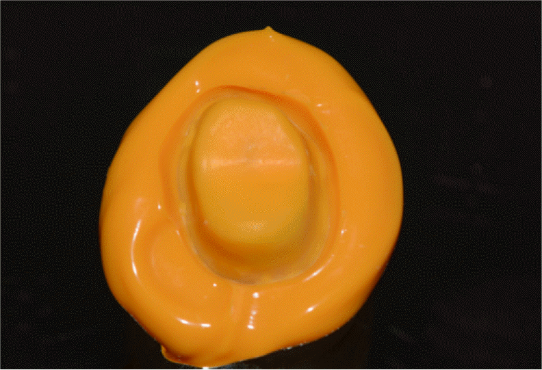 | Fig. 6.Silicone film representing the space between the abutment tooth and the zirconia prosthesis. |

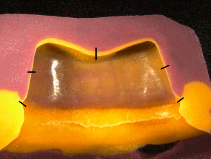 | Fig. 8.Schematic representation of the five predetermined measuring points in the cross-sectional cut of the replica. |
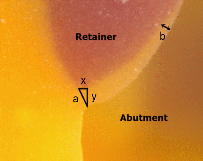 | Fig. 9.Microscopic cross-sectional photograph of a replica. Definition of measuring distances for marginal accuracy: x, horizontal marginal discrepancy; y, vertical marginal discrepancy; a, absolute marginal discrepancy; b, internal gap. |
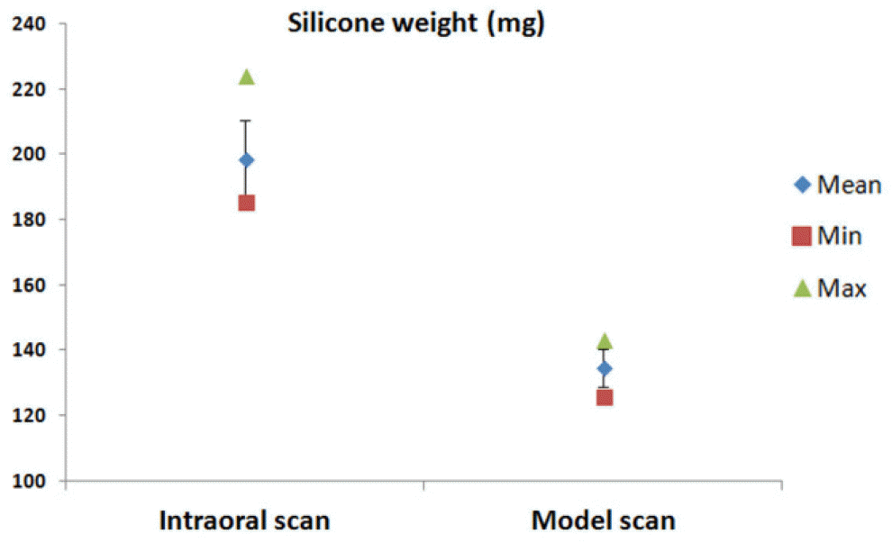 | Fig. 10.Means of silicone weight measurements for the intraoral scan group and model scan group (n = 10, for each). Error bars represent ± SD. |
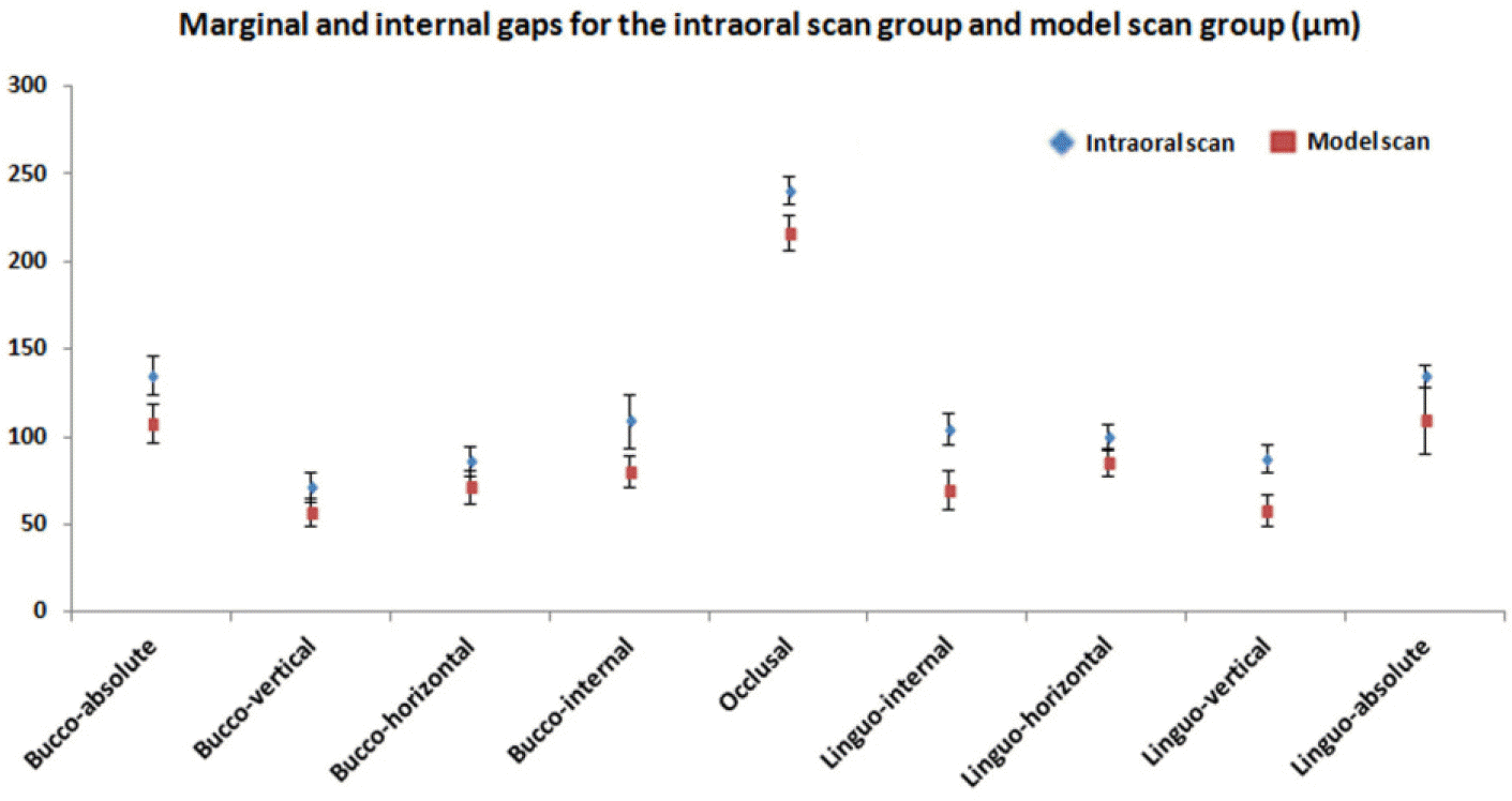 | Fig. 11.Means of marginal and internal gaps for the intraoral scan group and model scan groups (n = 10, for each). Error bars represent ± SD. |
Table 1.
Means ± SD value of silicone weight measurements for the intraoral scan group and model scan group (n = 10, for each), with independent t test results. Silicone weight (mg)
| Mean | SD | Min | Max | P value | |
|---|---|---|---|---|---|
| Intraoral scan | 198.59 | 11.93 | 185.30 | 224.00 | < .001 |
| Model scan | 134.57 | 5.86 | 125.70 | 143.00 |
Table 2.
Means ± SD value of the marginal and internal gaps for the intraoral scan group and model scan group (n = 10, for each), in ㎛, with independent t test result
 XML Download