Abstract
Gradual occlusal attrition is a normal process of aging. However, severe attrition causes pathogenic pulp, occlusal disharmony, functional disorder and esthetic problems. Alteration of vertical dimension should be considered for space regaining for tooth restoration, esthetic improvement or correction of occlusal relationship. Vertical dimension should be determined within the range of minimal invasive process satisfying patient's esthetic requirements and operator's functional goal. And patient's adaptation to newly determined vertical dimension should be assessed simultaneously. Deep overbite is not a simple problem of overbite, instead it is an usually complicated problem with anterior-posterior occlusal relationship. Considering these facts, appropriate restoration of edentulous part as well as improvement of anterior-posterior relationship should be performed to solve this fundamental problems. In this study, a 67 year-old male patient with many worn teeth and loss of posterior teeth was treated with removable partial denture at edentulous mandibular area to increase vertical dimension and fixed prostheses at dentulous maxillary and mandibular area. With these treatments, we attained a satisfactory result in functional and esthetic aspects as a report case. (J Korean Acad Prosthodont 2016;54:65-71)
Go to : 
REFERENCES
1.Dawson PE. Functional Occlusion: from TMJ to smile design. St. Louis; Mo: Mosby;2007. p. 430–52.
2.Lerner J. A systematic approach to full-mouth reconstruction of the severely worn dentition. Pract Proced Aesthet Dent. 2008. 20:81–7.
3.Small BW. Occlusal plane analysis using the Broadrick flag. Gen Dent. 2005. 53:250–2.
4.Briggs P., Bishop K. Fixed prostheses in the treatment of tooth wear. Eur J Prosthodont Restor Dent. 1997. 5:175–80.
5.Hemmings KW., Darbar UR., Vaughan S. Tooth wear treated with direct composite restorations at an increased vertical dimension: results at 30 months. J Prosthet Dent. 2000. 83:287–93.

6.Sato S., Hotta TH., Pedrazzi V. Removable occlusal overlay splint in the management of tooth wear: a clinical report. J Prosthet Dent. 2000. 83:392–5.

7.Dahl BL., Krogstad O. The effect of a partial bite-raising splint on the inclination of upper and lower front teeth. Acta Odontol Scand. 1983. 41:311–4.

8.Ramfjord SP., Blankenship JR. Increased occlusal vertical dimension in adult monkeys. J Prosthet Dent. 1981. 45:74–83.

9.Turner KA., Missirlian DM. Restoration of the extremely worn dentition. J Prosthet Dent. 1984. 52:467–74.

10.Hull CA., Junghans JA. A cephalometric approach to establishing the facial vertical dimension. J Prosthet Dent. 1968. 20:37–42.

11.Willis FM. Features of the face involved in full denture prosthesis. Dent Cosmos. 1935. 77:851–4.
12.Silverman MM. The speaking method in measuring vertical dimension. 1952. J Prosthet Dent. 2001. 85:427–31.
13.Rivera-Morales WC., Mohl ND. Restoration of the vertical dimension of occlusion in the severely worn dentition. Dent Clin North Am. 1992. 36:651–64.
14.Hemmings KW., Howlett JA., Woodley NJ., Griffiths BM. Partial dentures for patients with advanced tooth wear. Dent Update. 1995. 22:52–9.
15.Ibbetson RJ., Setchell DJ. Treatment of the worn dentition: 2. Dent Update. 1989. 16:300–2. 305–7.
16.Park JH., Jeong CM., Jeon YC., Lim JS. A study on the occlusal plane and the vertical dimension in Korean adults with natural dentition. J Korean Acad Prosthodont. 2005. 43:41–51.
17.Curtis TA., Langer Y., Curtis DA., Carpenter R. Occlusal considerations for partially or completely edentulous skeletal class II patients. Part I: Background information. J Prosthet Dent. 1988. 60:202–11.

Go to : 
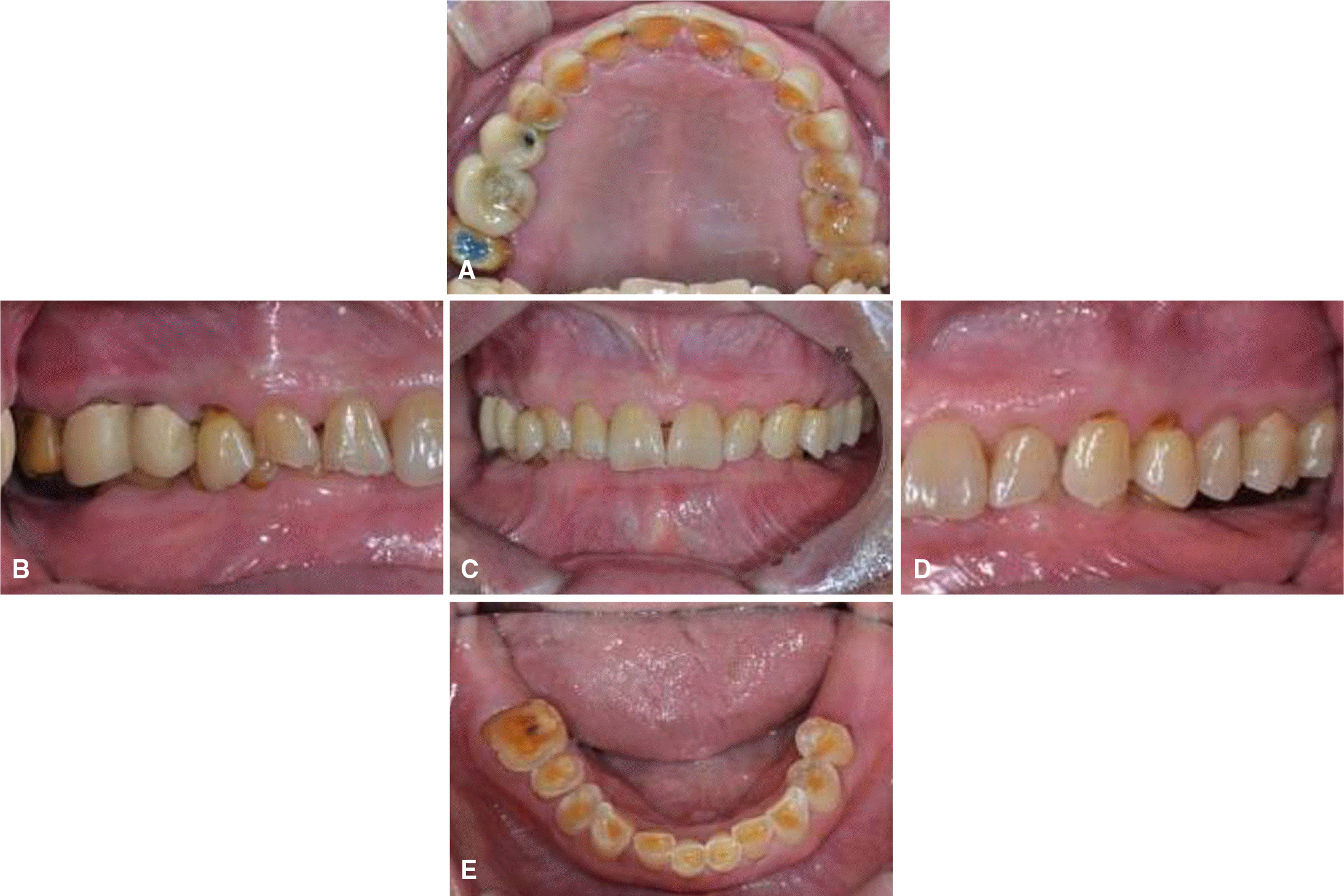
 | Fig. 1.Intraoral photograph before treatment. (A) Upper, (B) Right, (C) Frontal, (D) Left, (E) Lower. |
 | Fig. 3.TMJ series before treatment. (A) Rt. close, (B) Rt. opening, (C) Lt. close, (D) Lt. opening. |
 | Fig. 6.Cross articulation. (A) Mounting of provisional restoration, (B) Mounting of working cast, (C) Customized guide table. |


 PDF
PDF ePub
ePub Citation
 Print
Print


 XML Download