Abstract
Clinical therapy that combines full-mouth rehabilitation with immediate implantation and orthognathic surgery poses a challenge to prosthodontists. This clinical report describes a multidisciplinary approach to the diagnosis and treatment of a patient presenting with skeletal discrepancy and rampant caries. The results thus achieved indicate that full-mouth rehabilitation by fixed immediate and early loading implantation accompanied by orthognathic surgery can be a predictable and effective treatment procedure. (J Korean Acad Prosthodont 2016;54:57-64)
REFERENCES
1.Ravindran DM., Sudhakar U., Ramakrishnan T., Ambalavanan N. The efficacy of flapless implant surgery on soft-tissue profile comparing immediate loading implants to delayed loading implants: A comparative clinical study. J Indian Soc Periodontol. 2010. 14:245–51.

2.Williams AC., Shah H., Sandy JR., Travess HC. Patients' motivations for treatment and their experiences of orthodontic preparation for orthognathic surgery. J Orthod. 2005. 32:191–202.

3.Janson M., Janson G., Sant'Ana E., Tibola D., Martins DR. Orthognathic treatment for a patient with Class III malocclusion and surgically restricted mandible. Am J Orthod Dentofacial Orthop. 2009. 136:290–8.

4.Lindeboom JA., Tjiook Y., Kroon FH. Immediate placement of implants in periapical infected sites: a prospective randomized study in 50 patients. Oral Surg Oral Med Oral Pathol Oral Radiol Endod. 2006. 101:705–10.

5.Degidi M., Piattelli A., Carinci F. Immediate loaded dental implants: comparison between fixtures inserted in postextractive and healed bone sites. J Craniofac Surg. 2007. 18:965–71.
6.Pieri F., Aldini NN., Fini M., Corinaldesi G. Immediate occlusal loading of immediately placed implants supporting fixed restorations in completely edentulous arches: a 1-year prospective pilot study. J Periodontol. 2009. 80:411–21.

7.Raes F., Cooper LF., Tarrida LG., Vandromme H., De Bruyn H. A case-control study assessing oral-health-related quality of life after immediately loaded single implants in healed alveolar ridges or extraction sockets. Clin Oral Implants Res. 2012. 23:602–8.

8.Meizi E., Meir M., Laster Z. New-design dental implants: a 1-year prospective clinical study of 344 consecutively placed implants comparing immediate loading versus delayed loading and flapless versus full-thickness flap. Int J Oral Maxillofac Implants. 2014. 29:e14–21.

9.Chrcanovic BR., Albrektsson T., Wennerberg A. Immediately loaded non-submerged versus delayed loaded submerged dental implants: a meta-analysis. Int J Oral Maxillofac Surg. 2015. 44:493–506.

10.Robling AG., Turner CH. Mechanical signaling for bone modeling and remodeling. Crit Rev Eukaryot Gene Expr. 2009. 19:319–38.

11.Wang HL., Boyapati L. "PASS" principles for predictable bone regeneration. Implant Dent. 2006. 15:8–17.

12.Vogl S., Stopper M., Hof M., Wegscheider WA., Lorenzoni M. Immediate Occlusal versus Non-Occlusal Loading of Implants: A Randomized Clinical Pilot Study. Clin Implant Dent Relat Res. 2015. 17:589–97.

13.Lang NP., Pun L., Lau KY., Li KY., Wong MC. A systematic review on survival and success rates of implants placed immediately into fresh extraction sockets after at least 1 year. Clin Oral Implants Res. 2012. 23:39–66.
14.Tomasi C., Sanz M., Cecchinato D., Pjetursson B., Ferrus J., Lang NP., Lindhe J. Bone dimensional variations at implants placed in fresh extraction sockets: a multilevel multivariate analysis. Clin Oral Implants Res. 2010. 21:30–6.

15.Calvo-Guirado JL., Boquete-Castro A., Negri B., Delgado Ruiz R., Go′mez-Moreno G., Iezzi G. Crestal bone reactions to immediate implants placed at different levels in relation to crestal bone. A pilot study in Foxhound dogs. Clin Oral Implants Res. 2014. 25:344–51.
16.Caneva M., Salata LA., de Souza SS., Baffone G., Lang NP., Botticelli D. Influence of implant positioning in extraction sockets on osseointegration: histomorphometric analyses in dogs. Clin Oral Implants Res. 2010. 21:43–9.

17.De Rouck T., Collys K., Cosyn J. Single-tooth replacement in the anterior maxilla by means of immediate implantation and pro-visionalization: a review. Int J Oral Maxillofac Implants. 2008. 23:897–904.
18.Kobayashi T., Watanabe I., Ueda K., Nakajima T. Stability of the mandible after sagittal ramus osteotomy for correction of prognathism. J Oral Maxillofac Surg. 1986. 44:693–7.

19.Proffit WR., Phillips C., Dann C 4th., Turvey TA. Stability after surgical-orthodontic correction of skeletal Class III malocclusion. I. Mandibular setback. Int J Adult Orthodon Orthognath Surg. 1991. 6:7–18.
Fig. 1.
Intraoral photographs of patient before treatment. (A) Maxillary occlusal view, (B) Right side, (C) Frontal view, (D) Left side, (E) Mandibular occlusal view.
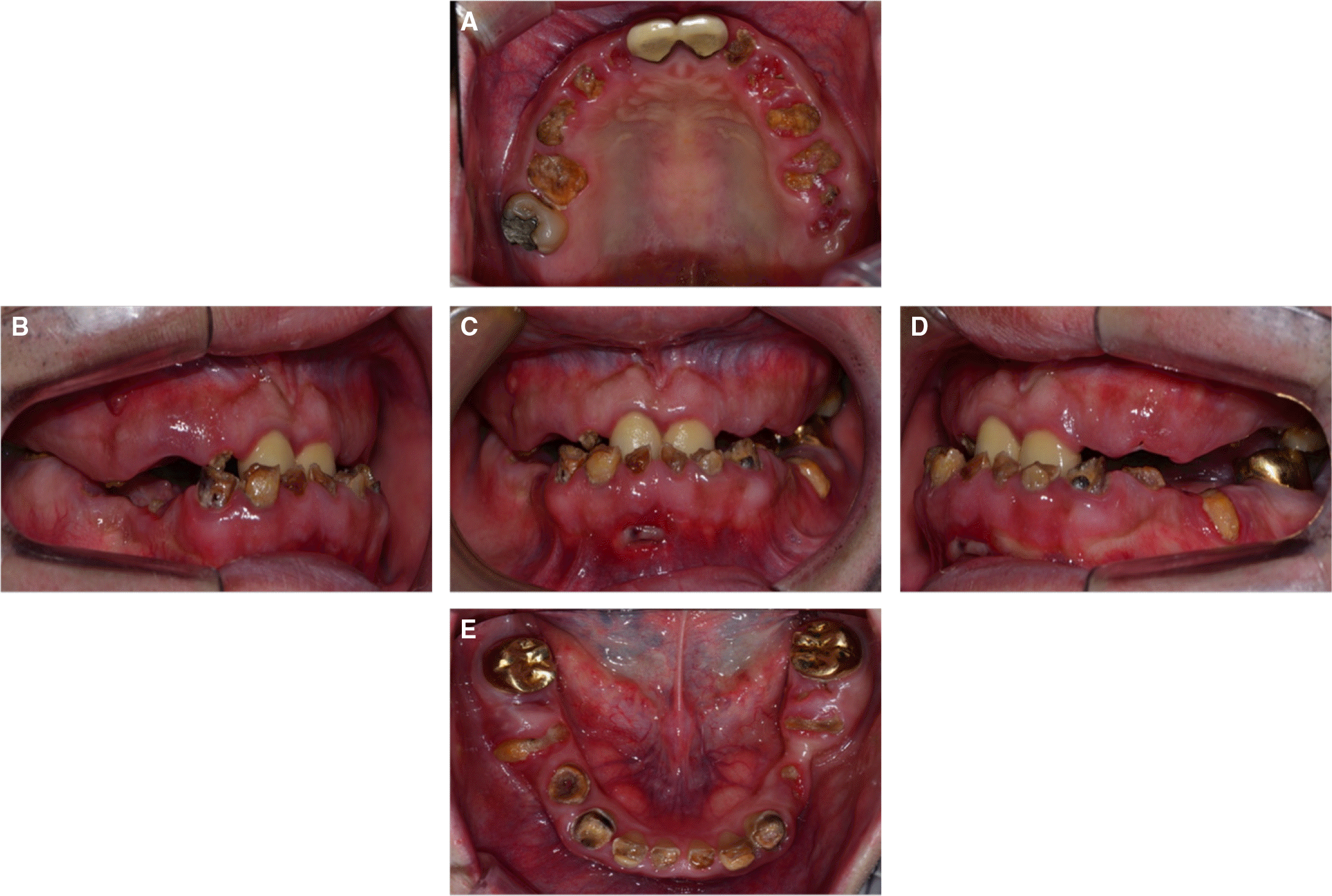
Fig. 4.
Diagnostic wax-up. The diagnostic wax-up was done in the ideal form. The occlusion was given canine guidance. (A) Right side, (B) Frontal view, (C) Left side.

Fig. 5.
Intraoral photographs at provisional prostheses delivery. (A) Maxillary occlusal view, (B) Right side, (C) Frontal view, (D) Left side, (E) Mandibular occlusal view.
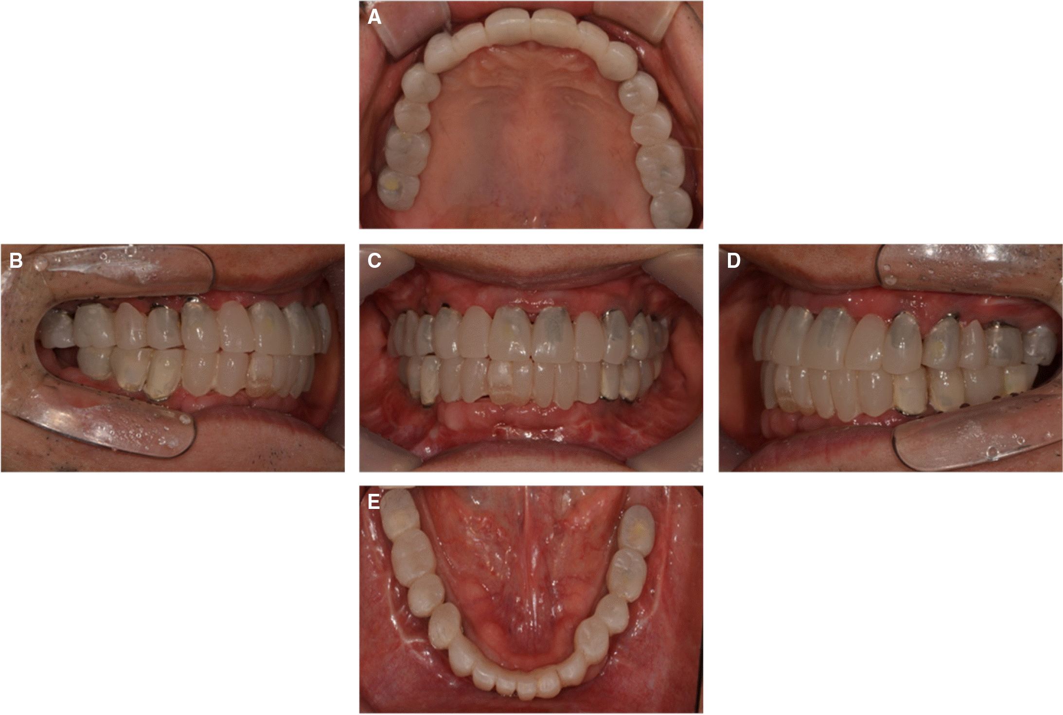
Fig. 6.
Intraoral photographs at definitive prostheses delivery. (A) Maxillary occlusal view, (B) Right side, (C) Frontal view, (D) Left side, (E) Mandibular occlusal view.
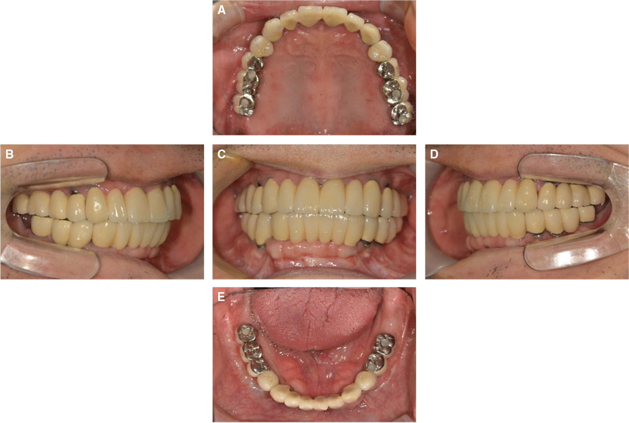
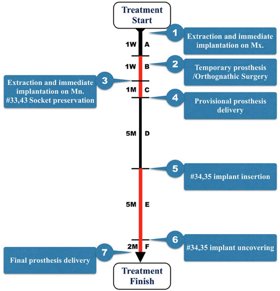
Fig. 9.
Overall treatment procedure. The overall treatment period was 15 months, from December 2012 to March 2014. The B, C, E, and F periods represent indispensable time. However, the A period could be reduced by immediate loading, and the D period could be reduced by positioning implantsin the #32 and 42 extraction sockets. (A, D periods: variable time, black line; B, C, E, F periods: indispensable time, red line; M: month(s))


 PDF
PDF ePub
ePub Citation
 Print
Print


 XML Download