Abstract
The implant prosthesis can be divided into the screw retained prosthesis and cement retained prosthesis. Each type has advantages as well as disadvantages which is unfavorable to maintain the implants. To overcome these drawbacks, T-screw system was developed. T-screw system which utilizes a lingual direction of the screw to retain the implant prosthesis, has advantages of retrievability of the prosthesis, passive fit, and possibility to form esthetic and functional occlusal surface. The prior prosthesis which utilized horizontal screws had difficulty in fabrication especially in the case of multiple units, and also limited use with all-ceramic prosthesis. In this case, fabricating the implant prosthesis by using the T-screw system showed superior results in easy maintenance, esthetics, and also functions. In addition, we are to report the method of using the T-screw system in implant prosthesis, such as multiple units of implant prosthesis and all ceramic prosthesis. (J Korean Acad Prosthodont 2016;54:423-30)
Go to : 
REFERENCES
1.Michalakis KX., Hirayama H., Garefis PD. Cement-retained versus screw-retained implant restorations: a critical review. Int J Oral Maxillofac Implants. 2003. 18:719–28.
2.Chee W., Felton DA., Johnson PF., Sullivan DY. Cemented versus screw-retained implant prostheses: which is better? Int J Oral Maxillofac Implants. 1999. 14:137–41.
3.Goodacre CJ., Bernal G., Rungcharassaeng K., Kan JY. Clinical complications with implants and implant prostheses. J Prosthet Dent. 2003. 90:121–32.

4.Kaufman EG., Coelho DH., Colin L. Factors influencing the retention of cemented gold castings. J Prosthet Dent. 1961. 11:487–502.

5.Al-Omari WM., Shadid R., Abu-Naba'a L., El Masoud B. Porcelain fracture resistance of screw-retained, cement-retained, and screw-cement-retained implant-supported metal ceramic posterior crowns. J Prosthodont. 2010. 19:263–73.

6.Misch CE. Screw-retained versus cement-retained implant-supported prostheses. Pract Periodontics Aesthet Dent. 1995. 7:15–8.
7.Chee W., Jivraj S. Screw versus cemented implant supported restorations. Br Dent J. 2006. 201:501–7.

8.Takeshita F., Suetsugu T., Asai Y., Nobayashi K. Various designs of ceramometal crown for implant restorations. Quintessence Int. 1997. 28:117–20.
9.Clausen GF. The lingual locking screw for implant-retained restorations-aesthetics and retrievability. Aust Prosthodont J. 1995. 9:17–20.
10.Chio A., Hatai Y. Restoration of two implants using custom abutments and transverse screw-retained zirconia crowns. American J Esthet Dent. 2012. 2:264–80.
11.Karunagaran S., Paprocki GJ., Wicks R., Markose S. A review of implant abutments-abutment classification to aid prosthetic selection. J Tenn Dent Assoc. 2013. 93:18–23.
12.Aparicio C. A new method for achieving passive fit of an interim restoration supported by Brånemark implants: a technical note. Int J Oral Maxillofac Implants. 1995. 10:614–8.
13.Valbao FP Jr., Perez EG., Breda M. Alternative method for retention and removal of cement-retained implant prostheses. J Prosthet Dent. 2001. 86:181–3.

14.Wilson TG Jr. The positive relationship between excess cement and peri-implant disease: a prospective clinical endoscopic study. J Periodontol. 2009. 80:1388–92.
Go to : 
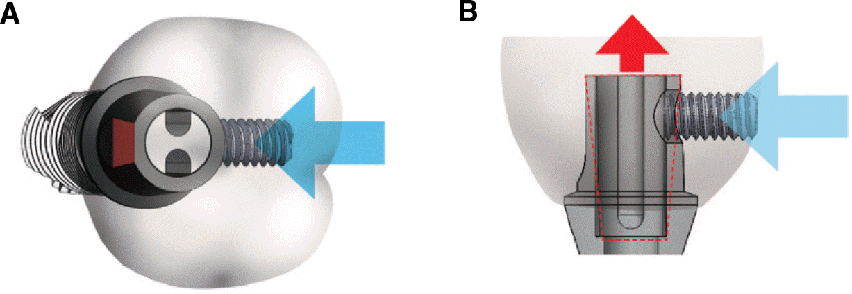
 | Fig. 1.Schematic diagram of the T-screw system. (A) Occlusal view, (B) Cross-sectional view. |
 | Fig. 2.Panoramic radiograph. (A) Pre-operative panoramic radiograph, (B) Post-operative panoramic radiograph. |
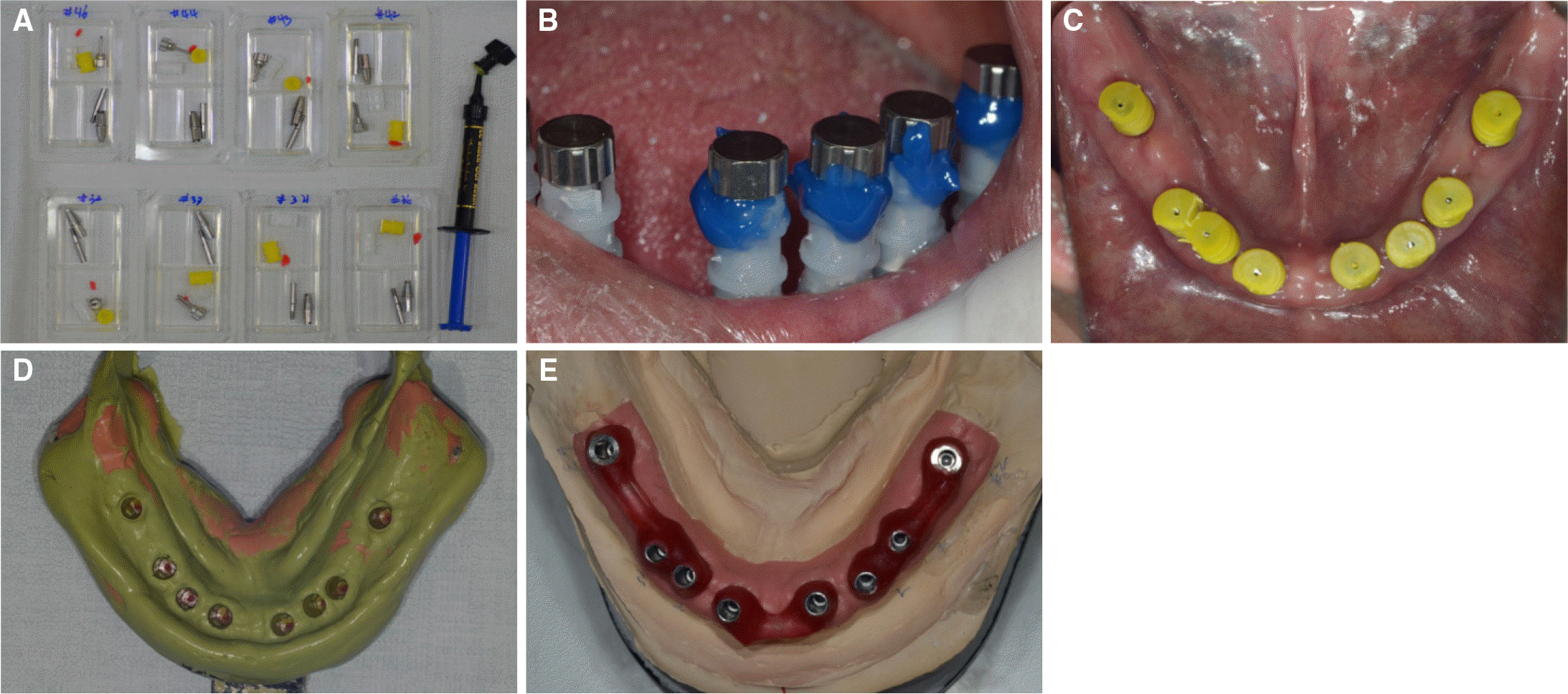
 | Fig. 3.Final impression taking. (A) Index cap and impression cap, (B) Index caps were used to duplicate internal thread of implants, (C) Connection of impression cap for taking impression, (D) Impression of Mandible, (E) Master cast of Mandible. |
 | Fig. 4.Registration of centric relation. (A, B) Connection of Lucia jig for neuromuscular reprogramming, (C) Registration of centric relation with Lucia jig and silicon bite material. |
 | Fig. 5.Try-in of framework. (A, B) Try-in of framework fabricated after connection of T-screw housing. (C) Centric relation registration with Lucia jig. |
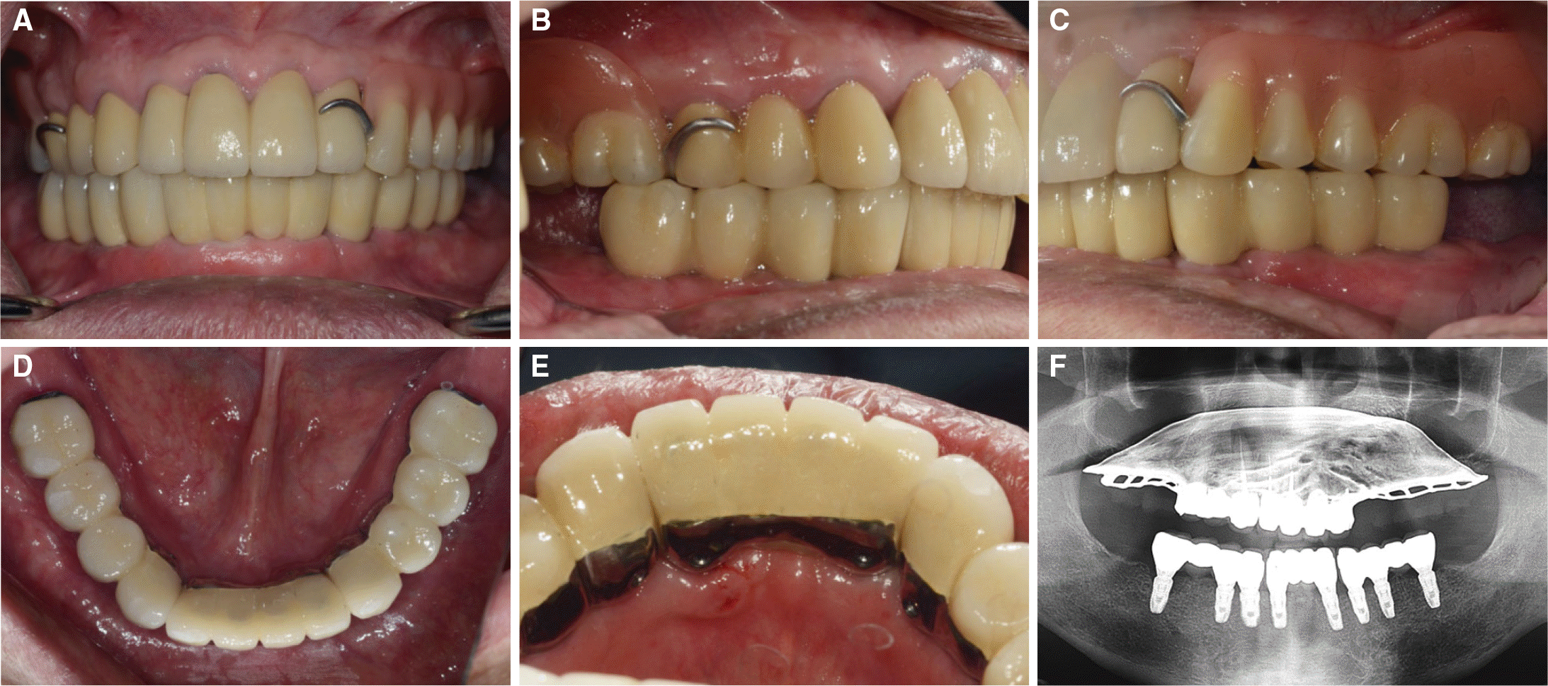
 | Fig. 6.Clinical pictures and panoramic radiograph after the placement of definitive prosthesis. (A) Frontal view, (B) Right buccal view, (C) Left buccal view, (D) Mandibular occlusal view, (E) T-screw located on lingual side, (F) Panoramic radiograph. |
 | Fig. 7.Panoramic radiograph. (A) Pre-operative panoramic radiograph, (B) Post-operative panoramic radiograph. |
 | Fig. 8.Final impression taking and fabrication of master cast. (A) SP indicator for duplicate internal thread of implants, (B) Marking the direction of ALIPS, (C) Connection of impression cap for taking impression, (D) Impression of mandibular arch, (E) Master cast connected with ALIPS, (F, G) Verifying the same direction of ALIPS. |


 PDF
PDF ePub
ePub Citation
 Print
Print


 XML Download