

 PDF
PDF ePub
ePub Citation
 Print
Print


Abstract
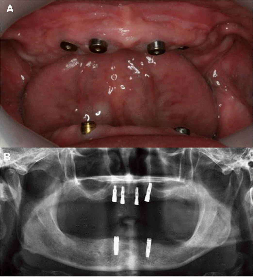
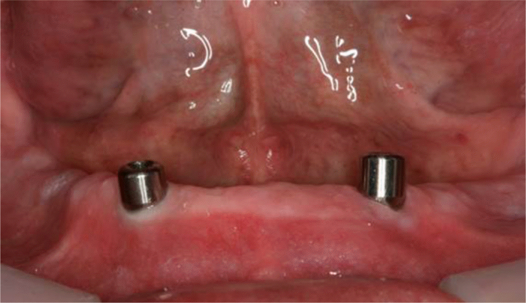
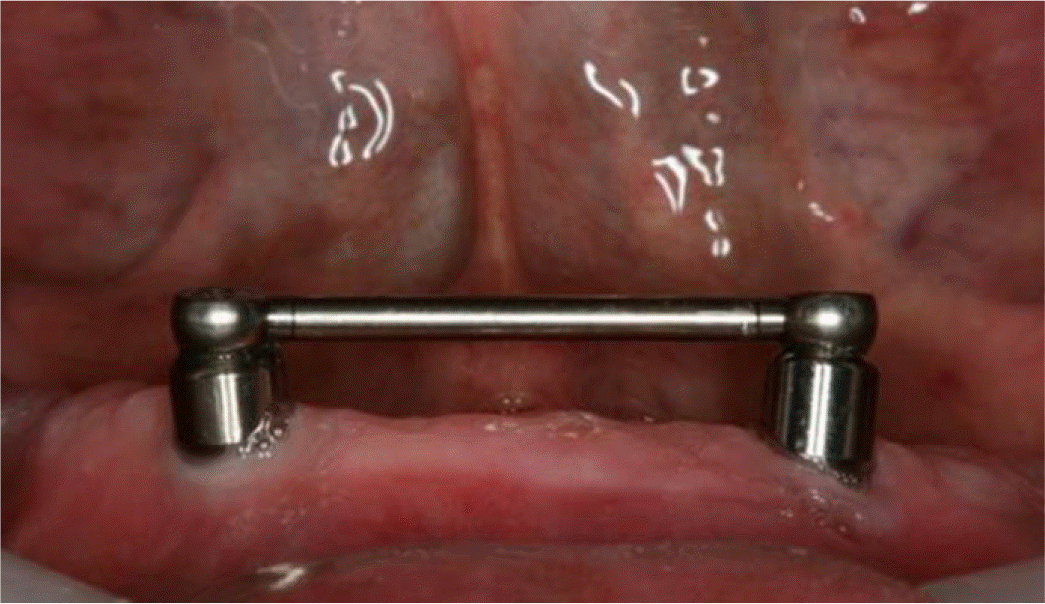
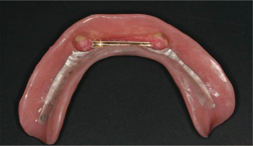
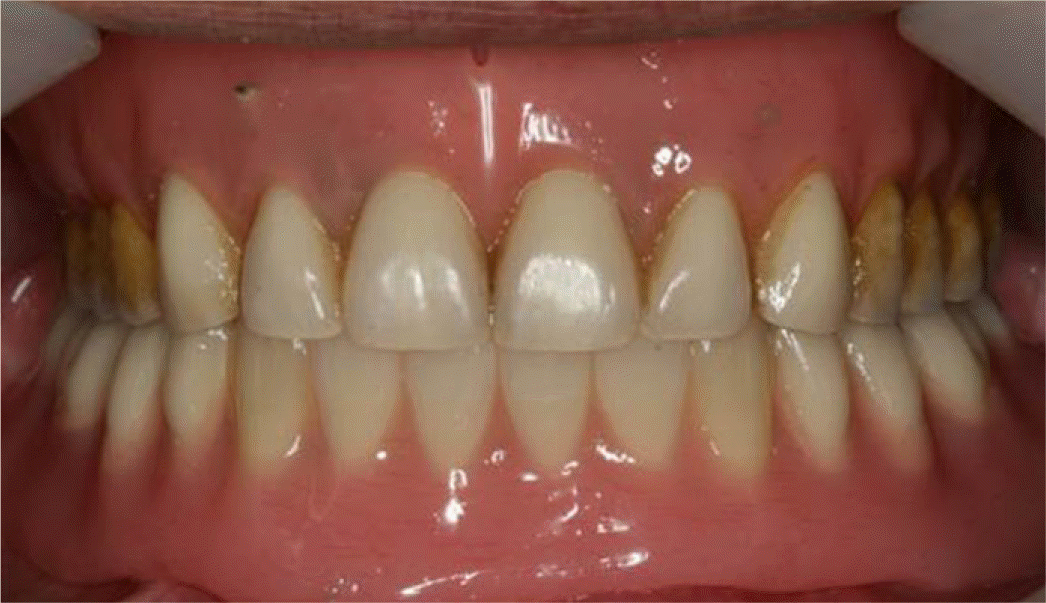
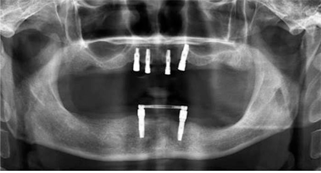
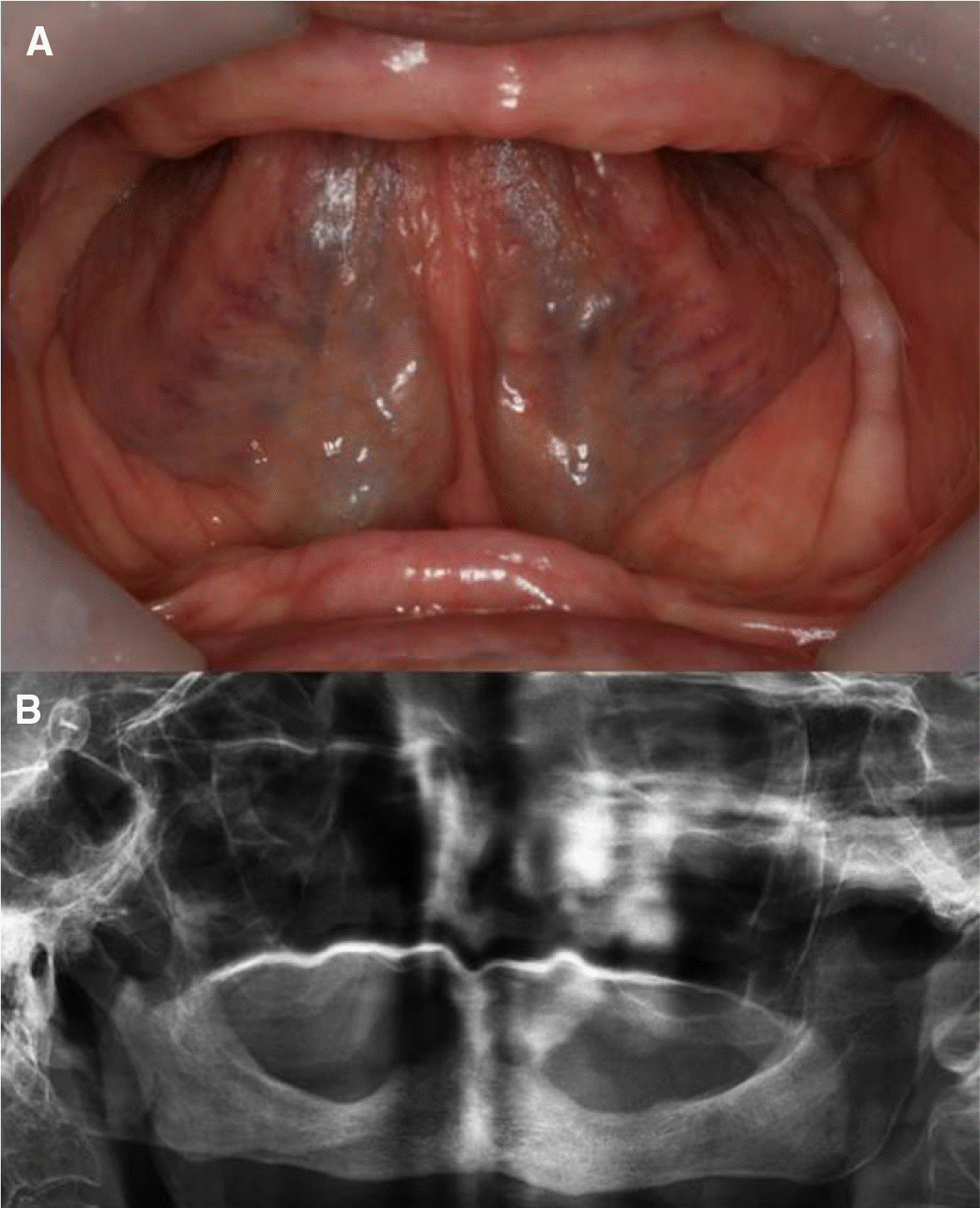
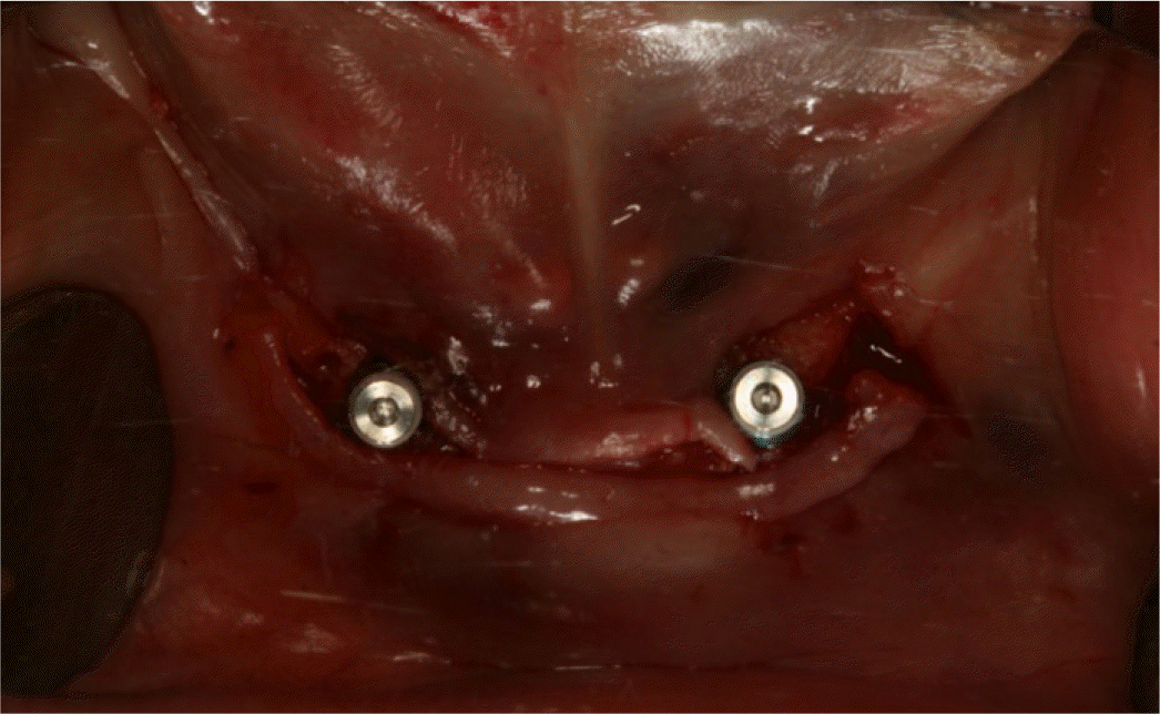
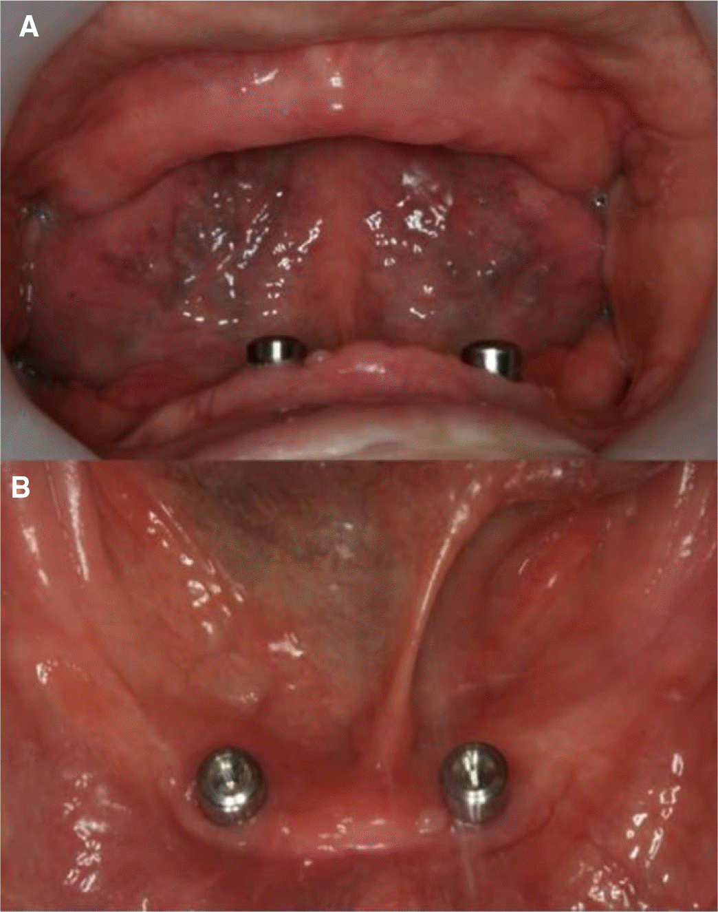
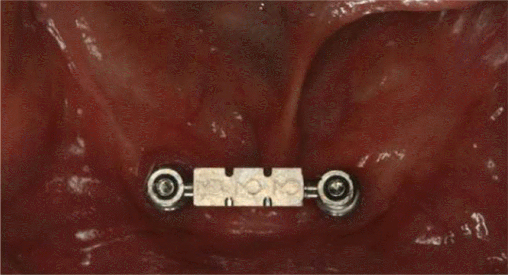
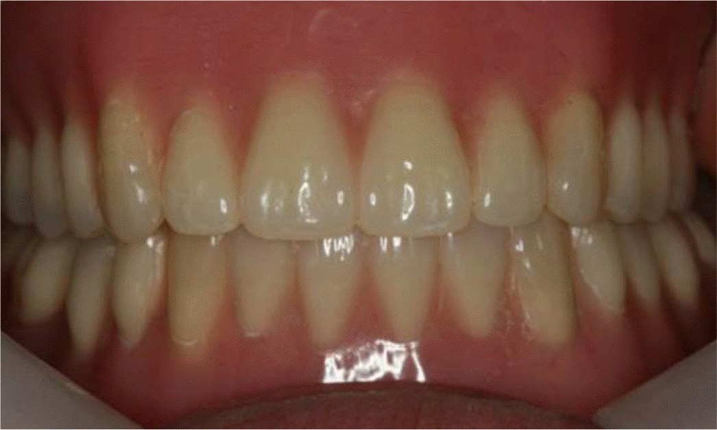
Edentulous patients with severe alveolar bone resorption have trouble with using traditional complete denture. In order to overcome these problems, implant-retained overdenture was developed. SFI-bar® system can save time and cost compared to other existing bar systems which need complicated laboratory procedures because it can be adjusted directly in a patient's mouth. A 55-year-old male, who had experienced a fractured lower old implant-retained overdenture, wanted a durable and painless denture. The fractured Locator® attachments were removed and edentulous mandible was restored with SFI-bar®. A 77-year-old female with a medical history of the Parkinson's disease and severely absorbed alveolar bone of mandible, wanted to wear a retentive mandibular denture without pain. After placing two implants in front of mental foramen, two adaptors were connected to two implants and a tube bar was connected to the adaptors. A female part fitted to the bar was attached to the new denture. These clinical reports describe two-implant-retained overdenture using the SFI-bar® system in mandibular edentulous patients. Since the patients were satisfied esthetically and functionally during 2 years' observation, we would like to report cases. (J Korean Acad Prosthodont 2016;54:41-8)
Go to : 
REFERENCES
1.Petropoulos VC., Smith W., Kousvelari E. Comparison of retention and release periods for implant overdenture attachments. Int J Oral Maxillofac Implants. 1997. 12:176–85.
2.Van Kampen F., Cune M., Van der Bilt A., Bosman F. Retention and postinsertion maintenance of bar-clip, ball and magnet attachments in mandibular implant overdenture treatment: an in vivo comparison after 3 months of function. Clin Oral Implants Res. 2003. 14:720–6.

3.Feine JS., Carlsson GE., Awad MA., Chehade A., Duncan WJ., Gizani S., Head T., Heydecke G., Lund JP., MacEntee M., Mericske-Stern R., Mojon P., Morais JA., Naert I., Payne AG., Penrod J., Stoker GT., Tawse-Smith A., Taylor TD., Thomason JM., Thomson WM., Wismeijer D. The McGill consensus statement on overdentures. Mandibular two-implant overdentures as first choice standard of care for edentulous patients. Gerodontology. 2002. 19:3–4.
4.Thomason JM., Kelly SA., Bendkowski A., Ellis JS. Two implant retained overdentures-a review of the literature supporting the McGill and York consensus statements. J Dent. 2012. 40:22–34.
5.Juan F. Martinez Lage Azorin, Gustavo Segura Andres, Joan Faus Lopez, Ruben Agustin Panadero. Rehabilitation with implant-supported overdentures in total edentulous patients: a review. J Clin Exp Dent. 2013. 5:e267–72.
6.Hong JW., Ahn SG., Leem DH., Seo JM. Immediate placement and functional loading of implants on canine with fixed partial denture for a patient having canine protected occlusion: a case report. J Adv Prosthodont. 2012. 4:52–6.
7.Stoumpis C., Kohal RJ. To splint or not to splint oral implants in the implant-supported overdenture therapy? A systematic literature review. J Oral Rehabil. 2011. 38:857–69.
8.Ha SR., Kim SH., Song SI., Hong ST., Kim GY. Implant-supported overdenture with prefabricated bar attachment system in mandibular edentulous patient. J Adv Prosthodont. 2012. 4:254–8.
Go to :
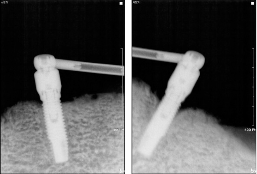
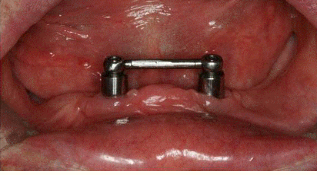
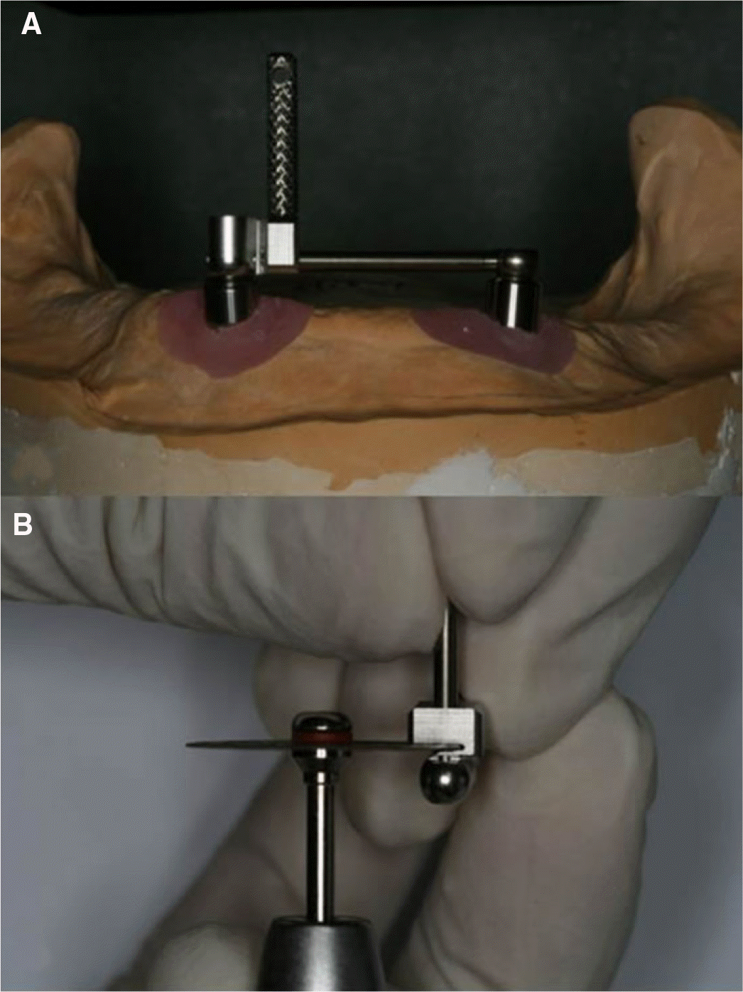 | Fig. 2.Customization of the bar. (A) After combining ball joint and tube bar on one side, the tube bar gauge was connected on the other side, (B) Tube bar was cut off at the gap of tube bar gauge with a disc. |

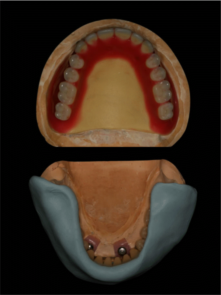
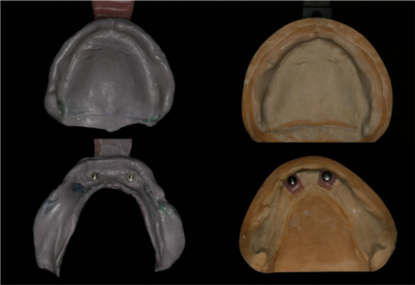 | Fig. 13.Final impression taking of maxilla and mandible with individual trays and corresponding definitive cast. |
 XML Download