

 PDF
PDF ePub
ePub Citation
 Print
Print


Abstract
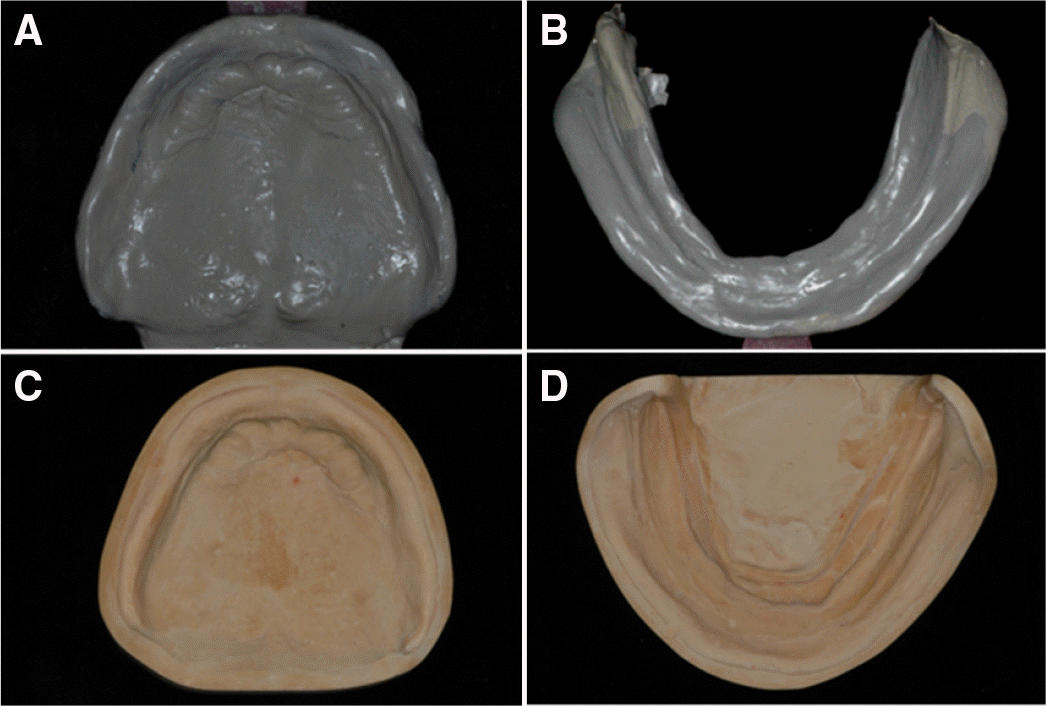
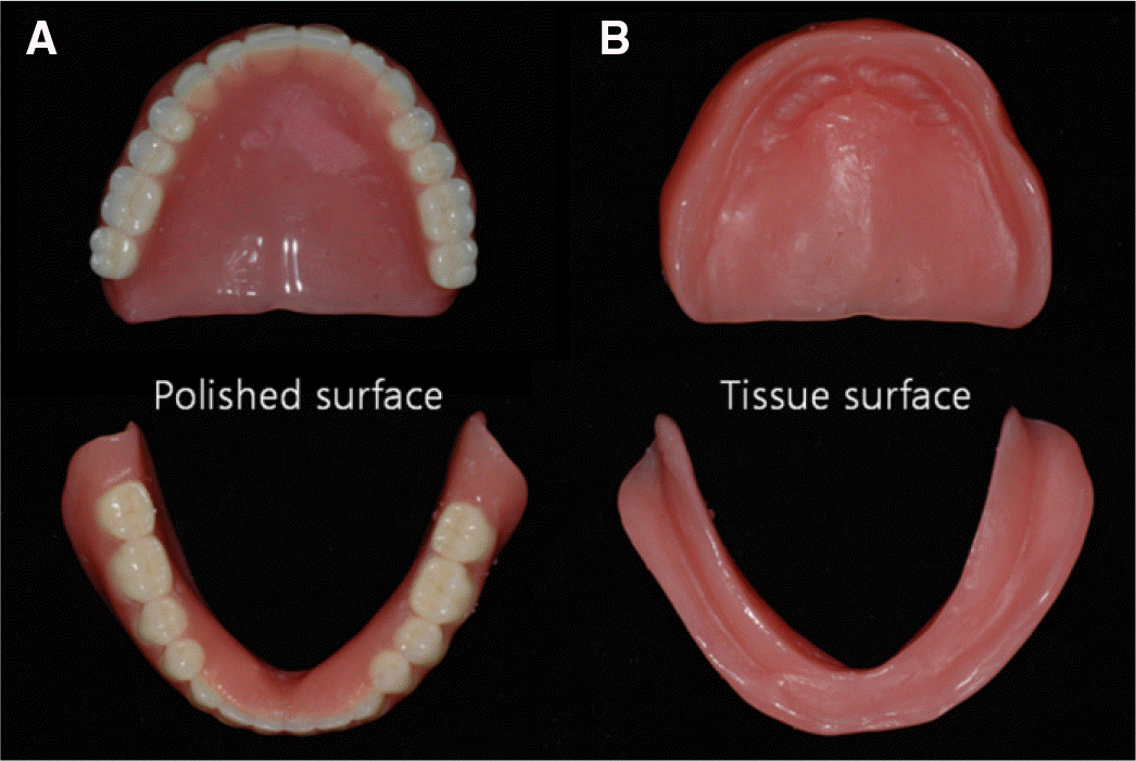
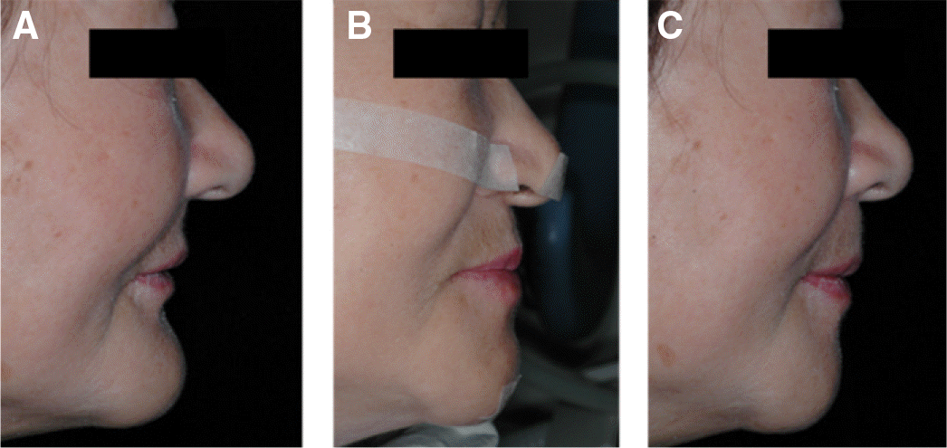
The neutral zone technique is an alternative approach for the construction of complete dentures on highly atrophic ridges with history of denture instability. This technique achieves two objectives. First, the teeth will not interfere with the normal muscle function, and second, the force exerted by the musculature against the denture is more favorable for stability and retention. In this case of a 78-years-old female patient with severely atrophic ridges who had been using unstable complete dentures, the neutral zone impression technique was used to increase the stability and the retention of dentures. The neutral zone for both arch was located with modeling compound. After the arrangement of artificial teeth within the neutral zone, the external impression was taken to determine the tissue surface. The final dentures showed enhanced stability and retention and the patient was satisfied with the new dentures with respect to functional and esthetic aspects. (J Korean Acad Prosthodont 2016;54:407-12)
Go to : 
REFERENCES
1.Fish EW. An analysis of the stabilising factors in full denture construction. Br Dent J. 1931. 52:559–70.
2.Beresin VE., Schiesser FJ. The neutral zone in complete and partial dentures. 2nd ed.St. Louis: CV Mosby;1978.
3.Fahmy FM., Kharat DU. A study of the importance of the neutral zone in complete dentures. J Prosthet Dent. 1990. 64:459–62.

4.Beresin VE., Schiesser FJ. The neutral zone in complete dentures. 1976. J Prosthet Dent. 2006. 95:93–100.
5.Beresin VE., Schiesser FJ. The neutral zone in complete dentures. J Prosthet Dent. 1976. 36:356–67.
6.Gahan MJ., Walmsley AD. The neutral zone impression revisited. Br Dent J. 2005. 198:269–72.
7.Basker RM., Harrison A., Ralph JP. A survey of patients referred to restorative dentistry clinics. Br Dent J. 1988. 164:105–8.
8.Atwood DA. Postextraction changes in the adult mandible as illustrated by microradiographs of midsagittal sections and serial cephalometric roentgenograms. J Prosthet Dent. 1963. 13:810–24.
9.Cagna DR., Massad JJ., Schiesser FJ. The neutral zone revisited: from historical concepts to modern application. J Prosthet Dent. 2009. 101:405–12.
10.Rehmann P., Zenginel M., Wostmann B. Alternative procedure to improve the stability of mandibular complete dentures: a modified neutral zone technique. Int J Prosthodont. 2012. 25:506–8.
11.Fahmi FM. The position of the neutral zone in relation to the alveolar ridge. J Prosthet Dent. 1992. 67:805–9.
Go to :

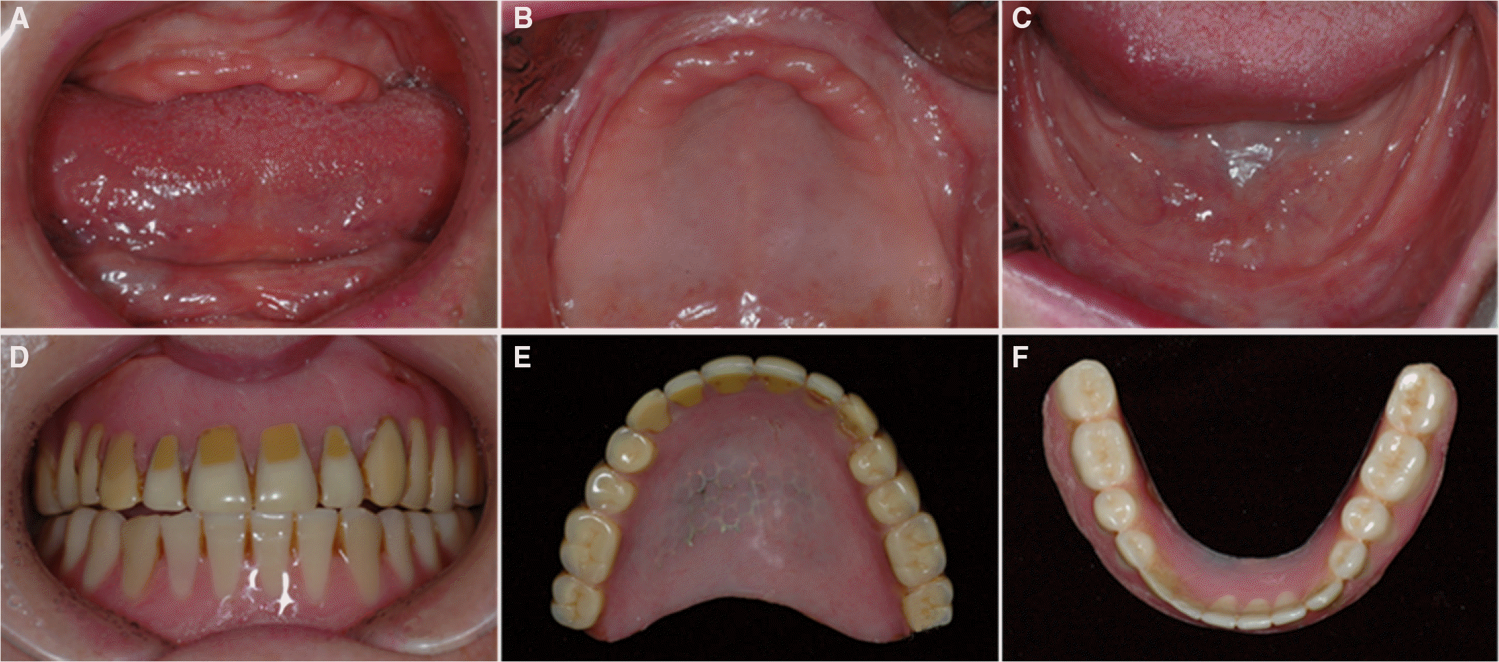 | Fig. 2.Initial intraoral photographs. Severe alveolar ridge resorption was observed on maxilla and mandible. (A) Frontal view, (B) occlusal view of maxilla, (C) occlusal view of mandible, (D) frontal view of old dentures, (E) occlusal view of upper denture, (F) occlusal view of lower denture. |
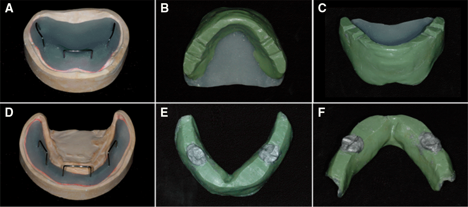 | Fig. 4.Upper acrylic resin base with retentive loops (A) and compound rim (B), (C) lower acrylic resin base with retentive loops (D) and compound rim (E), (F). |
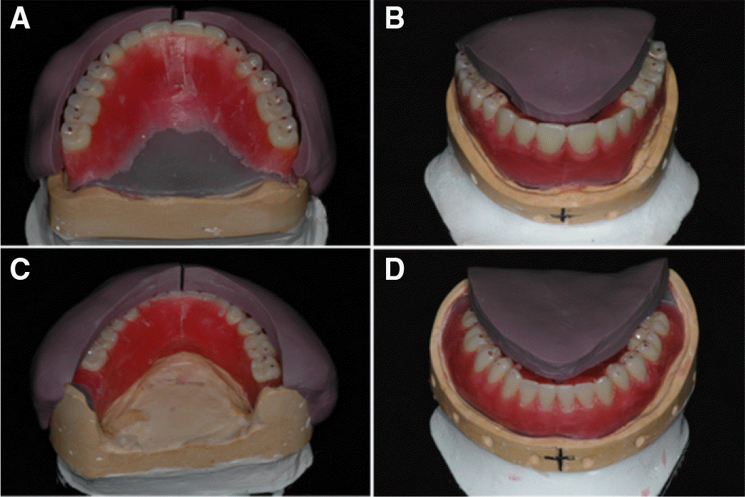 | Fig. 6.Artificial tooth arrangement according to the silicone putty index. Palatal view (A) and labial/buccal view (B) of upper denture tooth arrangement. Lingual view (C) and labial/buccal view (D) of lower denture tooth arrangement. |
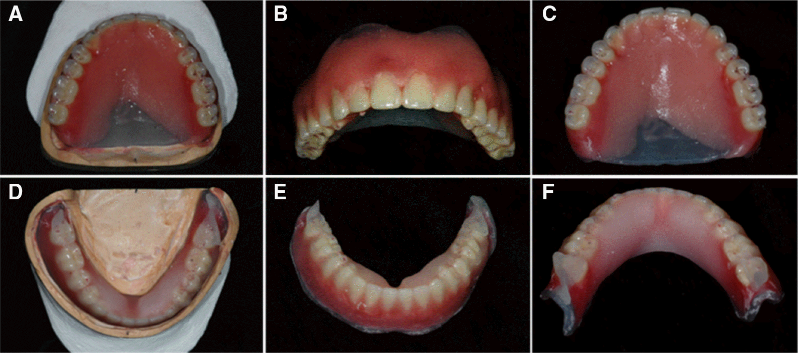 | Fig. 7.External impression on labial (A), buccal (B), palatal (C) surface of upper wax-denture using Coe-Soft (Coe-Soft, GC America, Chicago, IL, USA), External impression on labial (D), buccal (E), lingual (F) surface of lower wax-denture using Viscogel (Viscogel, Dentsply Ltd., Weybridge, UK). |
 XML Download