

 PDF
PDF ePub
ePub Citation
 Print
Print


Abstract
Purpose
The present study aims at researching the subjective satisfaction of patients who have experienced both conventional impression taking and digital impression taking to measure the possibility of wide clinical application of digital impression.
Materials and methods
The study surveyed 170 adult patients over the age of 20, between October 2015 and April 2016, who voluntarily consented to participation and who experienced both conventional impression and digital impression at five dental hospitals that use intraoral digital impression. A total of 128 surveys were used for data analysis, involving frequency analysis, multiple response frequency analysis, descriptive statistics, and contingency table analysis, with the significance level set at 0.05.
Results
Responses on the reason for taking impressions using the digital method appeared in the order of 'for implant treatment' (43.8%), 'for crown treatment' (30.5%), and 'for inlay treatment' (15.6%). Patients satisfaction was higher for digital impression taking than conventional impression taking (P<.05). As the preferred choice of impression, digital impression (60.2%) was higher than conventional impression (11.7%). Responses on the reason for choosing digital impression taking appeared in the order of 'no vomiting reflex' (35.1%), 'reliability of 3D digital scanning' (33.8%), and 'short time' (33.8%).
Go to : 
REFERENCES
1.Luthardt RG., Walter MH., Weber A., Koch R., Rudolph H. Clinical parameters influencing the accuracy of 1- and 2-stage impressions: a randomized controlled trial. Int J Prosthodont. 2008. 21:322–7.
2.Jo HY. Comparison of the accuracy with digital impression system and conventional impression technique. Graduate School of Yonsei University;2012.
3.Linke BA., Nicholls JI., Faucher RR. Distortion analysis of stone casts made from impression materials. J Prosthet Dent. 1985. 54:794–802.

4.Christensen GJ. The state of fixed prosthodontic impressions: room for improvement. J Am Dent Assoc. 2005. 136:343–6.
5.Lee GT. Accuracy of digitized dental model made from white light scanner according to different scanning method. Graduate school of Korea University;2013.
6.Kim SR. Comparison of parallel confocal laser scanning impression with conventional silicone impression regarding the marginal fitness and internal fitness of zirconia core. Graduate school of Korea University;2011.
7.Kim JH. In vitro study on accuracy and reliability of dental model based on the digital intraoral impression technique. Graduate school of Korea University;2012.
8.Pyo SW., Park YB., Kim JH., Moon HS., Lee KW. Maxillary anterior all ceramic restoration using digital impression and CAD/CAM. J Korean Acad Prosthodont. 2011. 49:263–9.
9.Hong YS., Park EJ., Kim SJ., Kim MR., Heo SJ., Park JM. Customized abutment and screw-type implant prostheses after cementation based on the digital intraoral impression technique. J Korean Acad Prosthodont. 2012. 50:67–73.
10.Christensen GJ. Impressions are changing: deciding on conventional, digital or digital plus in-office milling. J Am Dent Assoc. 2009. 140:1301–4.
11.Duret F., Preston JD. CAD/CAM imaging in dentistry. Curr Opin Dent. 1991. 1:150–4.
12.Kim JH. Evaluation of the intraoral scanning technique and the fitness of all-ceramic restoration in the digital workflow. Graduate school of Korea University;2014.
13.Choi JH., Lim YJ., Lee WJ., Han JS., Lee SP. Review of recent developments for intraoral scanners. J Dent Rehabil Appl Sci. 2015. 31:112–25.
14.Kim RW., Jang GW., Heo YR., Son MK. Understanding and application of digital impression in dentistry. Korean J Dent Mater. 2014. 41:253–61.
15.An S., Kim S., Choi H., Lee JH., Moon HS. Evaluating the marginal fit of zirconia copings with digital impressions with an intraoral digital scanner. J Prosthet Dent. 2014. 112:1171–5.
16.Lee WS., Kim WC., Kim HY., Kim WT., Kim JH. Evaluation of different approaches for using a laser scanner in digitization of dental impressions. J Adv Prosthodont. 2014. 6:22–9.
17.Abdel-Azim T., Zandinejad A., Elathamna E., Lin W., Morton D. The influence of digital fabrication options on the accuracy of dental implant-based single units and complete-arch frameworks. Int J Oral Maxillofac Implants. 2014. 29:1281–8.
18.Papaspyridakos P., Chen CJ., Gallucci GO., Doukoudakis A., Weber HP., Chronopoulos V. Accuracy of implant impressions for partially and completely edentulous patients: a systematic review. Int J Oral Maxillofac Implants. 2014. 29:836–45.
19.Anh JW., Park JM., Chun YS., Kim M., Kim M. A comparison of the precision of three-dimensional images acquired by 2 digital intraoral scanners: effects of tooth irregularity and scanning direction. Korean J Orthod. 2016. 46:3–12.
20.Kim J., Park JM., Kim M., Heo SJ., Shin IH., Kim M. Comparison of experience curves between two 3-dimensional intraoral scanners. J Prosthet Dent. 2016. 116:221–30.
21.Bae JW. A study on the color reproduction of digital intraoral scanner. Graduate school of Ewha Womans University;2014.
22.Bindl A., Mörmann WH. Clinical and SEM evaluation of all-ceramic chair-side CAD/CAM-generated partial crowns. Eur J Oral Sci. 2003. 111:163–9.
23.Seelbach P., Brueckel C., Wöstmann B. Accuracy of digital and conventional impression techniques and workflow. Clin Oral Investig. 2013. 17:1759–64.
24.Stevens DR., Flores-Mir C., Nebbe B., Raboud DW., Heo G., Major PW. Validity, reliability, and reproducibility of plaster vs digital study models: comparison of peer assessment rating and Bolton analysis and their constituent measurements. Am J Orthod Dentofacial Orthop. 2006. 129:794–803.
25.van der Meer WJ., Andriessen FS., Wismeijer D., Ren Y. Application of intraoral dental scanners in the digital workflow of implantology. PLoS One. 2012. 7:e43312.
26.Souza RO., özcan M., Pavanelli CA., Buso L., Lombardo GH., Michida SM., Mesquita AM., Bottino MA. Marginal and internal discrepancies related to margin design of ceramic crowns fabricated by a CAD/CAM system. J Prosthodont. 2012. 21:94–100.
27.Nedelcu RG., Persson AS. Scanning accuracy and precision in 4 intraoral scanners: an in vitro comparison based on 3-dimensional analysis. J Prosthet Dent. 2014. 112:1461–71.
28.Lee SJ., Gallucci GO. Digital vs. conventional implant impressions: efficiency outcomes. Clin Oral Implants Res. 2013. 24:111–5.
29.Park HR., Park JM., Chun YS., Lee KN., Kim M. Changes in views on digital intraoral scanners among dental hygienists after training in digital impression taking. BMC Oral Health. 2015. 15:151.
30.Kim KM. Likert scale. Korean J Fam Med. 2011. 32:1–2.
31.Wismeijer D., Mans R., van Genuchten M., Reijers HA. Patients' preferences when comparing analogue implant impressions using a polyether impression material versus digital impressions (Intraoral Scan) of dental implants. Clin Oral Implants Res. 2014. 25:1113–8.
32.Christensen GJ. Will digital impressions eliminate the current problems with conventional impressions? J Am Dent Assoc. 2008. 139:761–3.
33.Lee SJ., Macarthur RX 4th., Gallucci GO. An evaluation of student and clinician perception of digital and conventional implant impressions. J Prosthet Dent. 2013. 110:420–3.
Go to :
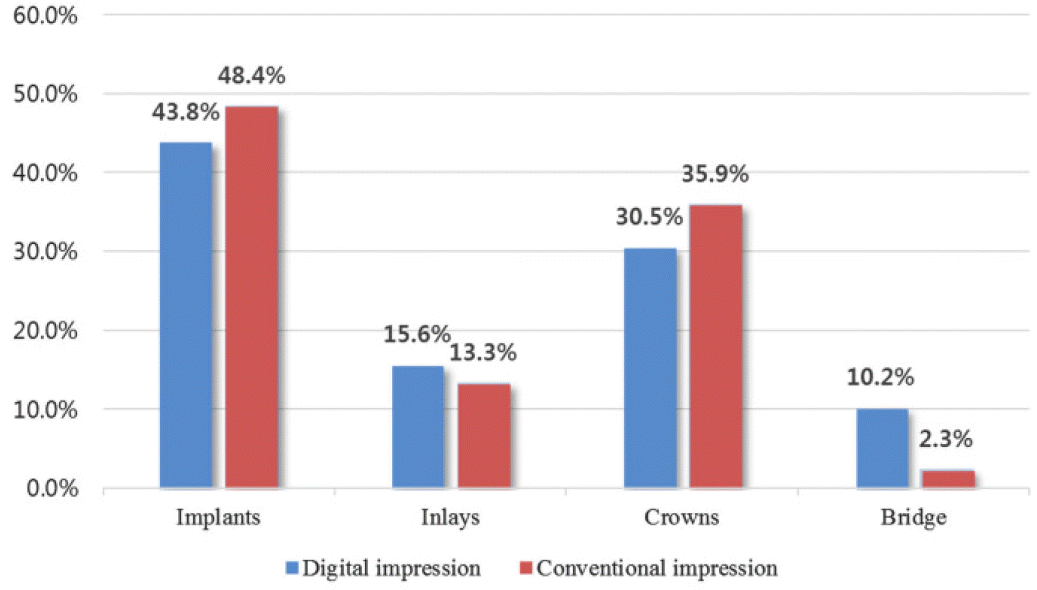 | Fig. 2.Clinical procedures with digital and conventional impression. The most frequent treatments using digital and conventional impressions were for the implant restorations. |
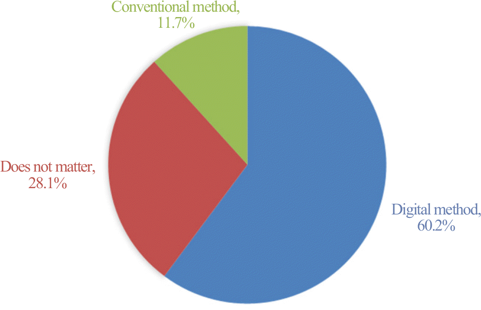 | Fig. 3.Preferred method for dental impression. The patients preferred digital impression to conventional method. |
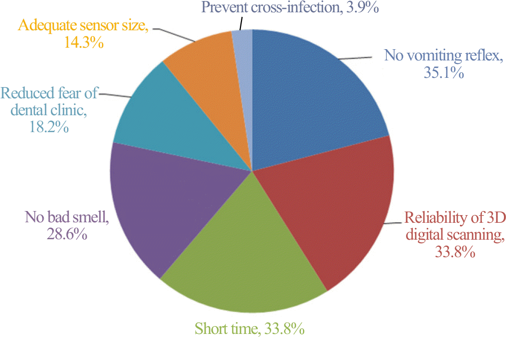
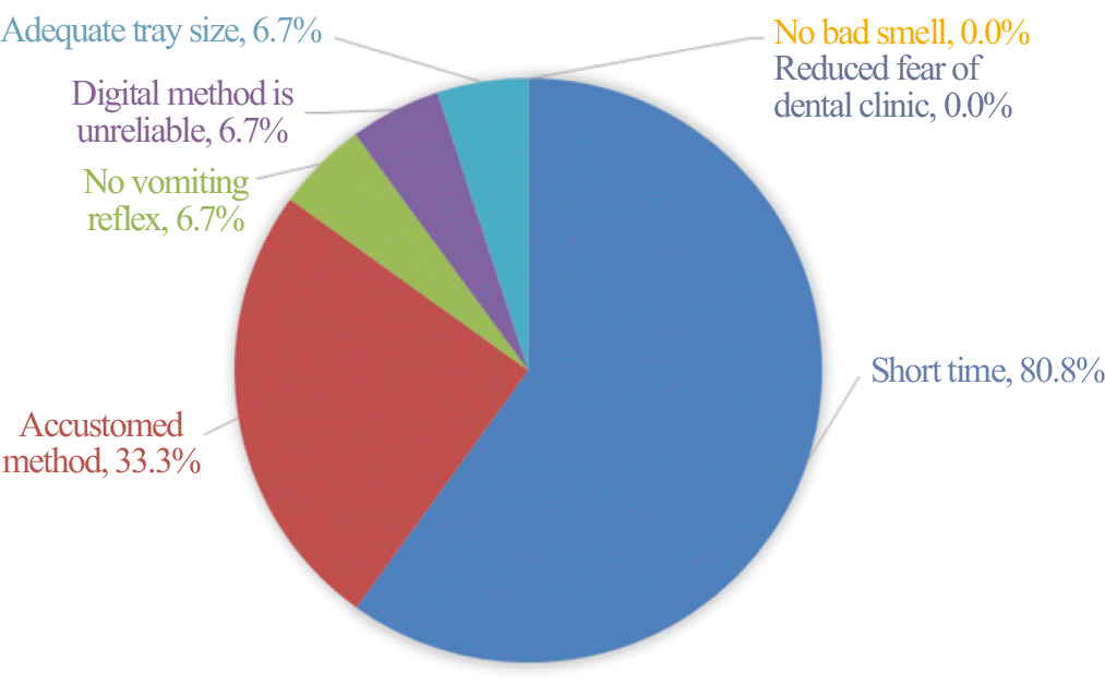
Table 1.
Patients' satisfaction for digital and conventional impression (Likert scale)
 XML Download