Abstract
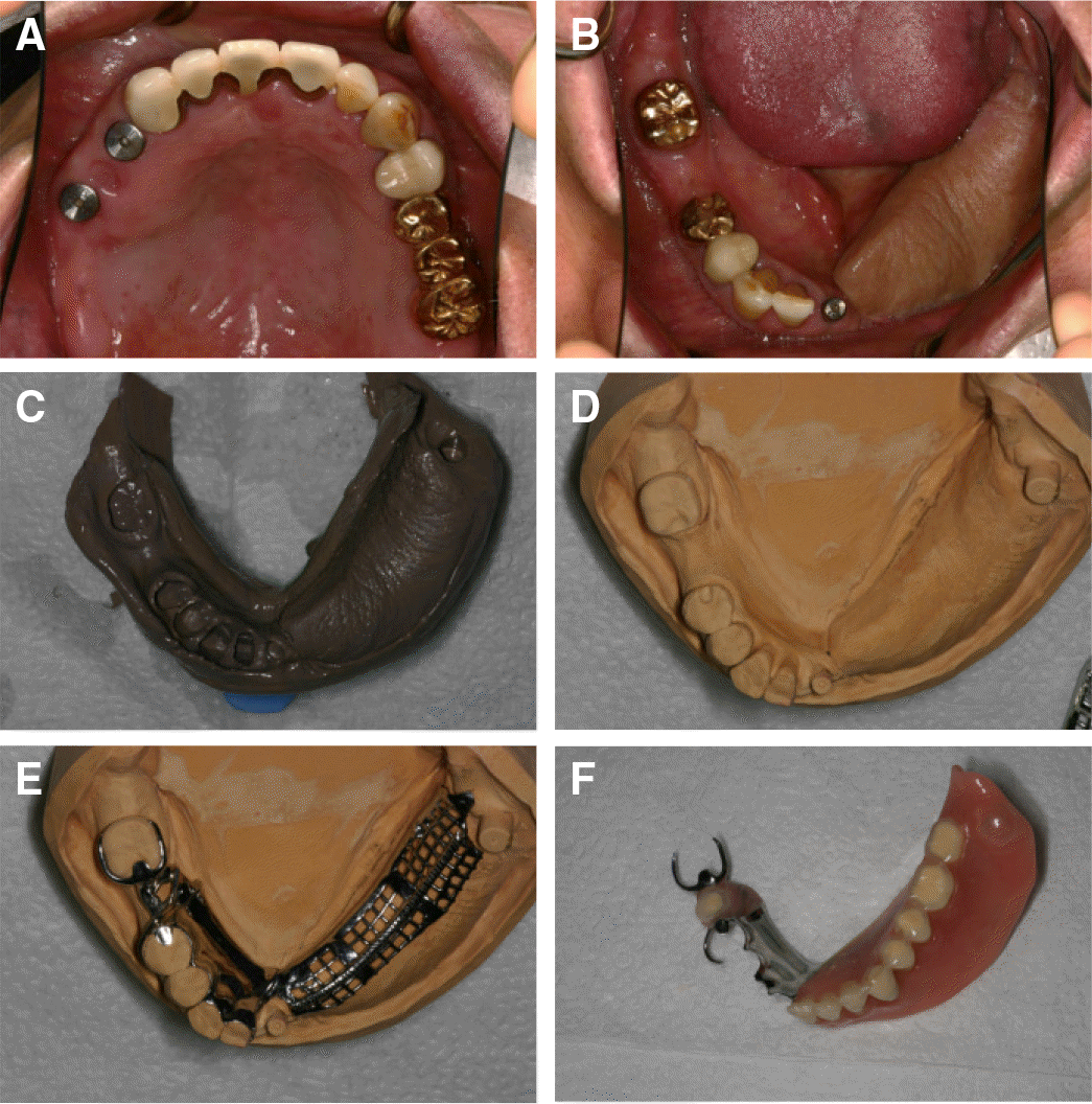
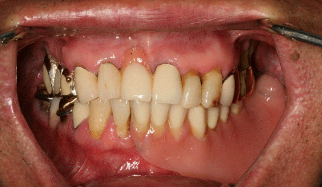
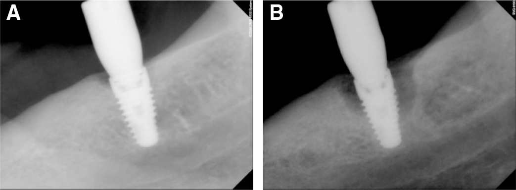
Mandible defects could be caused by congenital malformations, trauma, osteomyelitis, tumor resection. If large areas are included for reconstruction, those are primarily due to tumor resection defects. The large jaw defect results in a problem about mastication, swallowing, occlusion and phonetics, and poor esthetics causes a lot of inconvenience in daily life. It is almost impossible to be a part underwent mandibular resection completely reproduced, should be rebuilt artificially. This case is of a patient who was diagnosed with squamous cell carcinoma pT1N0M0, stage I in February 2004 and received surgery (combined mandibulectomy and neck dissection operation (COMMANDO) in oromaxillofacial surgery) in March 2004, by implant assisted removable partial denture. We could obtain good retention and stability through sufficient coverage and implant holding. Follow up period was about four years. Mandibular left third molar regions have been observed to have resorption of surrounding bone, and periodic check-ups are necessary conditions. (J Korean Acad Prosthodont 2016;54:280-5)
Go to : 
REFERENCES
1.Garrett N., Roumanas ED., Blackwell KE., Freymiller E., Abemayor E., Wong WK., Gerratt B., Berke G., Beumer J 3rd., Kapur KK. Efficacy of conventional and implant-supported mandibular resection prostheses: study overview and treatment outcomes. J Prosthet Dent. 2006. 96:13–24.

2.Teoh KH., Huryn JM., Patel S., Halpern J., Tunick S., Wong HB., Zlotolow IM. Implant prosthodontic rehabilitation of fibula free-flap reconstructed mandibles: a Memorial Sloan-Kettering Cancer Center review of prognostic factors and implant outcomes. Int J Oral Maxillofac Implants. 2005. 20:738–46.
3.Hupp JR. Tucker MR, Ellis E. Contemporary oral and maxillofacial surgery. 5th ed.St. Louis: CV Mosby;2005. p. 605–16.
4.Raoul G., Ruhin B., Briki S., Lauwers L., Haurou Patou G., Capet JP., Maes JM., Ferri J. Microsurgical reconstruction of the jaw with fibular grafts and implants. J Craniofac Surg. 2009. 20:2105–17.

5.Hotz G. Reconstruction of mandibular discontinuity defects with delayed nonvascularized free iliac crest bone grafts and endosseous implants: a clinical report. J Prosthet Dent. 1996. 76:350–5.

6.Disa JJ., Cordeiro PG. Mandible reconstruction with microvascular surgery. Semin Surg Oncol. 2000. 19:226–34.

7.Pogrel MA., Podlesh S., Anthony JP., Alexander J. A comparison of vascularized and nonvascularized bone grafts for reconstruction of mandibular continuity defects. J Oral Maxillofac Surg. 1997. 55:1200–6.

8.Leong EW., Cheng AC., Tee-Khin N., Wee AG. Management of acquired mandibular defects-prosthodontic considerations. Singapore Dent J. 2006. 28:22–33.
9.Baker A., McMahon J., Parmar S. Immediate reconstruction of continuity defects of the mandible after tumor surgery. J Oral Maxillofac Surg. 2001. 59:1333–9.
10.Adell R., Svensson B., Bågenholm T. Dental rehabilitation in 101 primarily reconstructed jaws after segmental resections-possibilities and problems. An 18-year study. J Craniomaxillofac Surg. 2008. 36:395–402.
11.Salinas TJ., Desa VP., Katsnelson A., Miloro M. Clinical evaluation of implants in radiated fibula flaps. J Oral Maxillofac Surg. 2010. 68:524–9.

Go to : 
 | Fig. 1.Panoramic radiograph at first visit and after marginal mandibulectomy. (A) radiograph at first visit on February 11, 2004, (B) radiograph after surgery on March 24, 2004. |
 | Fig. 2.Panoramic radiograph at bone graft and after removal of necrotic bone. (A) radiograph after iliac bone graft surgery on September 12, 2008, (B) radiograph after surgical removal of necrotic graft material. |


 PDF
PDF ePub
ePub Citation
 Print
Print


 XML Download