

 PDF
PDF ePub
ePub Citation
 Print
Print


Abstract
Implant has been an effective treatment option for the patients with oligodontia. However, it still remains unclear when the implant should be placed. Skeletal growth that can appear even after the growth period can lead to infraocclusion of the implant which can cause functional or esthetic complications. In order to minimize these problems, definitive restorations should be placed after the functional and esthetic rehabilitation is achieved through the use of provisional restorations. Definitive restorations made with monolithic zirconia were created by replicating provisional restorations by using the latest CAD/CAM technology. These definitive restorations were delivered to the patient and clinical observation after the treatment showed satisfactory result. (J Korean Acad Prosthodont 2016;54:28-34)
Go to : 
REFERENCES
1.Thilander B., Odman J., Gröndahl K., Friberg B. Osseointegrated implants in adolescents. An alternative in replacing missing teeth? Eur J Orthod. 1994. 16:84–95.
2.Oesterle LJ., Cronin RJ Jr., Ranly DM. Maxillary implants and the growing patient. Int J Oral Maxillofac Implants. 1993. 8:377–87.
3.Cronin RJ Jr., Oesterle LJ., Ranly DM. Mandibular implants and the growing patient. Int J Oral Maxillofac Implants. 1994. 9:55–62.
4.Fradeani M. Esthetic rehabilitation in fixed prosthodontics vol. I esthetic analysis: a systemic approach to prosthetic treatment. Quintessence publishing co., Ltd.;2004. p. 72–81.
5.Turner KA., Missirlian DM. Restoration of the extremely worn dentition. J Prosthet Dent. 1984. 52:467–74.

6.Kahler W. The cracked tooth conundrum: terminology, classification, diagnosis, and management. Am J Dent. 2008. 21:275–82.
7.Kwon KR., Woo YH., Choi DG. The study of relationship between sagittal condylar guide angle and incisal guide angle during mandibular protrusion in normal Korean. J Korean Acad Prosthodont. 1989. 27:11–36.
8.Rilo B., da Silva JL., Mora MJ., Santana U. Guidelines for occlusion strategy in implant-borne prostheses. A review. Int Dent J. 2008. 58:139–45.
9.Ash MM. Wheeler' s dental anatomy. 7th ed.Philadelphia: Saunders;1993. p. 150–93.
10.Hildebrand CN. Crown lengthening for optimum restorative success. Compend Contin Educ Dent. 2003. 24:620–2. 624–9.
11.Dawson PE. Determining the determinants of occlusion. Int J Periodontics Restorative Dent. 1983. 3:8–21.
12.Misch CE. Dental Implant Prosthetics,. 1st ed.New York: Elsevier;2003. p. 414–51.
13.Behrents RG. A treatise on the continuum of growth in the aging craniofacial skeleton. Michigan: UMI Dissertations Publishing;1984. p. 1–763.
Go to :
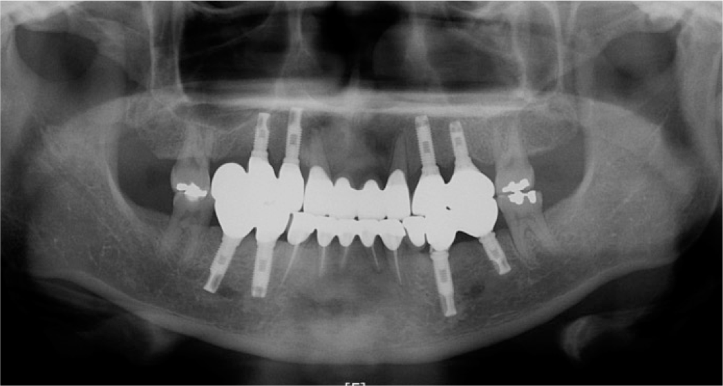
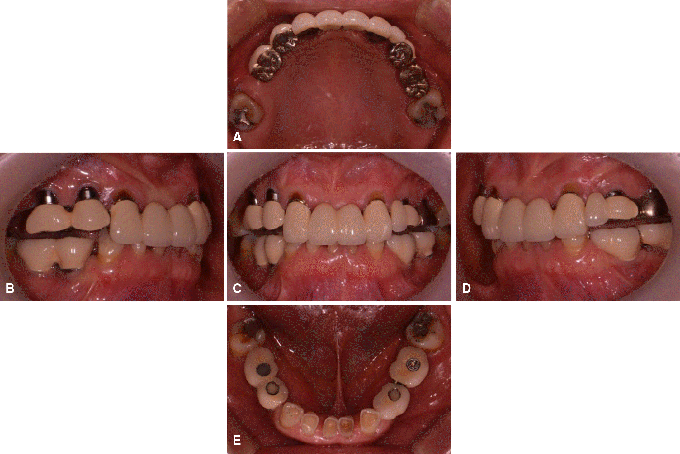 | Fig. 2.Pre-operative intraoral view showing implant infraocclusion and severe attrition on lower anterior teeth. (A) Occlusal view of maxilla, (B) Lateral view (right side), (C) Frontal view, (D) Lateral view (left side), (E) Occlusal view of mandible. |
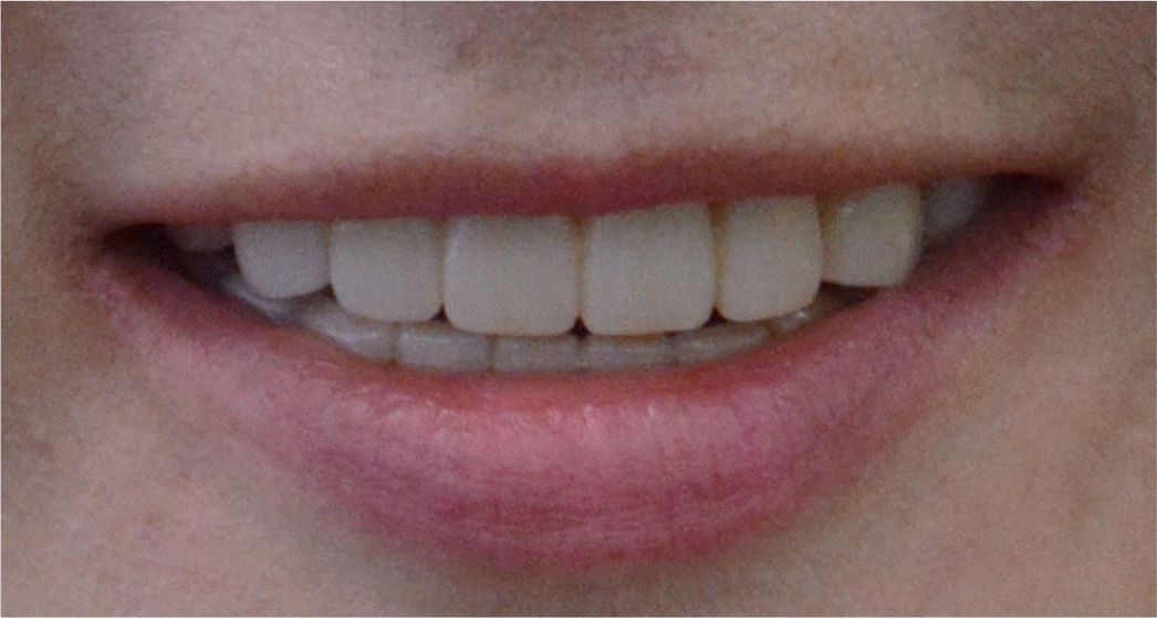
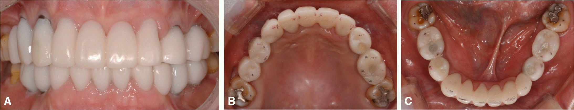 | Fig. 5.Provisional restoration. (A) Frontal view, (B, C) Occlusal view of maxilla and mandible (with occlusal dots). |
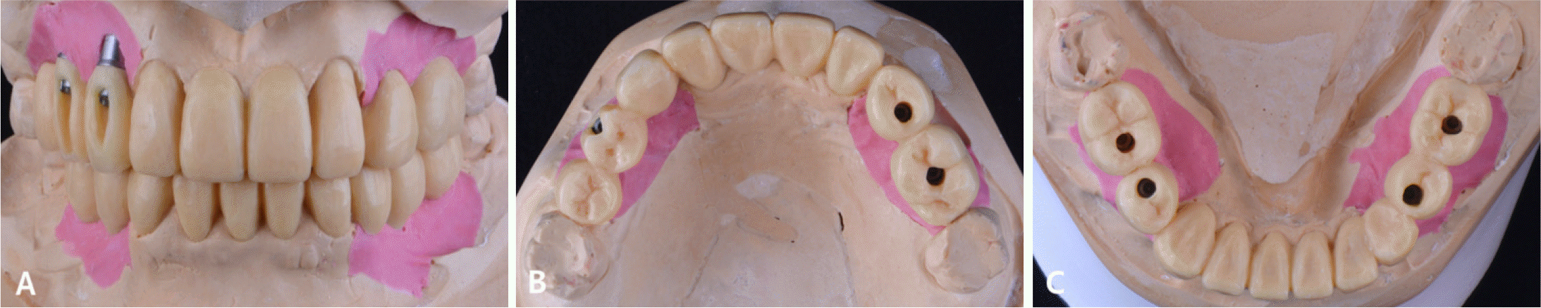
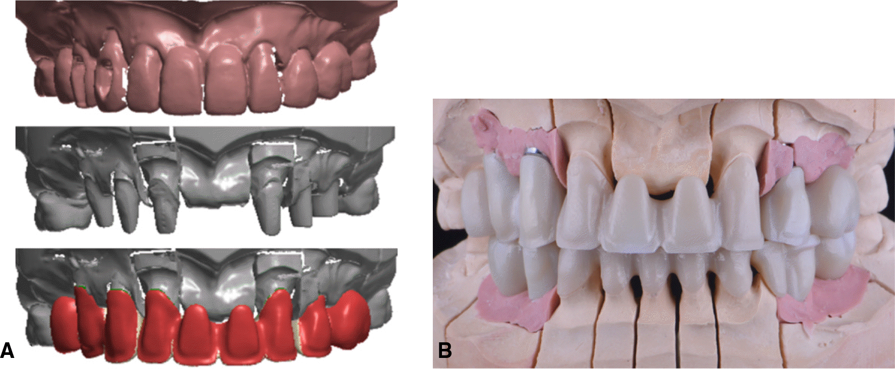 | Fig. 6.CAD/CAM procedure. (A) Maxillary superimposition and cutback, (B) Frontal view of maxilla and mandible (zirconia framework). |
 XML Download