Abstract
Severe tooth wear may lead to pathological changes of pulp, imbalance in occlusion as well as functional and esthetic problems. In this case, 34-year-old male came to the hospital because of generally worn dentition due to attrition and erosion. After evaluation, a full mouth restoration with elevation of the vertical dimension of occlusion was planned. After occlusion was stabilized by an occlusal stabilization appliance, centric relation position was recorded and subsequent provisional restorations were fabricated. After evaluation, a CAD-CAM (computer aided design-computer aided manufacturing) prosthetic restoration was carried out using monolithic zirconia. After 12 months of follow up observation, the patient was satisfied with function and esthetic appearance. (J Korean Acad Prosthodont 2016;54:273-9)
Go to : 
REFERENCES
3.Khan F., Young WG., Daley TJ. Dental erosion and bruxism. A tooth wear analysis from south east Queensland. Aust Dent J. 1998. 43:117–27.

4.Murphy T. Compensatory mechanisms in face height adjustment to functional tooth attrition. Aust Dent J. 2009. 4:312–23.
5.Berry DC., Poole DF. Attrition: possible mechanisms of compensation. J Oral Rehabil. 1976. 3:201–6.

7.Turner KA., Missirlian DM. Restoration of the extremely worn dentition. J Prosthet Dent. 1984. 52:467–74.

8.Ibbetson RJ., Setchell DJ. Treatment of the worn dentition: 2. Dent Update. 1989. 16:300–2. 305–7.
10.Khan F., Young WG., Law V., Priest J., Daley TJ. Cupped lesions of early onset dental erosion in young southeast Queensland adults. Aust Dent J. 2001. 46:100–7.

12.Brunton PA., McCord JF. An analysis of nasolabial angles and their relevance to tooth position in the edentulous patient. Eur J Prosthodont Restor Dent. 1993. 2:53–6.
13.Dawson PE. Functional occlusion: From TMJ to smile design. Elsevier inc.;2007. p. 430–52.
14.Abduo J., Lyons K. Clinical considerations for increasing occlusal vertical dimension: a review. Aust Dent J. 2012. 57:2–10.

15.Tinschert J., Natt G., Mautsch W., Augthun M., Spiekermann H. Fracture resistance of lithium disilicate-, alumina-, and zirconia-based three-unit fixed partial dentures: a laboratory study. Int J Prosthodont. 2001. 14:231–8.
16.Christel P., Meunier A., Heller M., Torre JP., Peille CN. Mechanical properties and short-term in-vivo evaluation of yttrium-oxide-partially-stabilized zirconia. J Biomed Mater Res. 1989. 23:45–61.
17.Miyazaki T., Nakamura T., Matsumura H., Ban S., Kobayashi T. Current status of zirconia restoration. J Prosthodont Res. 2013. 57:236–61.

18.Baldissara P., Llukacej A., Ciocca L., Valandro FL., Scotti R. Translucency of zirconia copings made with different CAD/CAM systems. J Prosthet Dent. 2010. 104:6–12.

Go to : 
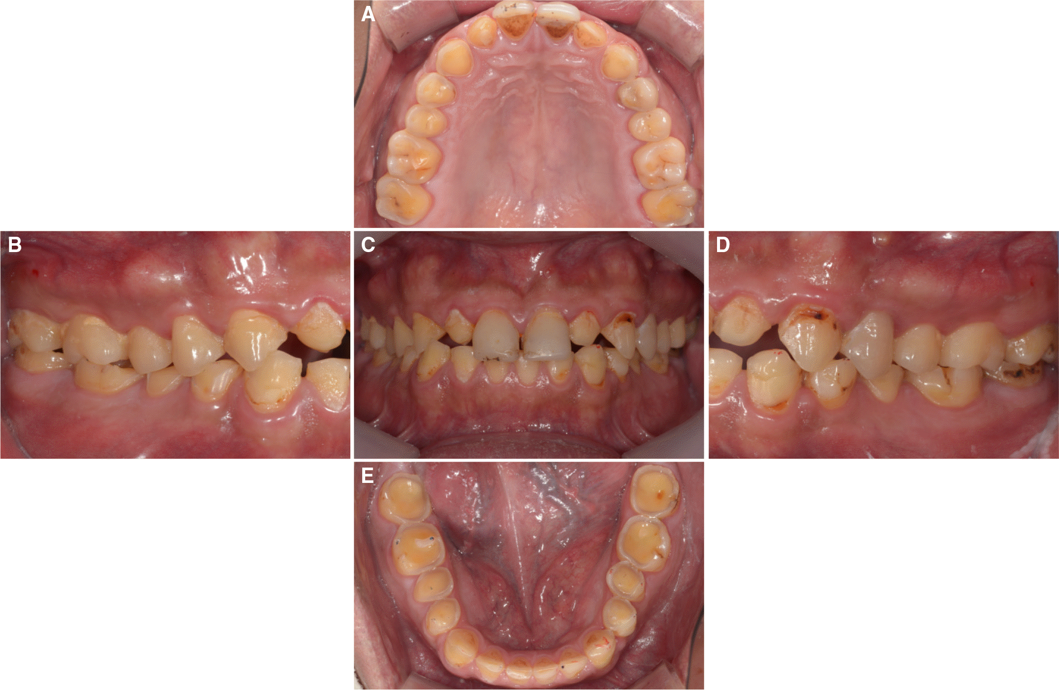 | Fig. 1.Initial intra-oral photographs. (A) Maxillary occlusal view, (B) Right lateral view, (C) Frontal view, (D) Left lateral view, (E) Mandibular occlusal view. |
 | Fig. 3.Initial TMJ series. (A) Right side on closed state, (B) Right side on opened state, (C) Left side on closed state, (D) Left side on opened state. |
 | Fig. 5.Diagnostic wax up. (A) Frontal view, (B) Maxillary occlusal view, (C) Mandibular occlusal view. |
 | Fig. 6.Provisional restoration delivery. (A) Frontal view, (B) Maxillary occlusal view, (C) Mandibular occlusal view. |
 | Fig. 7.(A) Full contour wax up on master cast. (B) Monolithic zirconia crown on master cast. |


 PDF
PDF ePub
ePub Citation
 Print
Print



 XML Download