Abstract
In edentulous mandible, implant supported overdenture was considered as a first treatment option. Konus type attachment supplies rigid support and cross arch stabilization so that more favorable force transmission and distribution can be attained. In the dentistry, computer aided design-computer aided manufacturing (CAD-CAM) system makes it possible to fabricate restorations with high precision and effectiveness. Recently, Palladium-silver (Pd-Ag) alloy which is millable has been developed. This article presents that application of CAD-CAM Konus type attachment can be provide satisfactory stability and function on four-implant supported mandibular overdenture. (J Korean Acad Prosthodont 2016;54:259-66)
Go to : 
REFERENCES
1.Zarb GA., Bolender CL., Eckert S., Fenton A., Jacob R., Mericske-Stern R. Prosthodontic treatment for edentulous patients. Complete dentures and implant supported prostheses. 12th ed.St. Louis: Mosby;2004.
2.Feine JS., Carlsson GE., Awad MA., Chehade A., Duncan WJ., Gizani S., Head T., Lund JP., MacEntee M., Mericske-Stern R., Mojon P., Morais J., Naert I., Payne AG., Penrod J., Stoker GT., Tawse-Smith A., Taylor TD., Thomason JM., Thomson WM., Wismeijer D. The McGill consensus statement on overdentures. Mandibular two-implant overdentures as first choice standard of care for edentulous patients. Montreal, Quebec, May 24-25, 2002. Int J Oral Maxillofac Implants. 2002. 17:601–2.
3.Thomason JM., Feine J., Exley C., Moynihan P., Müller F., Naert I., Ellis JS., Barclay C., Butterworth C., Scott B., Lynch C., Stewardson D., Smith P., Welfare R., Hyde P., McAndrew R., Fenlon M., Barclay S., Barker D. Mandibular two implant-supported overdentures as the first choice standard of care for edentulous patients—the York Consensus Statement. Br Dent J. 2009. 207:185–6.
4.Besimo C., Kempf B. In vitro investigation of various attachments for overdentures on osseointegrated implants. J Oral Rehabil. 1995. 22:691–8.

5.Meijer HJ., Kuiper JH., Starmans FJ., Bosman F. Stress distribution around dental implants: influence of superstructure, length of implants, and height of mandible. J Prosthet Dent. 1992. 68:96–102.

6.Breitman JB., Nakamura S., Freedman AL., Yalisove IL. Telescopic retainers: an old or new solution? A second chance to have normal dental function. J Prosthodont. 2012. 21:79–83.

7.Kim IS., Kang DW., Kim BO., Yoo KH. Design and fabrication of inner Konus crown using three dimensional computer graphics. J Korean Acad Prosthodont. 2000. 38:544–51.
8.Shimakura M., Nagata T., Takeuchi M., Nemoto T. Retentive force of pure titanium konus telescope crowns fabricated using CAD/CAM system. Dent Mater J. 2008. 27:211–5.
9.Miyazaki T., Hotta Y., Kunii J., Kuriyama S., Tamaki Y. A review of dental CAD/CAM: current status and future perspectives from 20 years of experience. Dent Mater J. 2009. 28:44–56.

10.Goodacre CJ. Palladium-silver alloys: a review of the literature. J Prosthet Dent. 1989. 62:34–7.

11.Huget EF., Civjan S. Status report on palladium-silver-based crown and bridge alloys. J Am Dent Assoc. 1974. 89:383–5.

12.Bergler M., Holst S., Blatz MB., Eitner S., Wichmann M. CAD/CAM and telescopic technology: design options for implant-supported overdentures. Eur J Esthet Dent. 2008. 3:66–88.
13.Beuer F., Schweiger J., Edelhoff D. Digital dentistry: an overview of recent developments for CAD/CAM generated restorations. Br Dent J. 2008. 204:505–11.

14.Kansu G., Aydin AK. Evaluation of the biocompatibility of various dental alloys: Part I-Toxic potentials. Eur J Prosthodont Restor Dent. 1996. 4:129–36.
15.Ceragemgiosys. [cited 2015 February]. Available from. http://www.ceragembiosys.com.
Go to : 
 | Fig. 2.Clinical photo after extraction and diagnostic mounting. (A) Maxillary occlusal view, (B) Mandibular occlusal view, (C) Evaluation of interocclusal space. |
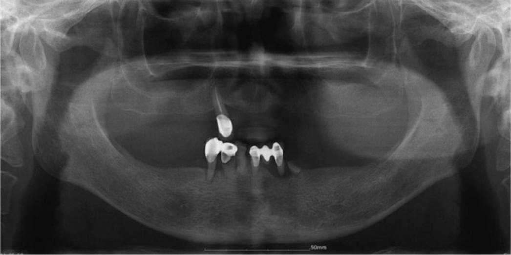
 | Fig. 3.Implantation. (A) Implantation and guided bone regeneration, (B) Panoramic radiograph after implantation. |
 | Fig. 4.2nd surgery and free gingiva graft. (A) Maxillary occlusal view, (B) Mandibular occlusal view. |
 | Fig. 5.Final impression on mandible. (A) Individual tray, (B) Connection of pick-up type impression copings, (C) Final impression taking. |
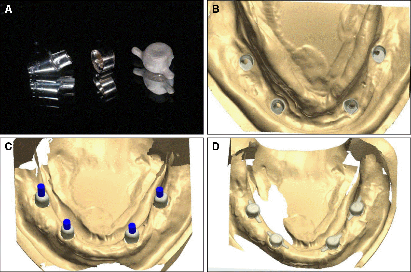
 | Fig. 6.Konus type attachment was manufactured with CAD-CAM system. (A) Implant abutment, inner crown and outer crown, (B) Implant abutment design, (C) Inner crown design, (D) Outer crown design. |
 | Fig. 7.CAD-CAM Konus type attachment check and try-in. (A) Attachment space re-evaluation with putty index, (B) Implant abutment and inner crown connection to the implant. |
 | Fig. 8.Metal framework check and pick up impression. (A) Pick-up impression taking of outer crown, (B) Outer crown and metal framework connection using laser welding, (C) Metal framework try-in. |
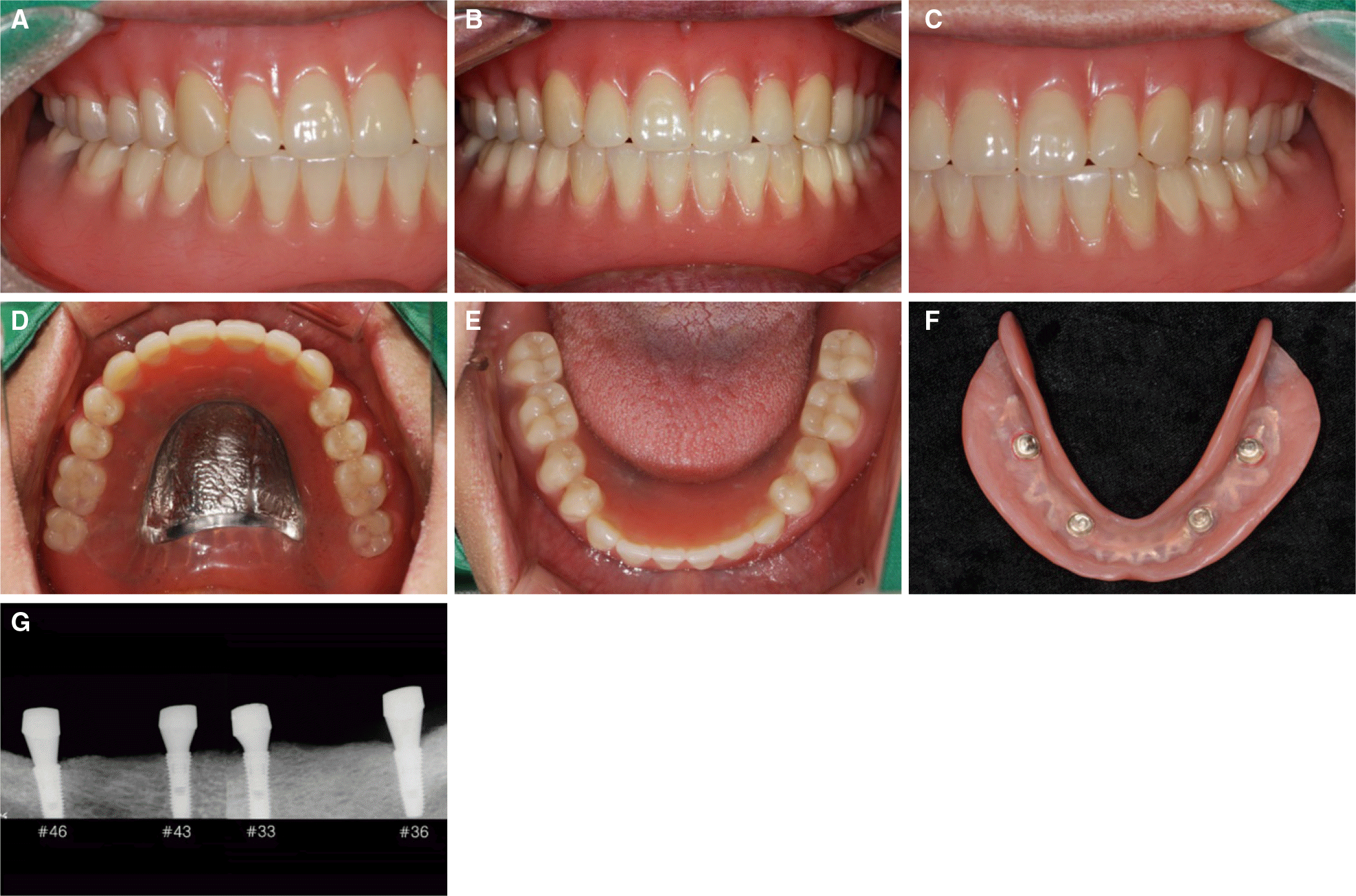
 | Fig. 9.Esthetics and function were restored with the definitive prosthesis. (A) Right lateral view, (B) Frontal view, (C) Left lateral view, (D) Maxillary occlusal view, (E) Mandibular occlusal view, (F) Inner surface of mandibular implant overdenture, (G) Periapical radiographic view after 6 months in use. |


 PDF
PDF ePub
ePub Citation
 Print
Print


 XML Download