

 PDF
PDF ePub
ePub Citation
 Print
Print


Abstract
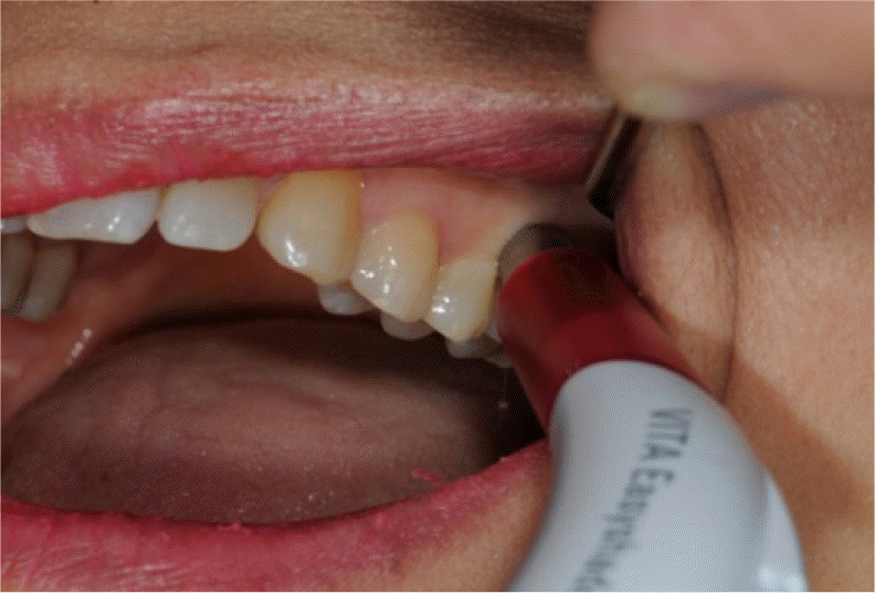
The development of translucent zirconia enabled clinicians to choose a monolithic zirconia crown as one treatment modality in the posterior dentition. Careful occlusal adjustments are recommended for monolithic zirconia crowns because grinding zirconia inevitably causes phase transformation, which may deteriorate mechanical properties. intraoral scanners enable the clinician to scan and superimpose a complete tooth structure before preparation onto the prepared abutment. This technique helps to reproduce the original tooth form and occlusion of the patient. In this case report, prostheses were fabricated for patients with cracked or fractured tooth by applying intraoral scanner, Computer aided design-computer aided manufacturing (CAD-CAM) and monolithic zirconia crown to reproduce the occlusion of original tooth and to minimize occlusal adjustment. The clinical results were satisfactory in both esthetic and functional aspects. (J Korean Acad Prosthodont 2016;54:253-8)
Go to : 
REFERENCES
1.Fukui K., Kaneuji A., Sugimori T., Ichiseki T., Kitamura K., Matsumoto T. Wear comparison between a highly cross-linked polyethylene and conventional polyethylene against a zirconia femoral head: minimum 5-year follow-up. J Arthroplasty. 2011. 26:45–9.
2.Oilo M., Kvam K., Gjerdet NR. Simulation of clinical fractures for three different all-ceramic crowns. Eur J Oral Sci. 2014. 122:245–50.

3.Triwatana P., Nagaviroj N., Tulapornchai C. Clinical performance and failures of zirconia-based fixed partial dentures: a review literature. J Adv Prosthodont. 2012. 4:76–83.
4.Miyazaki T., Nakamura T., Matsumura H., Ban S., Kobayashi T. Current status of zirconia restoration. J Prosthodont Res. 2013. 57:236–61.
5.Kohorst P., Junghanns J., Dittmer MP., Borchers L., Stiesch M. Different CAD/CAM-processing routes for zirconia restorations: influence on fitting accuracy. Clin Oral Investig. 2011. 15:527–36.
6.Fasbinder DJ., Poticny DJ. Accuracy of occlusal contacts for crowns with chairside CAD/CAM techniques. Int J Comput Dent. 2010. 13:303–16.
Go to :
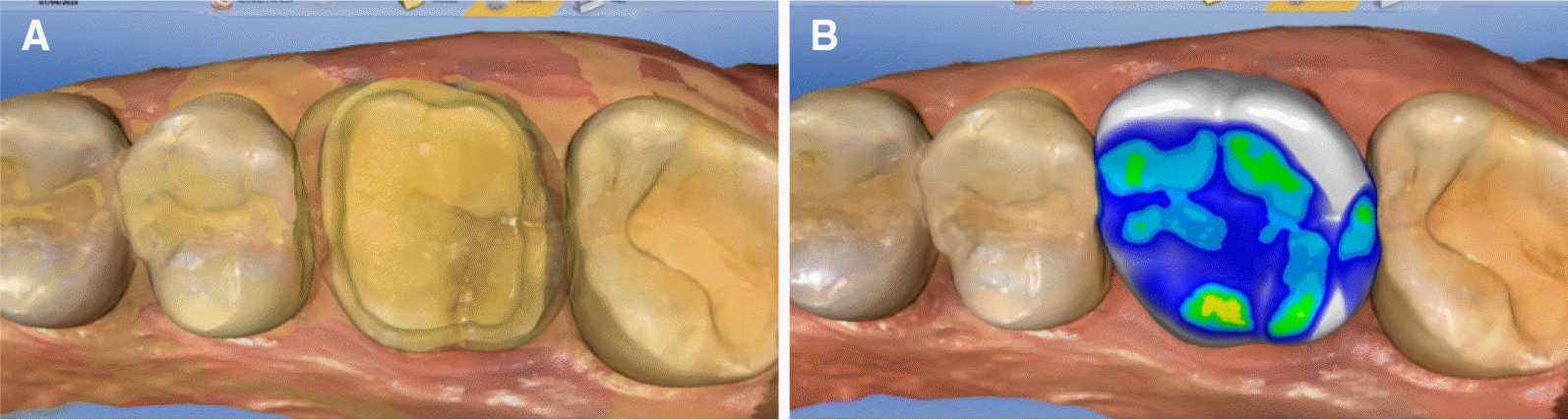 | Fig. 4.(A) Image of tooth before preparation was superimposed onto the abutment, (B) Designed crown using superimposition. |
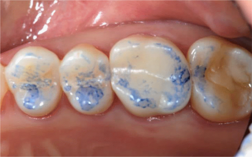
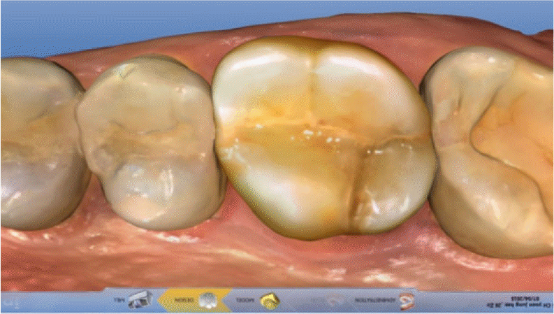
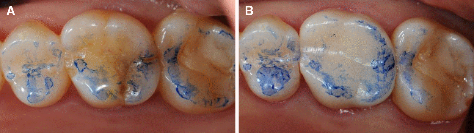
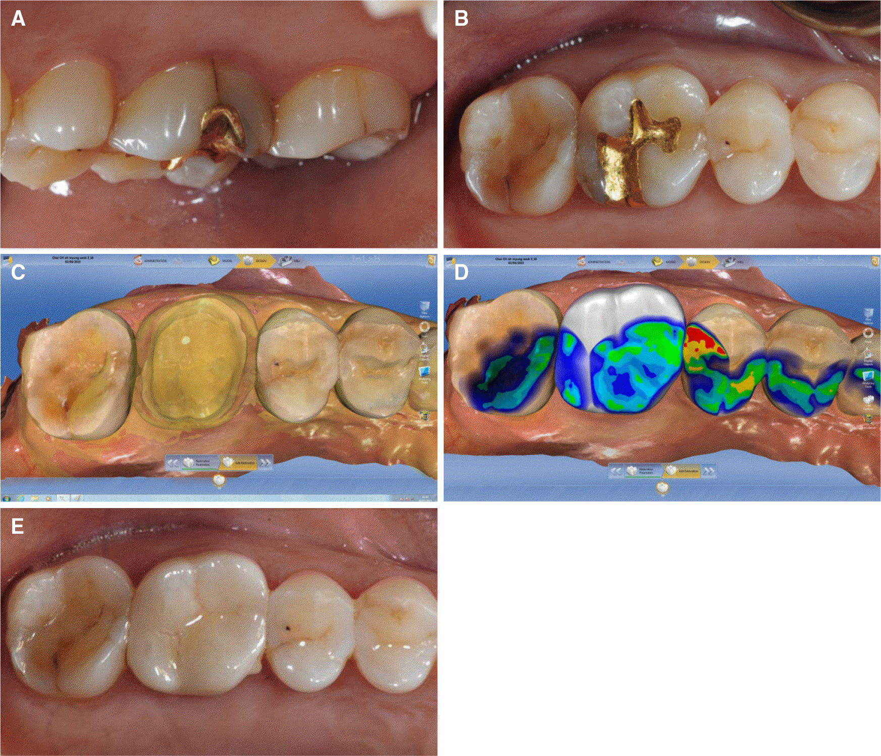 | Fig. 6.Cracked tooth case of maxillary right first molar. (A) A definite crack line was observed on the palatal surface of maxillary right first molar, (B) Occlusal view of maxillary right first molar before preparation, (C) Biogeneric copy of entire crown (superimposition), (D) The occlusion before tooth preparation was completely reproduced on the new restoration design by superimposition, (E) Delivery of zirconia crown. |
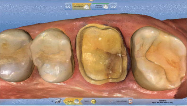
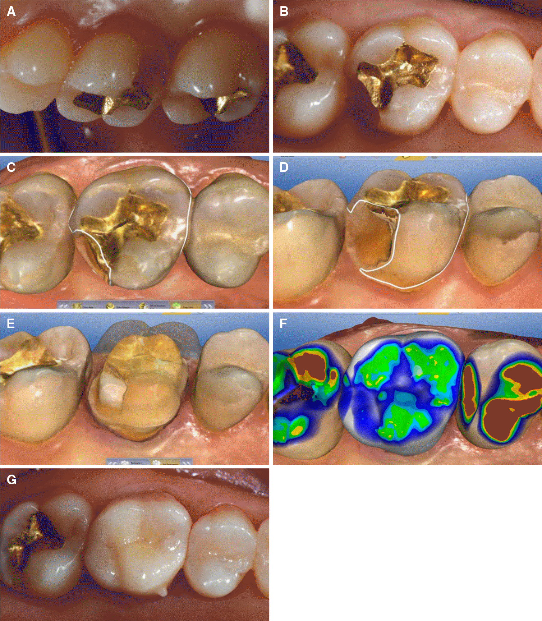 | Fig. 7.Crown fracture case of maxillary right first molar. (A) Crown fracture of distopalatal cusp on maxillary right first molar, (B) Occlusal view of maxillary right first molar before preparation, (C) Bio-copy excluding cusp of fractured area (occlusal view), (D) Bio-copy excluding cusp of fractured area (palatal view), (E) Superimposed images of the prepared abutment and biogeneric copy image excluding cusp fracture area, (F) Designed crown using double scanning, (G) Delivery of zirconia crown. |
 XML Download