

 PDF
PDF ePub
ePub Citation
 Print
Print


Abstract
In removable dental prostheses, it is important to minimize impairment of residual tissue caused by wearing dentures. There are two factors that harm residual tissue. The first is functional load bearing of remaining teeth and alveolar ridges and the second is the effect of poor oral hygiene. Double crown retained removable dental prostheses provide rigid support, and it may reduce impairment caused by load bearing of alveolar ridges. Also, dental plaque and oral deposits, which are attached to outer crowns and dentures, can be easily managed extra-orally. In addition, it is beneficial to the health of the marginal gingiva because inner crowns have easy access for oral hygiene. In this case, double crown retained removable dental prostheses were used for the partially edentulous patient with severe residual alveolar bone resorption and poor oral hygiene, and the result was clinically satisfactory in terms of functional, esthetical, and oral hygiene aspects. (J Korean Acad Prosthodont 2016;54:21-7)
REFERENCES
1.Langer A. Telescope retainers for removable partial dentures. J Prosthet Dent. 1981. 45:37–43.

2.Langer A. Tooth-supported telescope restorations. J Prosthet Dent. 1981. 45:515–20.
3.Bergman B. Periodontal reactions related to removable partial dentures: a literature review. J Prosthet Dent. 1987. 58:454–8.
4.Goswami R., Mahajan P., Siwach A., Gupta A. Telescopic over denture: Perio-prostho concern for advanced periodontitis. Contemp Clin Dent. 2013. 4:402–5.
5.Turner KA., Missirlian DM. Restoration of the extremely worn dentition. J Prosthet Dent. 1984. 52:467–74.
6.Geerts GA., Stuhlinger ME., Nel DG. A comparison of the accuracy of two methods used by pre-doctoral students to measure vertical dimension. J Prosthet Dent. 2004. 91:59–66.
7.Shetty S., Zargar NM., Shenoy K., Rekha V. Occlusal plane location in edentulous patients: a review. J Indian Prosthodont Soc. 2013. 13:142–8.
8.Bayer S., Stark H., Golz L., Keilig L., Kraus D., Hansen A., Enkling N. Telescopic crowns: extra-oral and intra-oral retention force measurement—in vitro/in vivo correlation. Gerodontology. 2012. 29:e340–7.
9.Wenz HJ., Lehmann KM. A telescopic crown concept for the restoration of the partially edentulous arch: the Marburg double crown system. Int J Prosthodont. 1998. 11:541–50.
10.Bergman B., Ericson A., Molin M. Long-term clinical results after treatment with conical crown-retained dentures. Int J Prosthodont. 1996. 9:533–8.
11.Bergman B., Hugoson A., Olsson CO. Caries, periodontal and prosthetic findings in patients with removable partial dentures: a ten-year longitudinal study. J Prosthet Dent. 1982. 48:506–14.
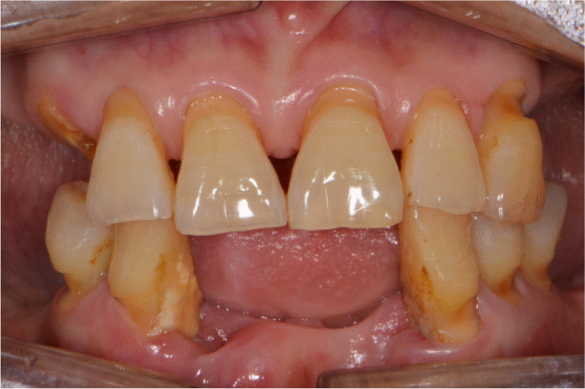
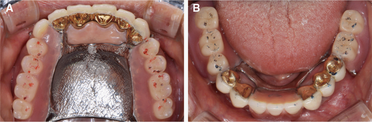
Fig. 3.
Teeth preparation with putty index. (A) Maxillary teeth preparation, (B, C) Mandibular teeth preparation.
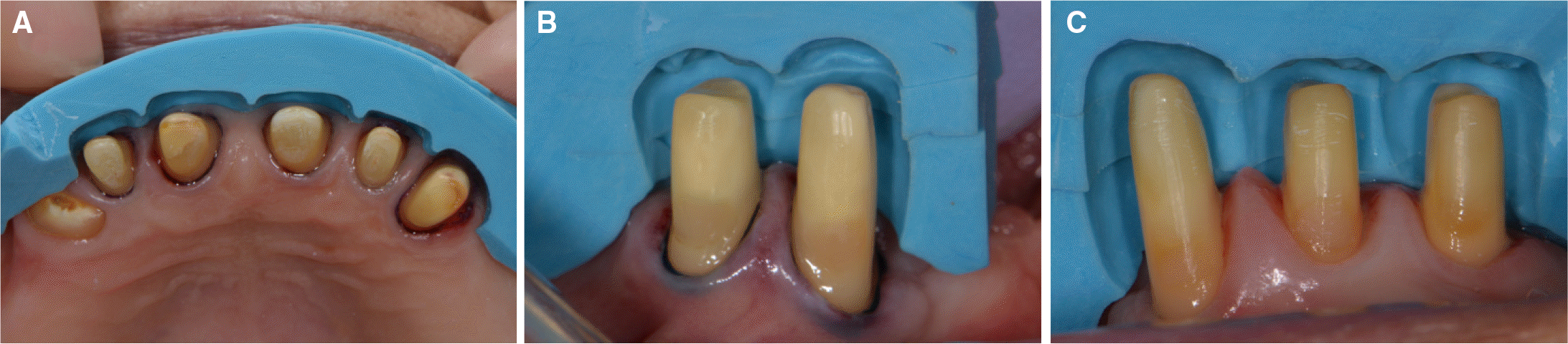
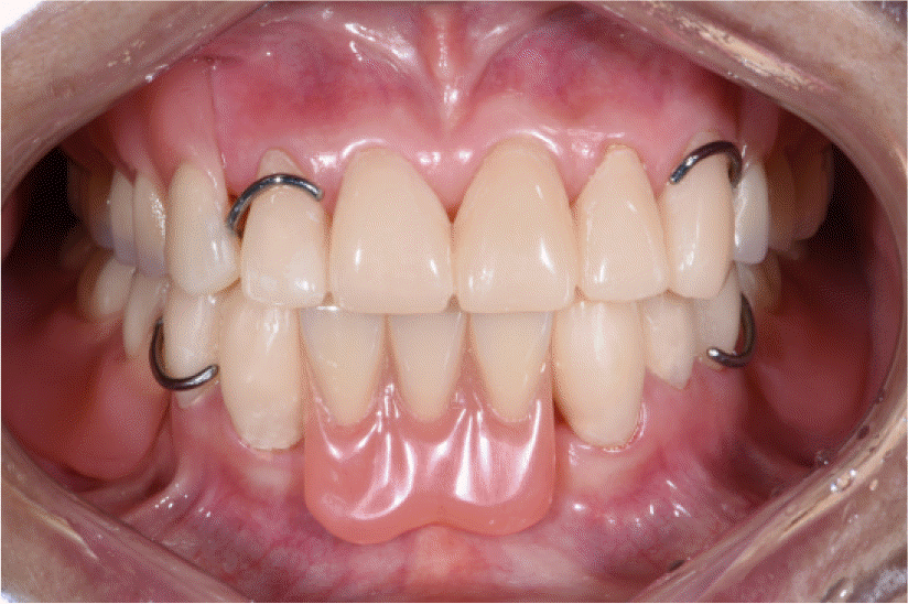
Fig. 5.
Transcranial radiograph of left temporomandibular joint. (A) At first visit, (B) 10 weeks after interim prostheses delivery.
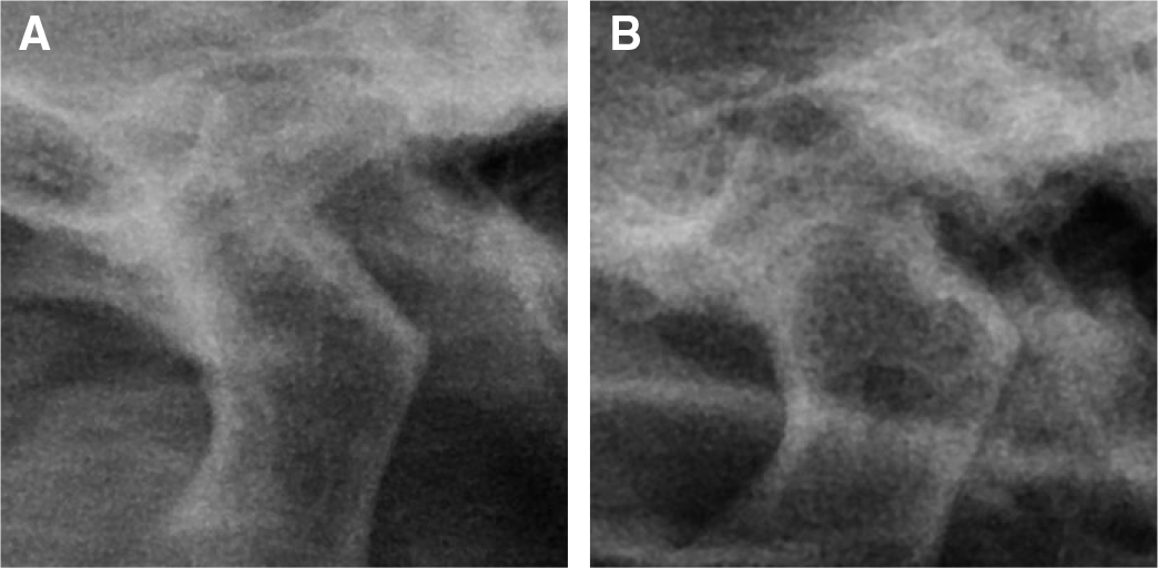
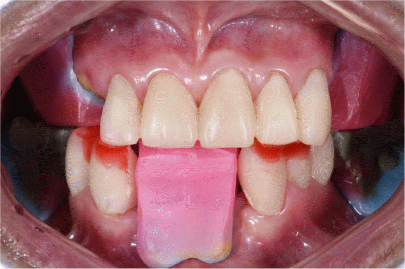
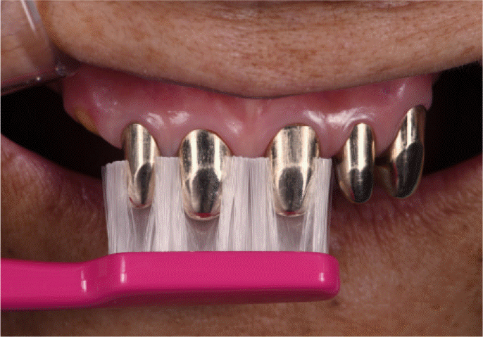
Fig. 7.
Retention force measurement of double crown. (A) Steel wire fixed in position in pattern resin filled into inner crown, (B) The retention force was measured using digital force gauge.
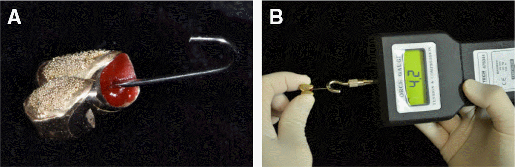
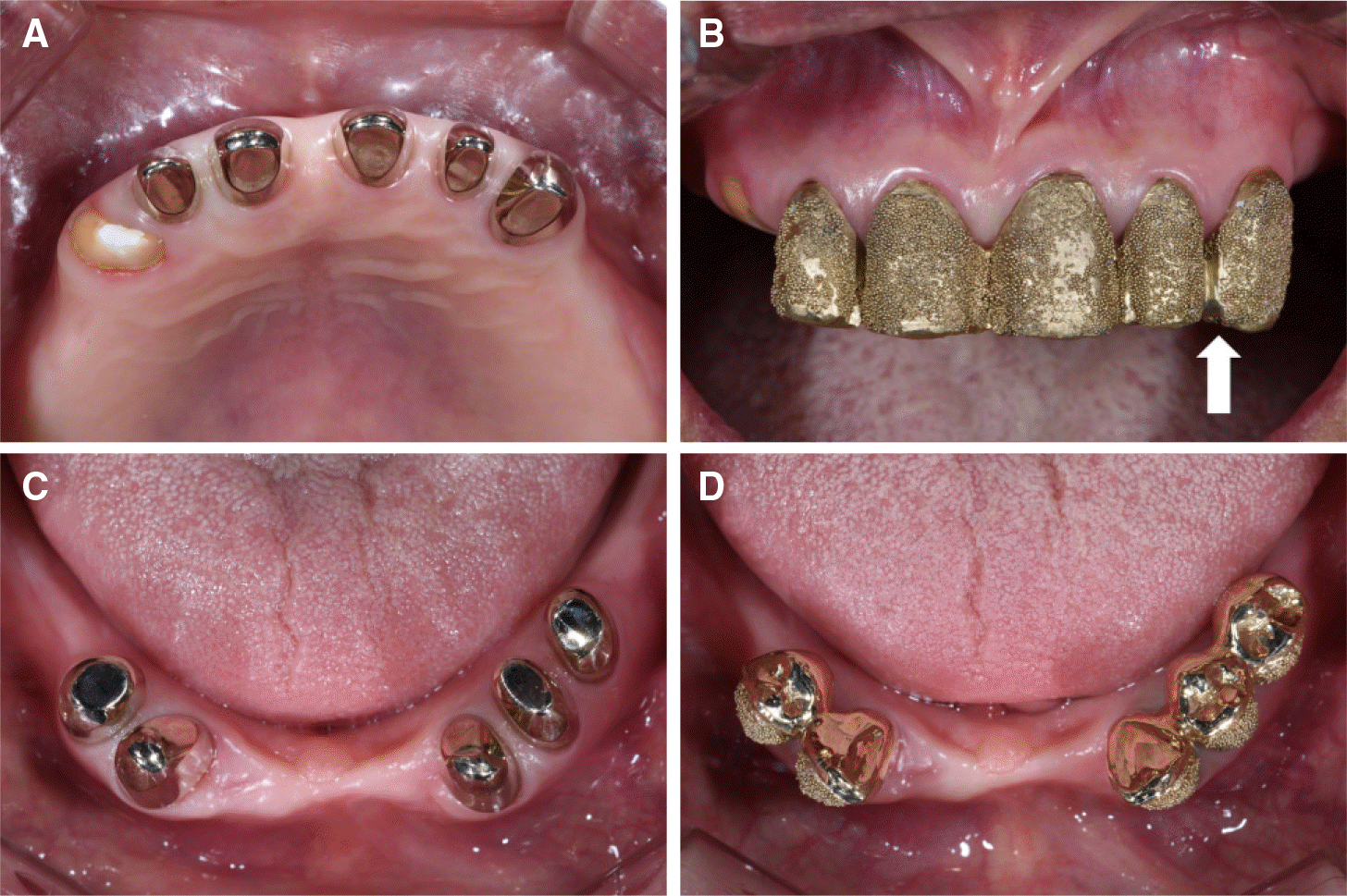
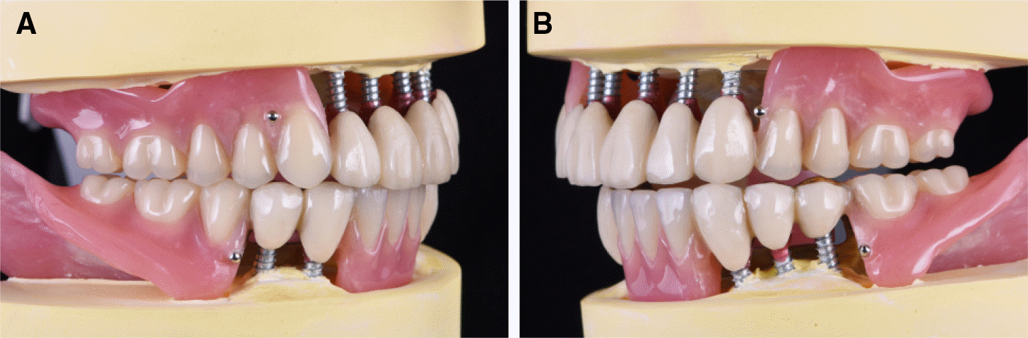
Fig. 8.
Inner crown and outer crown try-in. (A) Maxillary inner crown try-in, (B) Maxillary outer crown try-in. Cut and soldering performed on mesial side of left maxillary canine (arrow), (C) Mandibular inner crown try-in, (D) Mandibular outer crown try-in.
 XML Download