

 PDF
PDF ePub
ePub Citation
 Print
Print


Abstract
Purpose
This study was performed in order to assess the effect of the surface treatment methods and the use of bonding agent on the shear bond strength (SBS) between the aged CAD-CAM (computer aided design-computer aided manufacturing) hybrid materials and added composite resin.
Materials and methods
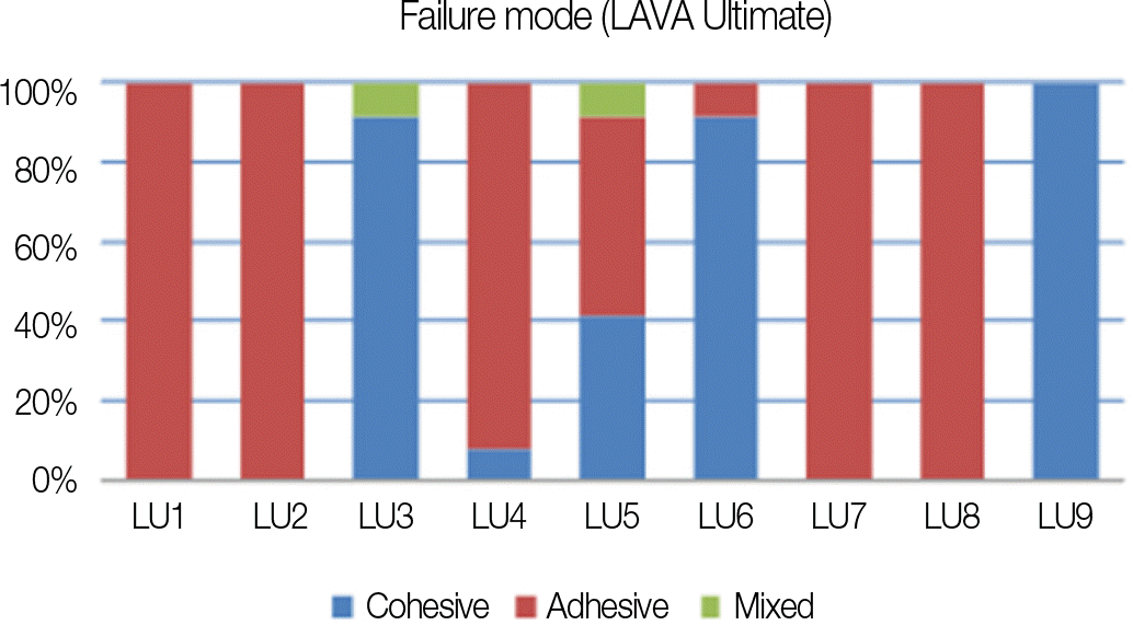
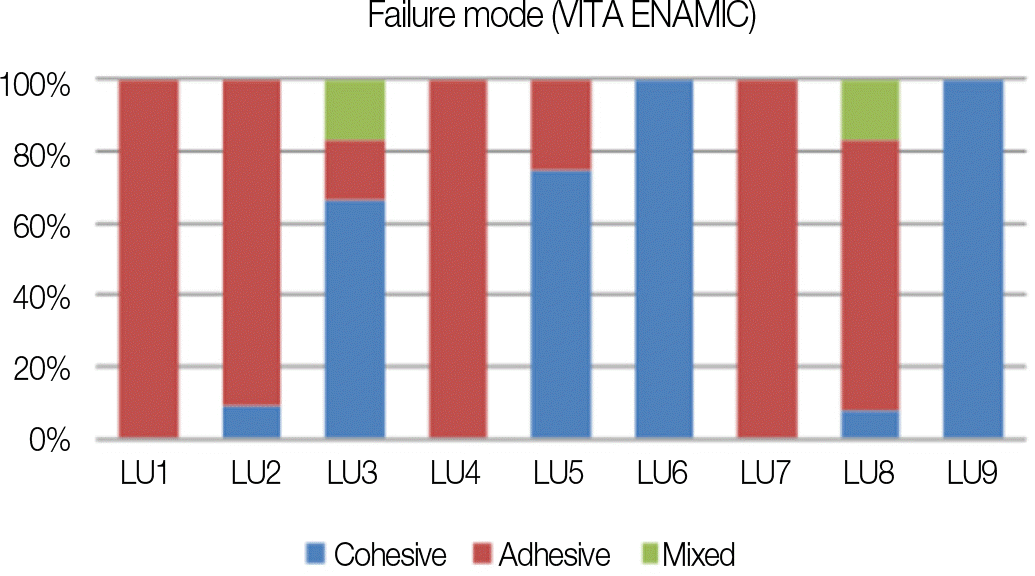
LAVA Ultimate (LU) and VITA ENAMIC (VE) specimens were age treated by submerging in a 37℃ water bath filled with artificial saliva (Xerova solution) for 30 days. The surface was ground with #220 SiC paper then the specimens were divided into 9 groups according to the combination of the surface treatment (no treatment, grinding, air abrasion with aluminum oxide, HF acid) and bonding agents (no bonding, Adper Single Bond 2, Single Bond Universal). Each group had 10 specimens. Specimens were repaired (added) using composite resin (Filtek Z250), then all the specimens were stored for 7 days in room temperature distilled water. SBS was measured and the fractured surfaces were observed with a scanning electron microscope (SEM). One-way ANOVA and Scheffe test were used for statistical analysis (α =.05).
Results
Mostly groups with bonding agent treatment showed higher SBS than groups without bonding agent. Among the groups without bonding agent the groups with aluminum oxide treatment showed higher SBS. However there was no significant difference between groups except two subgroups within LU group, which revealed a significant increase of SBS when Single Bond Universal was used on the ground LU specimen.
Go to : 
REFERENCES
1.He LH., Purton D., Swain M. A novel polymer infiltrated ceramic for dental simulation. J Mater Sci Mater Med. 2011. 22:1639–43.

2.Kim HC. Ceramic materials for chair side CAD/CAM. J Korean Acad Esthet Dent. 2014. 23:16–26.
3.Kurbad A., Kurbad S. A new, hybrid material for minimally invasive restorations in clinical use. Int J Comput Dent. 2013. 16:69–79.
4.Rocca GT., Bonnafous F., Rizcalla N., Krejci I. A technique to improve the esthetic aspects of CAD/CAM composite resin restorations. J Prosthet Dent. 2010. 104:273–5.
5.Frankenberger R., Hartmann VE., Krech M., Krämer N., Reich S., Braun A., Roggendorf M. Adhesive luting of new CAD/CAM materials. Int J Comput Dent. 2015. 18:9–20.
6.Lee JY., Im EB. A shear bond strength of resin cements bonded to pressable porcelain with various surface treatments. J Korean Acad Prosthodont. 2003. 41:379–86.
7.Kang HS., Choi HY. A study on the bond strength of repair resin to the surface treated composite resins. J Korean Acad Cons Dent. 1995. 20:487–507.
8.Amaral R., Ozcan M., Bottino MA., Valandro LF. Microtensile bond strength of a resin cement to glass infiltrated zirconia-reinforced ceramic: the effect of surface conditioning. Dent Mater. 2006. 22:283–90.
9.Lee YG., Moon SR., Cho YG. Effect of cutting instruments on the dentin bond strength of a self-etch adhesive. J Korean Acad Cons Dent. 2010. 35:13–9.
10.Lee CW., Kim JW., Lee SH. A study on microleakage of composite resin after surface treatment. J Korean Acad Pediatr Dent. 1998. 25:103–15.
11.Ozcan M. Evaluation of alternative intra-oral repair techniques for fractured ceramic-fused-to-metal restorations. J Oral Rehabil. 2003. 30:194–203.
12.Stawarczyk B., Krawczuk A., Ilie N. Tensile bond strength of resin composite repair in vitro using different surface preparation conditionings to an aged CAD/CAM resin nanoceramic. Clin Oral Investig. 2015. 19:299–308.
13.Daniels MW., Francis LF. Silane adsorption behavior, microstructure, and properties of glycidoxypropyltrimethoxysi-lane-modified colloidal silica coatings. J Colloid Interface Sci. 1998. 205:191–200.
14.Ozcan M. The use of chairside silica coating for different dental applications: a clinical report. J Prosthet Dent. 2002. 87:469–72.
15.Egilmez F., Ergun G., Cekic-Nagas I., Vallittu PK., Ozcan M., Lassila LV. Effect of surface modification on the bond strength between zirconia and resin cement. J Prosthodont. 2013. 22:529–36.
16.Swift EJ Jr., Cloe BC., Boyer DB. Effect of a silane coupling agent on composite repair strengths. Am J Dent. 1994. 7:200–2.
17.Brosh T., Pilo R., Bichacho N., Blutstein R. Effect of combinations of surface treatments and bonding agents on the bond strength of repaired composites. J Prosthet Dent. 1997. 77:122–6.
18.Hisamatsu N., Atsuta M., Matsumura H. Effect of silane primers and unfilled resin bonding agents on repair bond strength of a prosthodontic microfilled composite. J Oral Rehabil. 2002. 29:644–8.
19.Ru ME. Influence of artificial saliva contamination on bonding of dentin adhesives to dentin. J Korean Acad Cons Dent. 1992. 17:383–97.
20.Flores S., Charlton DG., Evans DB. Repairability of polyacid-modified composite resin. Oper Dent. 1995. 20:191–6.
21.Lauvahutanon S., Takahashi H., Shiozawa M., Iwasaki N., Asakawa Y., Oki M., Finger WJ., Arksornnukit M. Mechanical properties of composite resin blocks for CAD/CAM. Dent Mater J. 2014. 33:705–10.
22.Frankenberger R., Hartmann VE., Krech M., Krämer N., Reich S., Braun A., Roggendorf M. Adhesive luting of new CAD/CAM materials. Int J Comput Dent. 2015. 18:9–20.
Go to :
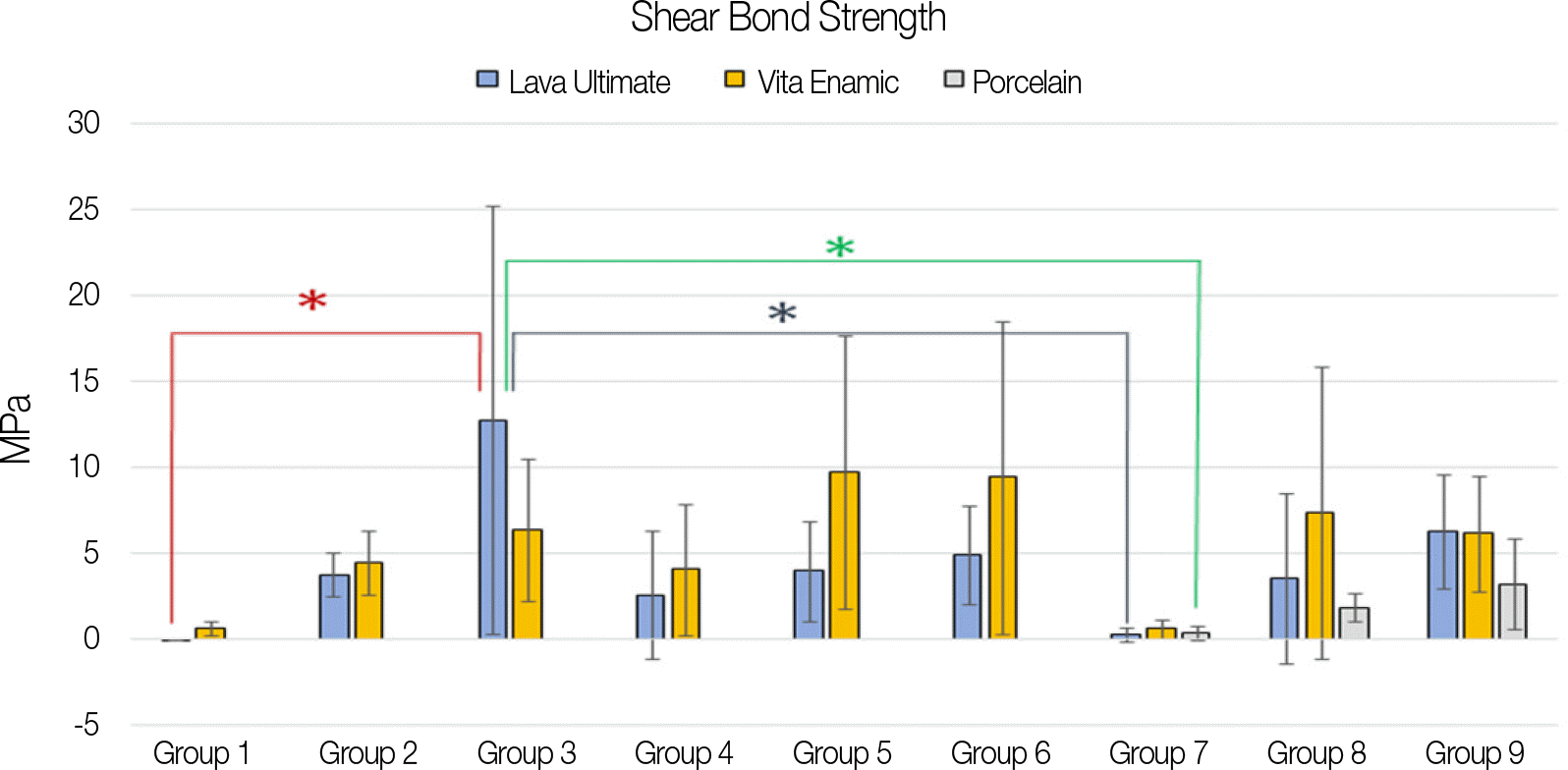 | Fig. 2.Means and standard deviations of shear bond strength of experimental groups with marks denoting statistical significance (∗ denotes P < .05). |
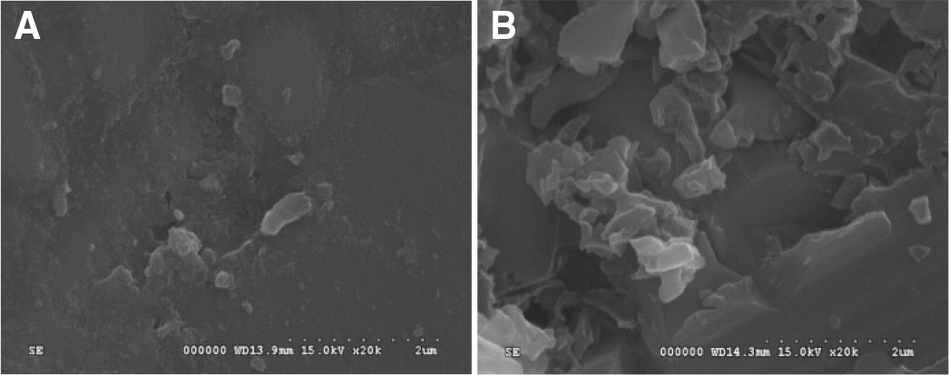 | Fig. 6.Scanning electron microscopic photomicrograph of polished surface of specimens before aging (×20,000 magnification). (A) LAVA Ultimate, (B) VITA ENAMIC. |
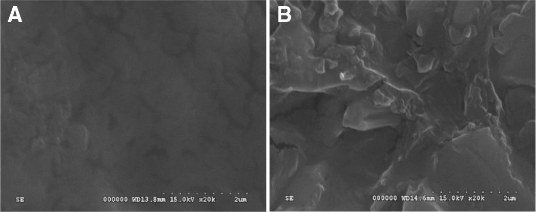 | Fig. 7.Scanning electron microscopic photomicrograph of specimens after artificial aging by submerging in artificial saliva for 1 month (×20,000 magnification). (A) LAVA Ultimate, (B) VITA ENAMIC. |
 | Fig. 8.Scanning electron microscopic photomicrograph of specimens after #220 SiC paper grinding (×20,000 magnification). (A) LAVA Ultimate, (B) VITA ENAMIC. |
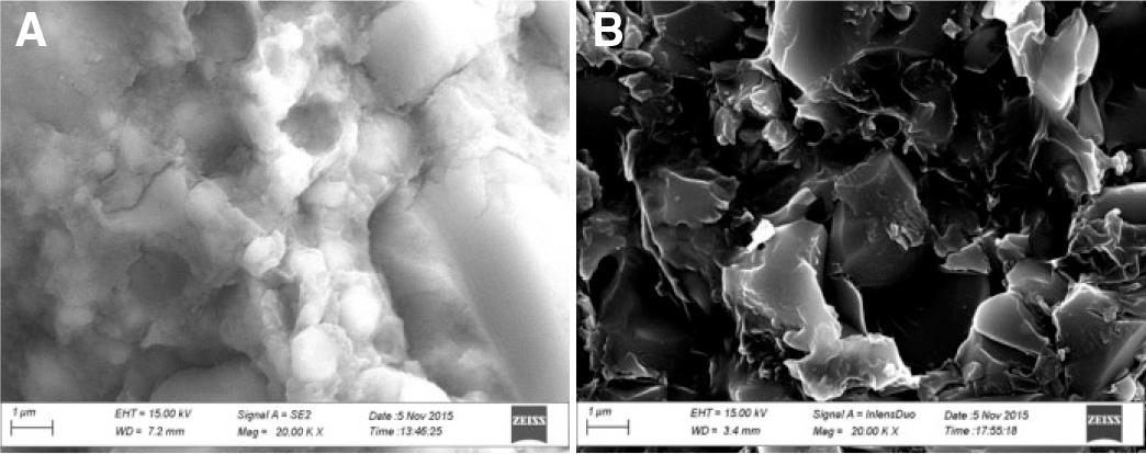
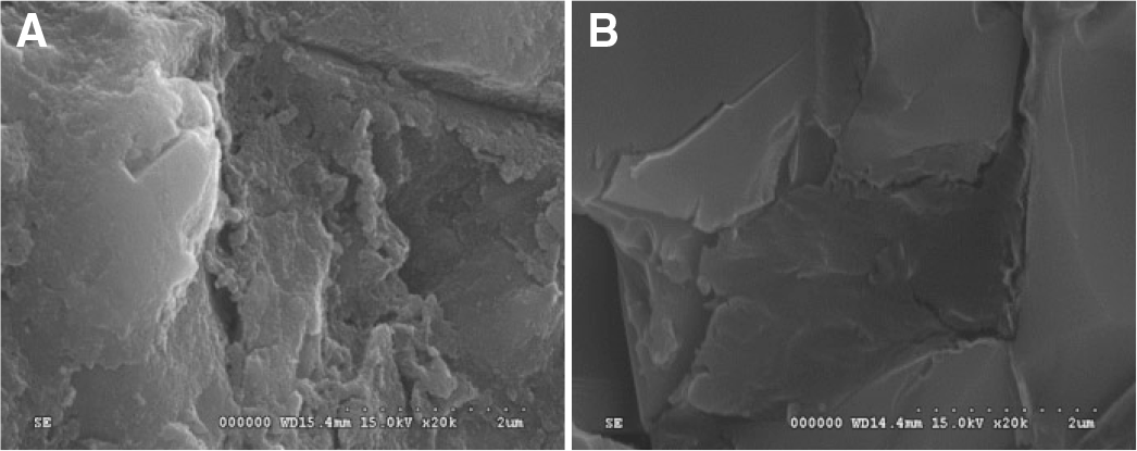 | Fig. 9.Scanning electron microscopic photomicrograph of specimens after aluminum oxide abrasion and ultrasonic cleaning (×20,000 magnification). (A) LAVA Ultimate, (B) VITA ENAMIC. |
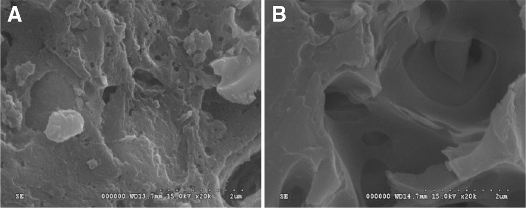 | Fig. 10.Scanning electron microscopic photomicrograph of specimens after HF acid etching for 1 minute (×20,000 magnification). (A) LAVA Ultimate, (B) VITA ENAMIC. |
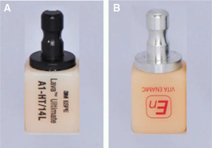
Table 1.
Base and repair material in this study
Table 2.
Material compositions used for surface treatment in this study
Table 3.
Composition of artificial saliva (Xerova solution) per 100 mL
| Calcium Chloride Hydrate | 15 mg |
| Carboxymethylcellulose Sodium | 1 g |
| Dibasic Potassium Phosphate | 34 mg |
| D-Sorbitol | 3 g |
| Magnesium Chloride | 5 mg |
| Potassium Chloride | 120 mg |
| Sodium Chloride | 84 mg |
Table 4.
Summary of surface treatment protocols for each group
Table 5.
Descriptive statistics for the shear bond strength test (Unit: MPa)
|
Material/surfaces treatment |
Group 1 | Group 2 | Group 3 | Group 4 | Group 5 | Group 6 | Group 7 | Group 8 | Group 9 |
|---|---|---|---|---|---|---|---|---|---|
|
SiC paper no.220 Aluminum oxide |
|||||||||
| Non |
Non S.B.2 |
S.B.U | Non | S.B.2 | S.B.U | Non |
HF acid S.B.2 |
S.B.U | |
| LU | LU1 | LU2 | LU3 | LU4 | LU5 | LU6 | LU7 | LU8 | LU9 |
| 0.01 | 3.75 | 12.76 | 2.59 | 3.96 | 4.95 | 0.27 | 3.57 | 6.28 | |
| (± 0.01) | (± 1.26) | (± 12.48)∗ | (± 3.72) | (± 2.88) | (± 2.86) | (± 0.43) | (± 4.96) | (± 3.34) | |
| VE | VE1 | VE2 | VE3 | VE4 | VE5 | VE6 | VE7 | VE8 | VE9 |
| 0.61 | 4.47 | 6.4 | 4.06 | 9.77 | 9.42 | 0.62 | 7.36 | 6.16 | |
| (± 0.42) | (± 1.88) | (± 4.14) | (± 3.84) | (± 7.95)∗ | (± 9.11) | (± 0.54) | (± 8.53) | (± 3.34) | |
| Po | Po7 | Po8 | Po9 | ||||||
| 0.37 | 1.84 | 3.19 | |||||||
| (± 0.44) | (± 0.82) | (± 2.65)∗ | |||||||
 XML Download