

 PDF
PDF ePub
ePub Citation
 Print
Print


Abstract
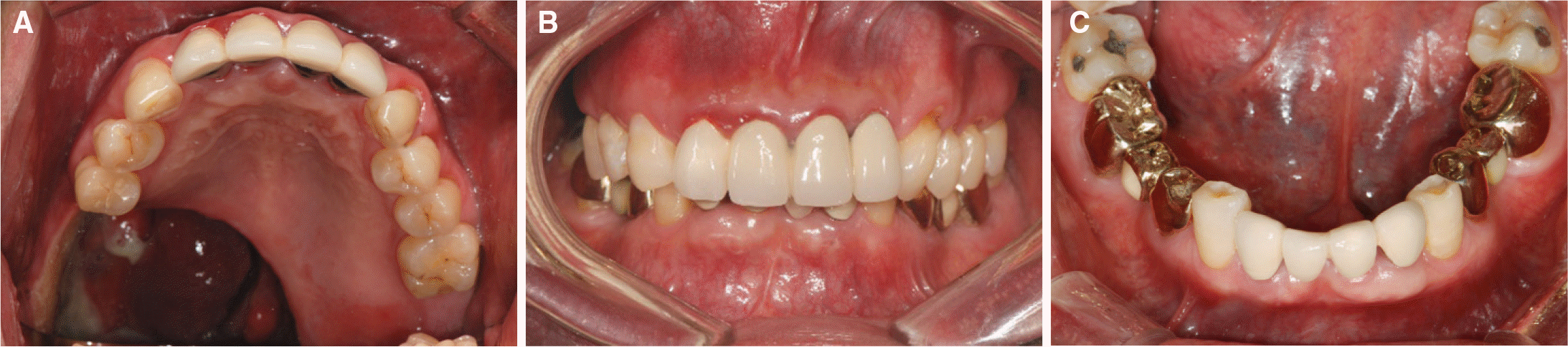
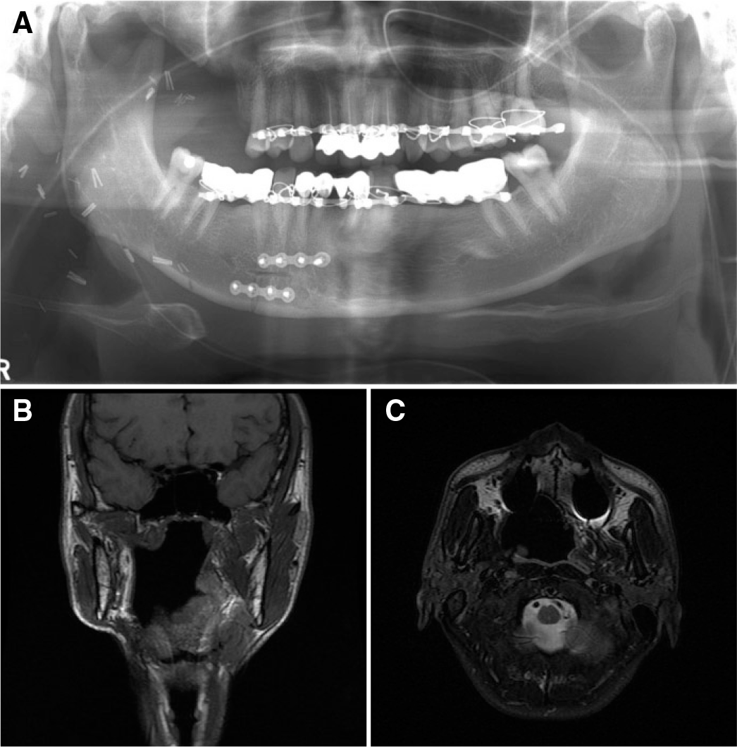
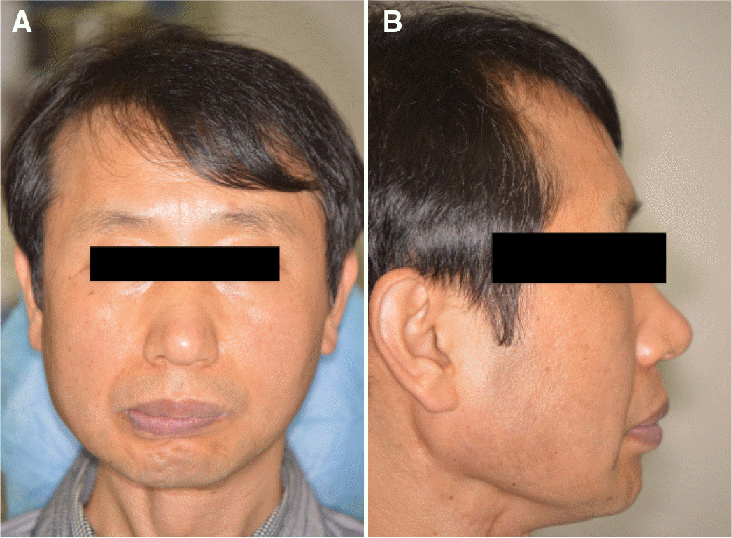
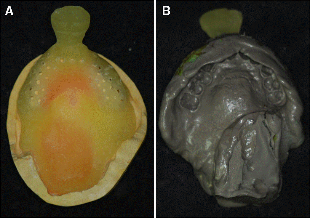
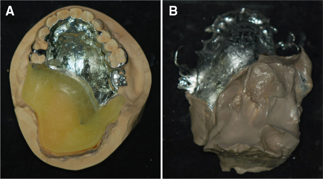
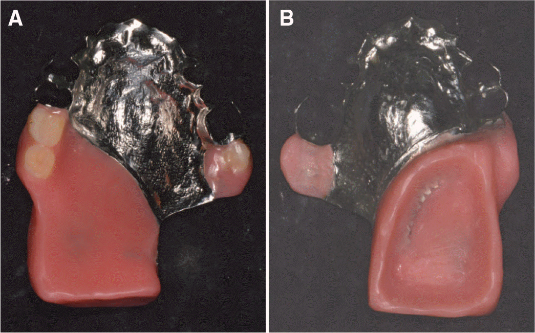
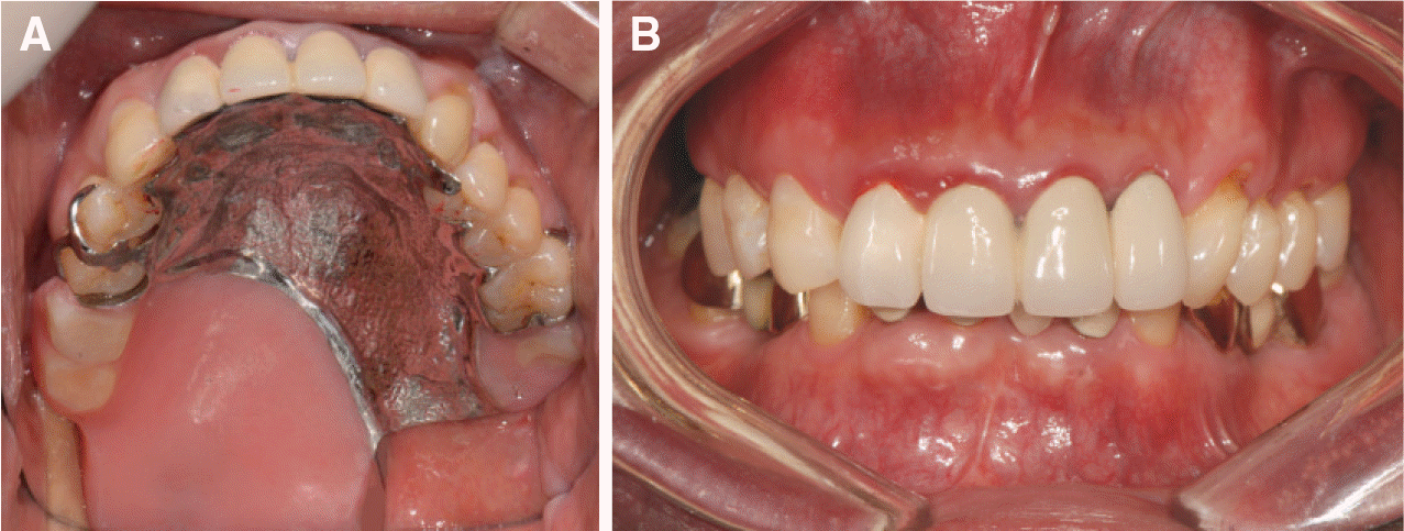
Patients undergone partial maxillectomy experience post-operative masticatory, phonetic, and swallowing difficulties. They also encounter social and psychological challenges due to changes in their facial appearances. Thus, functional and esthetic recovery through maxillofacial prosthesis becomes significant for these patients. The objective of an appropriate obturator is to restore palate and improve phonetic and swallowing ability by separating the oral cavity, nasal cavity, maxillary sinus, and nasopharynx. In this case report, an obturator was fabricated for a patient who had partial resection from the maxillary posterior region to the pharynx due to squamous cell carcinoma. The purpose of this case study is to describe the results because the patient was successfully improved both functionally and esthetically. (J Korean Acad Prosthodont 2016;54:167-71)
REFERENCES
1.Tang JA., Rieger JM., Wolfaardt JF. A review of functional outcomes related to prosthetic treatment after maxillary and mandibular reconstruction in patients with head and neck cancer. Int J Prosthodont. 2008. 21:337–54.
2.Lang BR., Bruce RA. Presurgical maxillectomy prosthesis. J Prosthet Dent. 1967. 17:613–9.

3.Ackerman AJ. The prosthetic management of oral and facial defects following cancer surgery. J Prosthet Dent. 1955. 5:413–32.
4.Beumer J., Marunick MT., Esposito SJ. Maxillofacial rehabilitation: Prosthodontic and surgical management of cancer-related, acquired, and congenital defects of the head and neck. 3rd ed.Chicago: Quintessence Publishing;2011.
5.Desjardins RP. Obturator prosthesis design for acquired maxillary defects. J Prosthet Dent. 1978. 39:424–35.
6.Kaires AK. Effect of partial denture design on bilateral force distribution. J Prosthet Dent. 1956. 6:373–85.
7.Oral K., Aramany MA., McWilliams BJ. Speech intelligibility with the buccal flange obturator. J Prosthet Dent. 1979. 41:323–8.
 XML Download