

 PDF
PDF ePub
ePub Citation
 Print
Print


Abstract
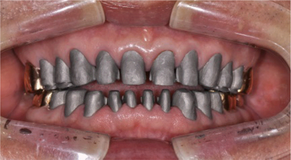
Although physiologic abrasion in normal range need not to be corrected, when hard tissue of teeth are worn abnormally fast, it can cause severe damage and destroy esthetics and, functional structure of occlusion consequently. To establish a correct occlusal plane and space for the patient with worn dentition, it is necessary to increase vertical dimension. However, actual occlusal vertical dimension remains unhanged with compensation for the increase of alveolar bone height equivalent to the decrease of teeth length. A 74-year-old male presented with worn dentition and fractured tooth. Based on the assessment of OVD including clinical findings, full-mouth rehabilitation without increase of OVD was planned. This case presents that a satisfactory clinical result was achieved by restoring the worn dentition without changing occlusal vertical dimension. (J Korean Acad Prosthodont 2016;54:160-6)
REFERENCES
1.Dawson PE. Evaluation, diagnosis and treatment of occlusal problems. 2nd ed.St. Louis: Mosby;1989.
2.Turner KA., Missirlian DM. Restoration of the extremely worn dentition. J Prosthet Dent. 1984. 52:467–74.

3.Murphy T. Compensatory mechanisms in facial height adjustment to functional tooth attrition. Aust Dent J. 1959. 4:312–23.
4.Hemmings KW., Howlett JA., Woodley NJ., Griffiths BM. Partial dentures for patients with advanced tooth wear. Dent Update. 1995. 22:52–9.
5.Ibbetson RJ., Setchell DJ. Treatment of the worn dentition: 2. Dent Update. 1989. 16:300–2. 305–7.
6.Park JH., Jeong CM., Jeon YC., Lim JS. A study on the occlusal plane and the vertical dimension in Korean adults with natural dentition. J Korean Acad Prosthodont. 2005. 43:41–51.
7.Jensen WO. Occlusion for the Class II jaw relations patient. J Prosthet Dent. 1990. 64:432–4.
8.Willis FM. Features of the face involved in full denture prosthesis. Dent Cosmos. 1935. 77:851–4.
9.Berry DC., Poole DF. Attrition: possible mechanisms of compensation. J Oral Rehabil. 1976. 3:201–6.
10.Dawson PE. Functional occlusion: from TMJ to smile design. St. Louis; Mo: Mosby;2007. p. 430–52.
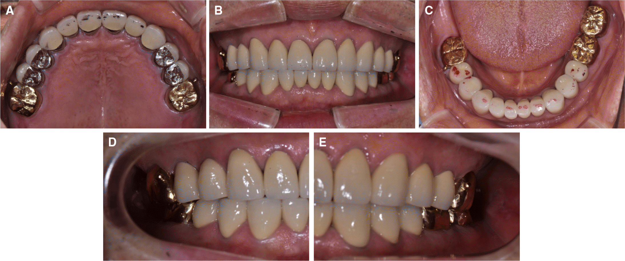
Fig. 1.
Initial intraoral photograph. (A) Maxillary occlusal view, (B) Frontal view, (C) Mandibular occlusal view, (D) Right side view, (E) Left side view.
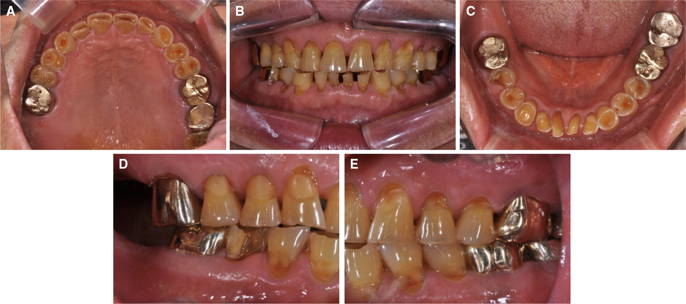
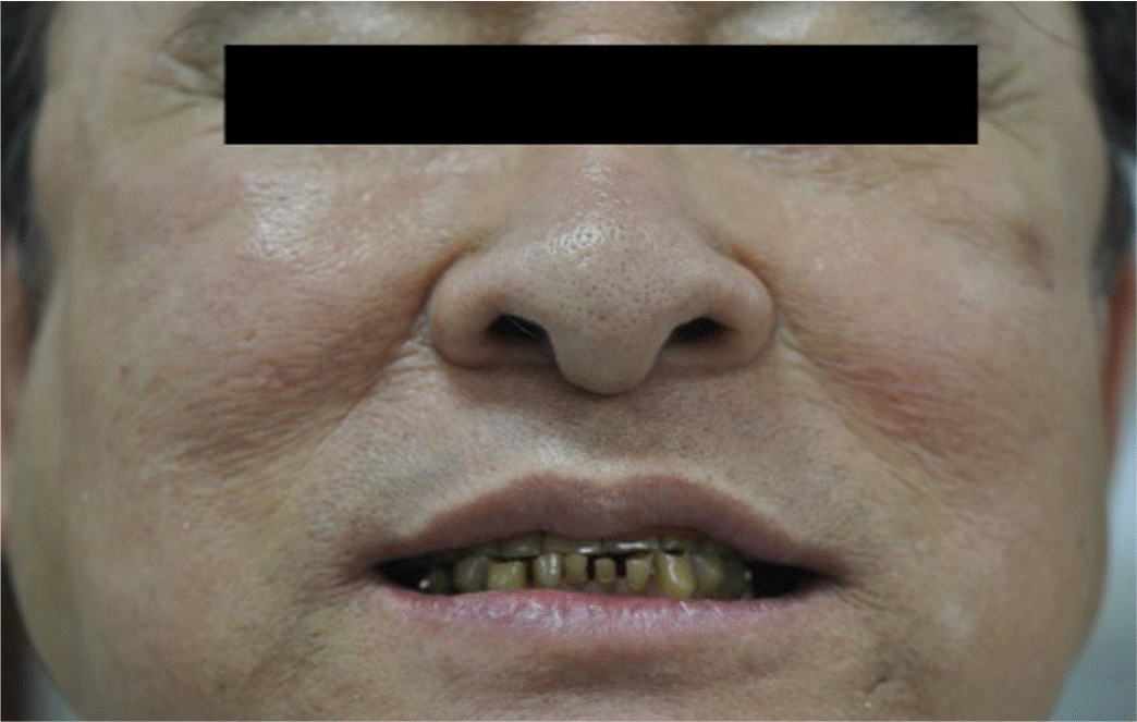
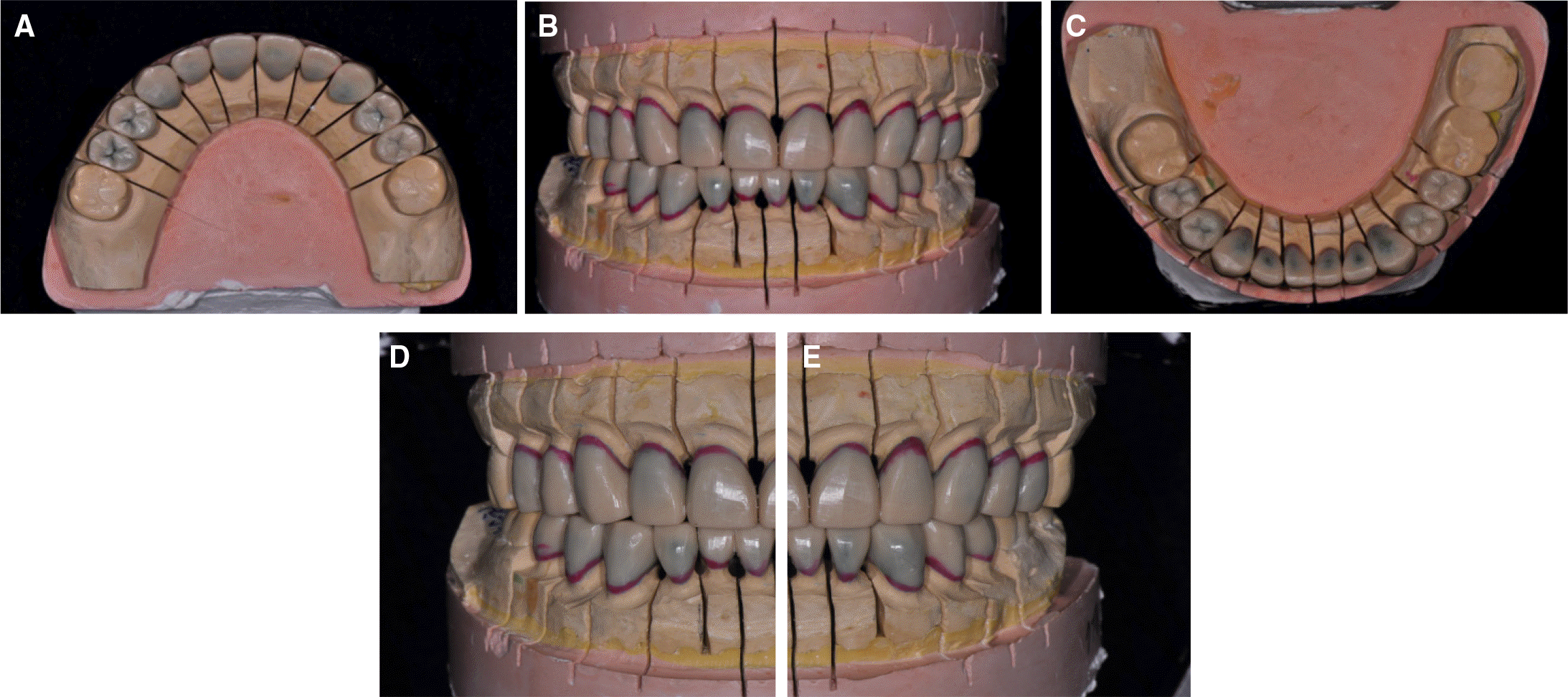
Fig. 6.
Provisional restorations. (A) Maxillary occlusal view, (B) Frontal view, (C) Mandibular occlusal view, (D) Right side view, (E) Left side view.
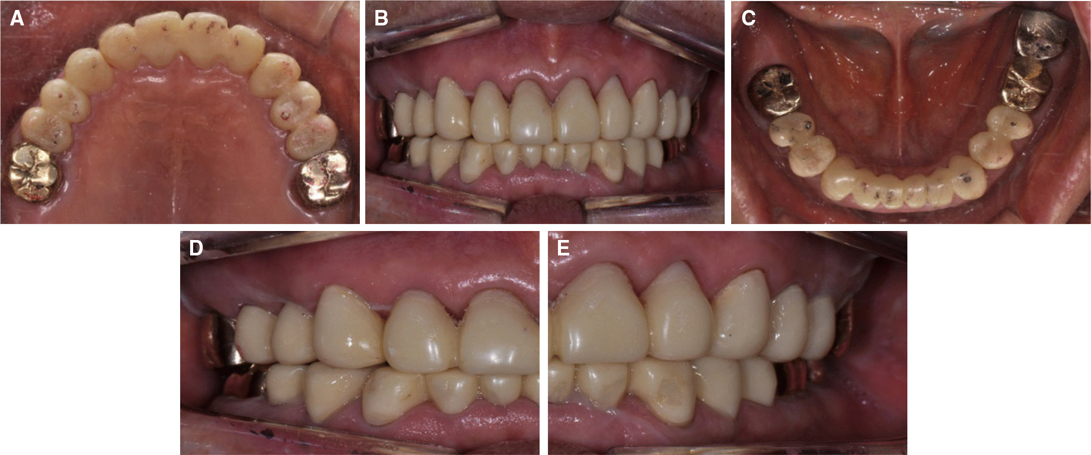
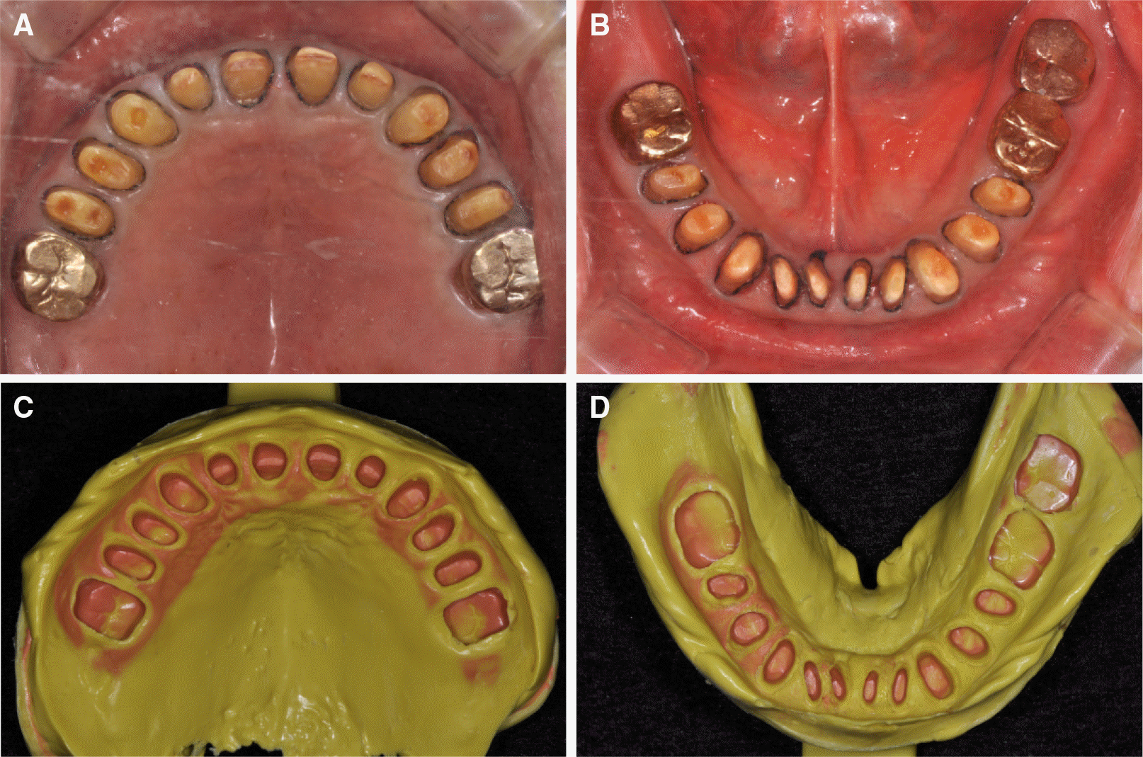
Fig. 7.
Final preparation and impression taking. (A) Maxillary preparation, (B) Mandibular preparation, (C) Maxillary impression, (D) Mandibular impression.
 XML Download