Abstract
Implant fixed prosthesis for the complete edentulous maxilla provides significant benefits in the aspects of functions and esthetics compared with the conventional denture. Implant supported fixed prosthesis are totally supported by implant, and thus stabilizes the prosthesis to the maximum degree as possible. Also, the improved retention and stability of fixed prosthesis enhance patients’ psychological and psychosocial health. This clinical presentation describes a maxillary full arch implant-supported fixed prosthesis in complete maxillary edentulous patient who showed vertical and horizontal alveolar bone resorption in the anterior ridge. To rehabilitate the esthetics and proper lip support, the zirconia framework was fabricated and the pink porcelain was veneered to reproduce the natural gingival tissue. After 9 months of follow up, the restorations were maintained without complications and the patient was satisfied with the restoration both functionally and esthetically. (J Korean Acad Prosthodont 2016;54:152-9)
Go to : 
REFERENCES
1.Misch CE. Contemporary Implant Dentistry. 3rd ed.St. Louis: CV Mosby;2009. p. 3–25.
2.Feine JS., Carlsson GE., Awad MA., Chehade A., Duncan WJ., Gizani S., Head T., Lund JP., MacEntee M., Mericske-Stern R., Mojon P., Morais J., Naert I., Payne AG., Penrod J., Stoker GT., Tawse-Smith A., Taylor TD., Thomason JM., Thomson WM., Wismeijer D. The McGill consensus statement on overdentures. Mandibular two-implant overdentures as first choice standard of care for edentulous patients. Montreal, Quebec, May 24-25, 2002. Int J Oral Maxillofac Implants. 2002. 17:601–2.
3.Drago C., Carpentieri J. Treatment of maxillary jaws with dental implants: guidelines for treatment. J Prosthodont. 2011. 20:336–47.

4.Misch CE. Contemporary Implant Dentistry. 3rd ed.St. Louis: CV Mosby;2009. p. 92–104.
5.Wheeler RC. The permanent maxillary central incisors. In Wheeler RC (ed): Dental Anatomy, Physiology and Occlusion. 5th ed.Philadelphia: Saunders;1974. p. 144.
6.Seibert JS. Reconstruction of deformed, partially edentulous ridges, using full thickness onlay grafts. Part I. Technique and wound healing. Compend Contin Educ Dent. 1983. 4:437–53.
7.Misch CE. Contemporary Implant Dentistry. 3rd ed.St. Louis: CV Mosby;2009. p. 367–88.
8.Misch CE. Dental Implant Prosthetics. 2nd ed.Elsevier: Mosby;2014. p. 829–73.
Go to : 
 | Fig. 1.Pretreament intraoral view. (A) frontal view, (B) upper occlusal view (C) upper occlusal view with old RPD. |
 | Fig. 2.Panoramic radiograph (A) pretreatment panoramic radiograph (B) With radiolographic stent 3 months after extracton of #14, 15, 24, 25. |
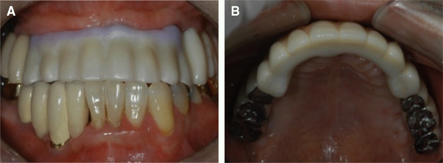
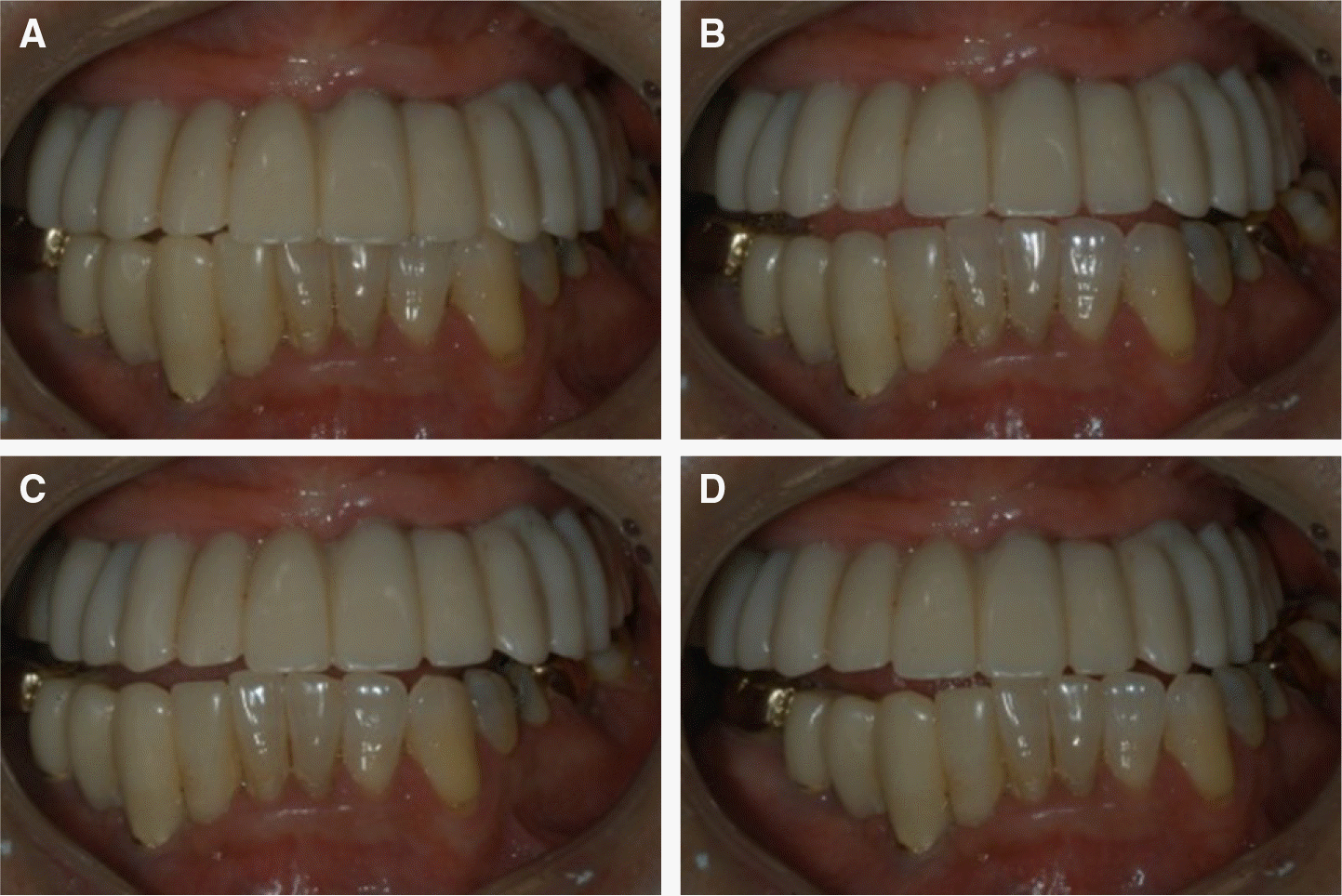 | Fig. 3.Screw retained interim implant prosthesis. (A) Maximum intercuspation, (B) Protrusive movement, (C) Right lateral movement, (D) Left lateral movement. |
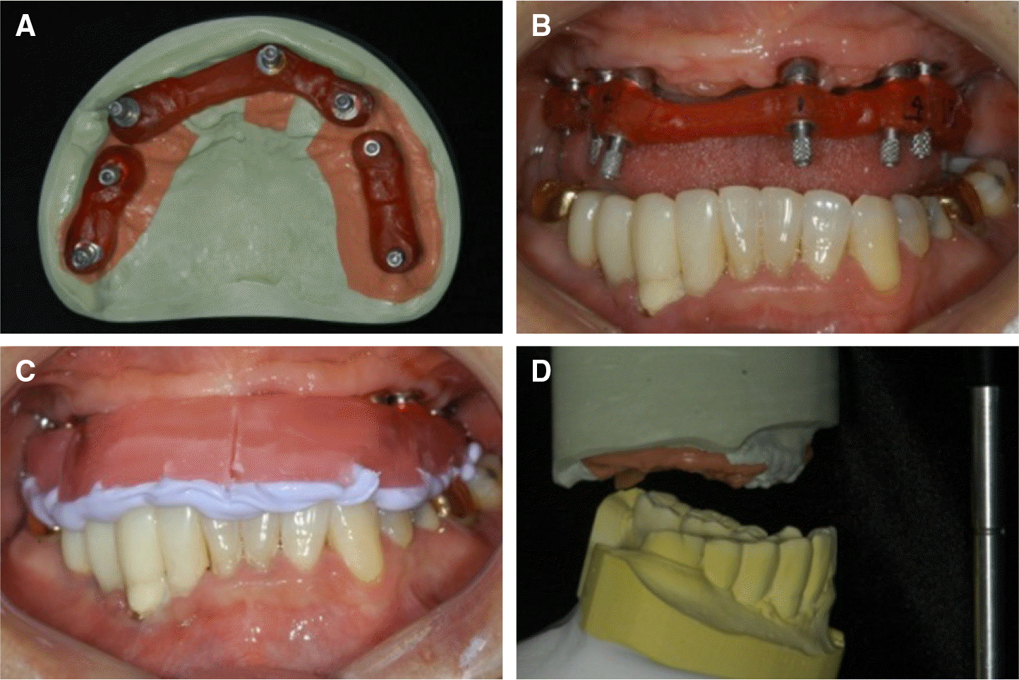 | Fig. 4.(A) Fabrication of repositioning jig by splinting pick up impression copings with pattern resin on the master cast, (B) Repositiong jigs are connected to check the accuracy of implant position, (C) Registration of interocclusal relationship using repositioning jig & interim prosthesis, (D) Mounted definitive cast. |
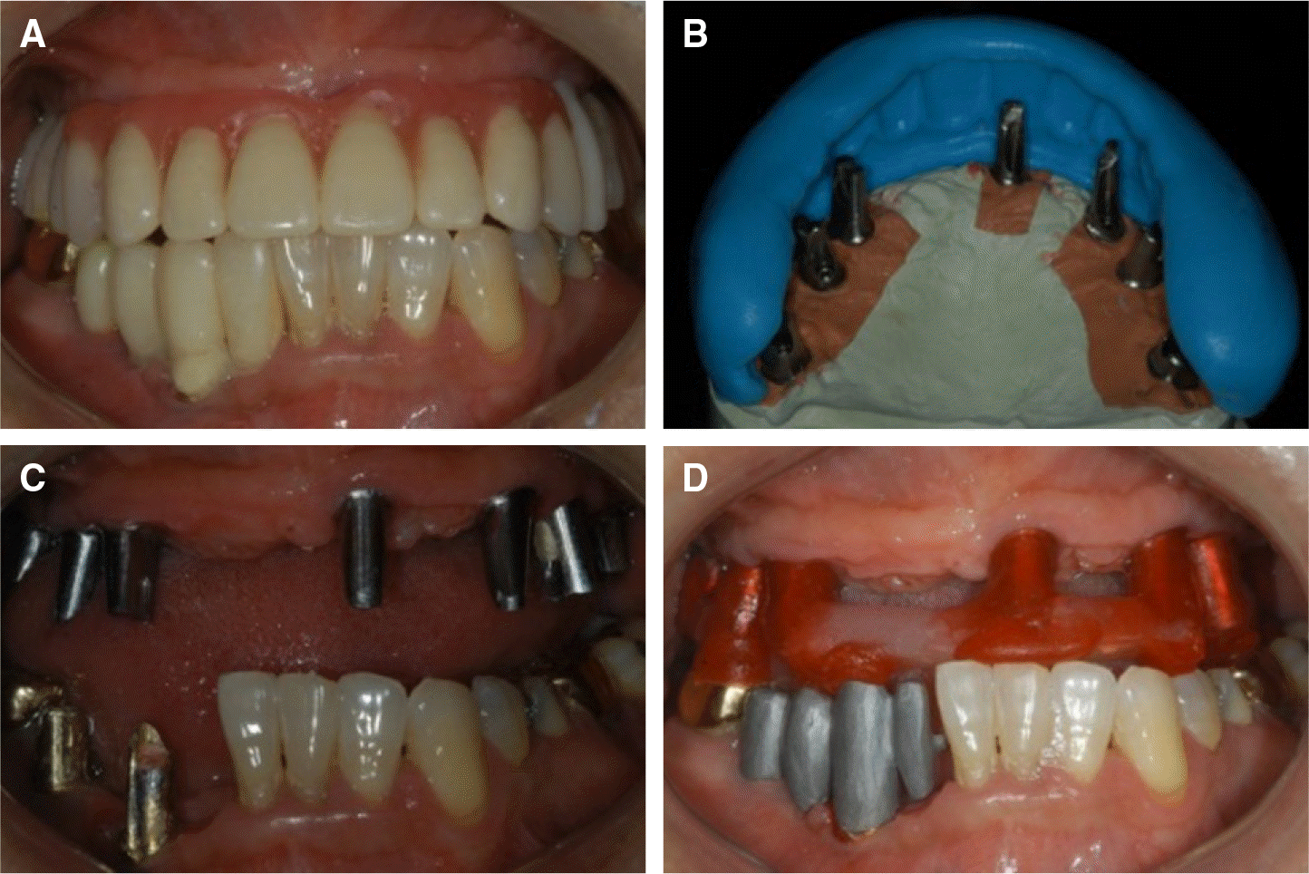 | Fig. 5.Fabrication of customized titanium abutment. (A) Esthetic try-in for abutment fabrication, (B) Putty index for fabrication of abutments with proper position and angulation, (C) Placement of the customized titanium abutment by repositioning jig, (D) Bite registration using pattern resin. |
 | Fig. 7.Definitive restoration. (A) Maximum intercuspation, (B) Protrusive movement, (C) Right lateral movement, (D) Left lateral movement. |
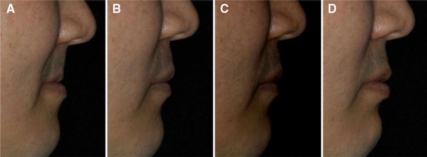 | Fig. 8.Facial profile. (A) pretreatment, (B) during wearing a denture, (C) esthetic try-in, (D) post treatment. |


 PDF
PDF ePub
ePub Citation
 Print
Print



 XML Download