

 PDF
PDF ePub
ePub Citation
 Print
Print


Abstract
Purpose
The aim of this study was to know whether there is significant difference of peri-implant bone density according to the state of antagonist region.
Materials and methods
51 patients who had implant operation in Daejeon Dental Hospital of Wonkwang University participated in this study and total of 51 implants were analyzed. Implants were classified depending on opposing antagonist region, gender, age and location of jaw. The opposing antagonist region was divided into four groups; natural tooth, implant, pontic and edentulous region. Fractal analysis was performed using two periapical radiographs; one after implant placement and the other after 10 weeks following prosthetic restoration. The analysis was done by Image J. The data was statistically analyzed using one-way ANOVA and Tukey multiple comparison test.
Results
The mean value of fractal difference was 0.009 ± 0.048 with opposing natural tooth, 0.026 ± 0.080 with opposing implant, 0.025 ± 0.068 with opposing pontic and 0.093 ± 0.171 with opposing edentulous area. There was a statistically significant difference in fractal value between opposing implant and opposing edentulous state. And there was no statistically significant difference according to age, gender and location of jaw.
Conclusion
There was no statistically significant difference between 3 groups except opposing edentulous region and there was a statistically significant difference between opposing implant and edentulous region. And there was no statistically significant difference according to age, gender and location of jaw. (J Korean Acad Prosthodont 2016;54:14-20)
REFERENCES
1.Pearson OM., Lieberman DE. The aging of Wolff' s “law”: ontogeny and responses to mechanical loading in cortical bone. Am J Phys Anthropol. 2004. 39:63–99.
2.Traini T., Degidi M., Iezzi G., Artese L., Piattelli A. Comparative evaluation of the peri-implant bone tissue mineral density around unloaded titanium dental implants. J Dent. 2007. 35:84–92.

3.Gazit D., Ehrlich J., Kohen Y., Bab I. Effect of occlusal (mechanical) stimulus on bone remodelling in rat mandibular condyle. J Oral Pathol. 1987. 16:395–8.
4.Carlsson L., Röstlund T., Albrektsson B., Albrektsson T. Removal torques for polished and rough titanium implants. Int J Oral Maxillofac Implants. 1988. 3:21–4.
5.Ivanoff CJ., Sennerby L., Johansson C., Rangert B., Lekholm U. Influence of implant diameters on the integration of screw implants. An experimental study in rabbits. Int J Oral Maxillofac Surg. 1997. 26:141–8.
6.Wennerberg A., Albrektsson T., Andersson B., Krol JJ. A histomorphometric and removal torque study of screw-shaped titanium implants with three different surface topographies. Clin Oral Implants Res. 1995. 6:24–30.
7.Johansson C., Albrektsson T. Integration of screw implants in the rabbit: a 1-year follow-up of removal torque of titanium implants. Int J Oral Maxillofac Implants. 1987. 2:69–75.
8.Johansson CB., Sennerby L., Albrektsson T. A removal torque and histomorphometric study of bone tissue reactions to commercially pure titanium and Vitallium implants. Int J Oral Maxillofac Implants. 1991. 6:437–41.
9.Denissen H., Verhey H., de Blieck J., Corten F., Klein C., van Lingen A. Dual X-ray absorptiometry for alveolar bone: precision of peri-implant mineral measurements ex vivo. J Periodontal Res. 1996. 31:265–70.
10.Ellwood R., Horner K., Alexander S., Davies R. A digital subtraction radiography investigation of upper first molar proximal bone density changes in adolescents. J Periodontal Res. 1998. 33:172–7.
11.Bassi F., Procchio M., Fava C., Schierano G., Preti G. Bone density in human dentate and edentulous mandibles using computed tomography. Clin Oral Implants Res. 1999. 10:356–61.
12.Peitgen HO., Jurgens H., Saupe D. Chaos and fractals: new frontiers of science. New York: Springer-Verlag;1992.
13.Weinans H., Huiskes R., Grootenboer HJ. The behavior of adaptive bone-remodeling simulation models. J Biomech. 1992. 25:1425–41.
14.Feik SA., Storey E., Ellender G. Stress induced periosteal changes. Br J Exp Pathol. 1987. 68:803–13.
15.Rubin CT., Lanyon LE. Regulation of bone mass by mechanical strain magnitude. Calcif Tissue Int. 1985. 37:411–7.
16.Urdaneta RA., Daher S., Lery J., Emanuel K., Chuang SK. Factors associated with crestal bone gain on single-tooth locking-taper implants: the effect of nonsteroidal anti-inflammatory drugs. Int J Oral Maxillofac Implants. 2011. 26:1063–78.
17.Urdaneta RA., Leary J., Panetta KM., Chuang SK. The effect of opposing structures, natural teeth vs. implants on crestal bone levels surrounding single-tooth implants. Clin Oral Implants Res. 2014. 25:e179–88.
18.Ruttimann UE., Webber RL., Hazelrig JB. Fractal dimension from radiographs of peridental alveolar bone. A possible diagnostic indicator of osteoporosis. Oral Surg Oral Med Oral Pathol. 1992. 74:98–110.
19.Lynch JA., Hawkes DJ., Buckland-Wright JC. A robust and accurate method for calculating the fractal signature of texture in macroradiographs of osteoarthritic knees. Med Inform (Lond). 1991. 16:241–51.
20.Ulm C., Kneissel M., Schedle A., Solar P., Matejka M., Schneider B., Donath K. Characteristic features of trabecular bone in edentulous maxillae. Clin Oral Implants Res. 1999. 10:459–67.
21.Geraets WG., van der Stelt PF. Fractal properties of bone. Dentomaxillofac Radiol. 2000. 29:144–53.
22.Hong SW., Lee JI., Cho HW. Fractal Analysis of Peri-Implant Bone Mineral Density before and after Functional Loading on Implant. J Dent Rehabil Appl Sci. 2011. 27:359–70.
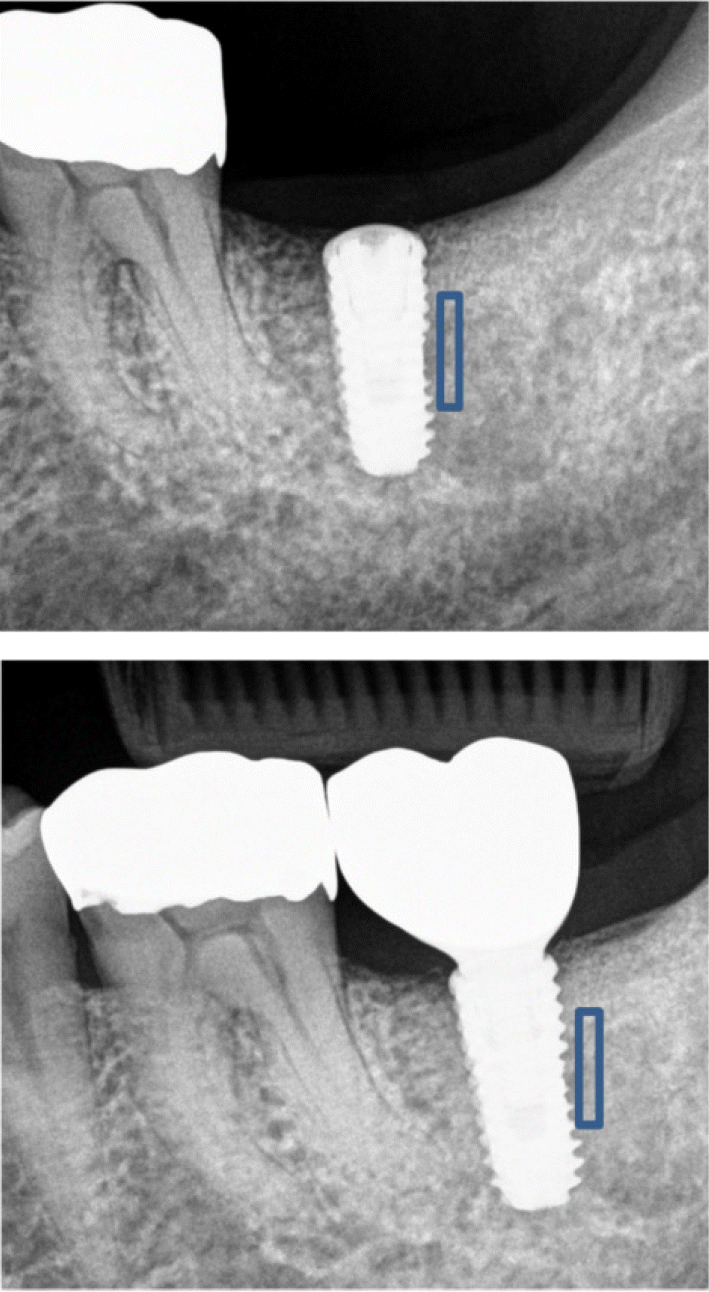
Fig. 2.
Fractal image processing procedures (A. Region of interest: 48 × 426 pixel, B. Add Gaussian filter 35 to region of interest, C. Subtracted (B) from (A), D. Adjust brightness to (C), E. Binary image, F. Erosion image, G. Dilation image, H. Skeletonized image).
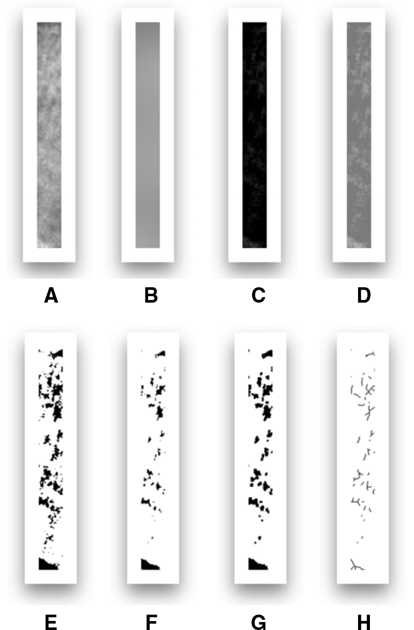
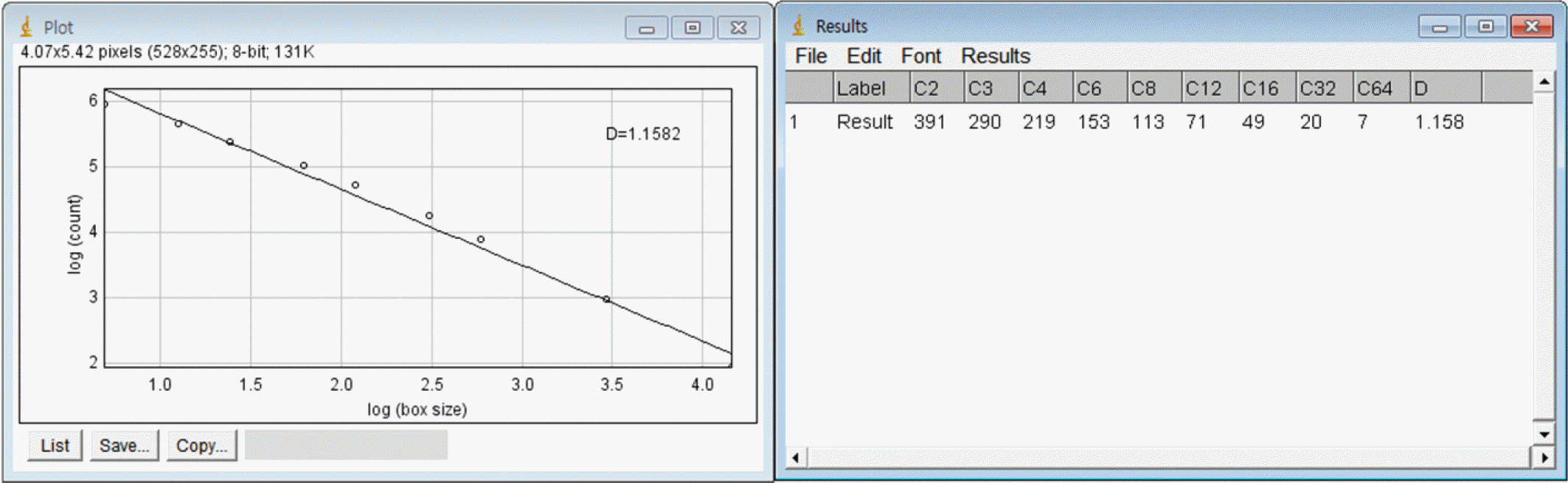
Fig. 4.
Comparison of mean fractal dimension value and standard deviation according to opposing antagonist.
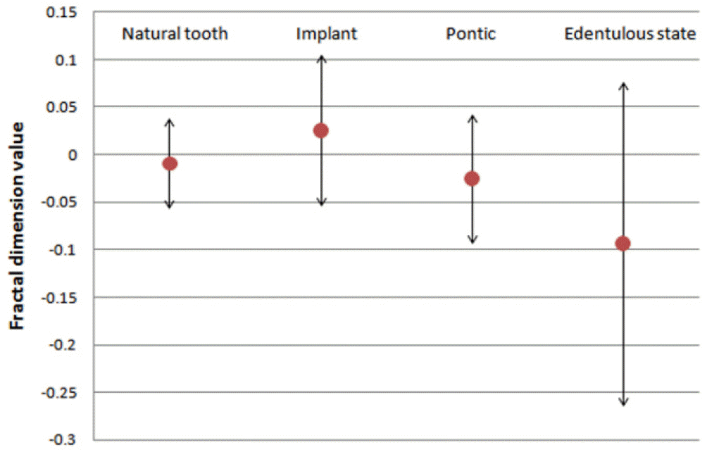
Table 1.
Gender, age and location distribution of study subjects
| n | % | ||
|---|---|---|---|
| Gender | Female | 23 | 45.1 |
| Male | 28 | 54.9 | |
| Age | < 50 | 11 | 21.6 |
| 50 ≤ | 40 | 78.4 | |
| Location | Maxilla | 21 | 41.2 |
| Mandible | 30 | 58.8 |
 XML Download