

 PDF
PDF ePub
ePub Citation
 Print
Print


Abstract
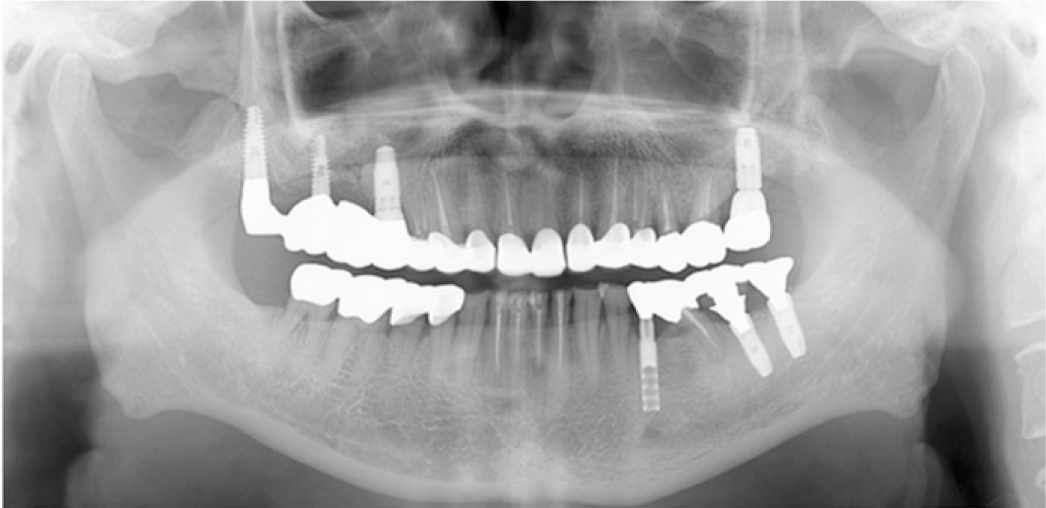
The progressive attrition of teeth is a normal process by aging. However, excessive tooth wear with decreased vertical dimension of occlusion and collapse of occlusal plane may cause pathologic pulpal condition, occlusal disharmony, functional disorders and esthetic problems. The change of vertical dimension is required in case that there is no sufficient clearance for restoration or in case that the occlusal relationship is modified. For gaining the vertical dimension, a careful diagnosis is essential prior to starting the restoration treatment. After evaluating adaptation of neuromuscular system of patient during provisional phase, the final restorations can be fabricated. In this case, a 78 year old male with severely worn down dentition was treated. To improve the esthetic appearance and to achieve the ideal occlusal relationship, the full mouth rehabilitation with minimal increase of vertical dimension is planned and diagnostic wax-up was performed at the increased vertical dimension. After evaluation of provisional restorations for 12 weeks, final restorations were fabricated and routine clinical assessments were made. After 1 year, the restorations with newly established occlusal scheme are well maintained without significant complications and esthetically and functionally satisfactory results were obtained. (J Korean Acad Prosthodont 2016;54:132-9)
Go to : 
REFERENCES
1.Dawson PE. Functional occlusion - From TMJ to smile design. Mosby;2006. p. 430–3.
2.Murphy T. Compensatory mechanisms in facial height adjustment to functional tooth attrition. Aust Dent J. 1959. 4:312–23.

3.Murphy TR. The progressive reduction of tooth cusps as it occurs in natural attrition. Dent Fract Dent Rec. 1968. 19:8–14.
4.Turner KA., Missirlian DM. Restoration of the extremely worn dentition. J Prosthet Dent. 1984. 52:467–74.
5.Silverman MM. The speaking method in measuring vertical dimension. 1952. J Prosthet Dent. 2001. 85:427–31.
6.Silverman MM. Determination of vertical dimension by phonetics. J Prosthet Dent. 1956. 6:467–71.
7.Williamson EH., Caves SA., Edenfield RJ., Morse PK. Cephalometric analysis: comparisons between maximum intercuspation and centric relation. Am J Orthod. 1978. 74:672–7.
8.Willis FM. Features of the face involved in full denture prosthesis. Dent Cosmos. 1935. 77:851–4.
9.Bloom DR., Padayachy JN. Increasing occlusal vertical dimension-why, when and how. Br Dent J. 2006. 200:251–6.
10.Abduo J. Safety of increasing vertical dimension of occlusion: a systematic review. Quintessence Int. 2012. 43:369–80.
11.Rivera-Morales WC., Mohl ND. Relationship of occlusal vertical dimension to the health of the masticatory system. J Prosthet Dent. 1991. 65:547–53.
12.Abduo J., Lyons K. Clinical considerations for increasing occlusal vertical dimension: a review. Aust Dent J. 2012. 57:2–10.
13.Carlsson GE., Ingervall B., Kocak G. Effect of increasing vertical dimension on the masticatory system in subjects with natural teeth. J Prosthet Dent. 1979. 41:284–9.
14.Johansson A. A cross-cultural study of occlusal tooth wear. Swed Dent J Suppl. 1992. 86:1–59.
15.Bindl A., Lüthy H., Mörmann WH. Thin-wall ceramic CAD/CAM crown copings: strength and fracture pattern. J Oral Rehabil. 2006. 33:520–8.
16.Kim JH., Park JH., Park YB., Moon HS. Fracture load of zirconia crowns according to the thickness and marginal design of coping. J Prosthet Dent. 2012. 108:96–101.
17.Park JH., Park S., Lee K., Yun KD., Lim HP. Antagonist wear of three CAD/CAM anatomic contour zirconia ceramics. J Prosthet Dent. 2014. 111:20–9.
18.Yin L., Song XF., Song YL., Huang T., Li J. An overview of in vitro abrasive finishing & CAD/CAM of bioceramics in restorative dentistry. Int J Mach Tool Manu. 2006. 46:1013–26.
19.Amer R., Kürklü D., Kateeb E., Seghi RR. Three-body wear potential of dental yttrium-stabilized zirconia ceramic after grinding, polishing, and glazing treatments. J Prosthet Dent. 2014. 112:1151–5.
20.Mitov G., Heintze SD., Walz S., Woll K., Muecklich F., Pospiech P. Wear behavior of dental Y-TZP ceramic against natural enamel after different finishing procedures. Dent Mater. 2012. 28:909–18.
21.Sailer I., Fehér A., Filser F., Gauckler LJ., Lüthy H., Hämmerle CH. Five-year clinical results of zirconia frameworks for posterior fixed partial dentures. Int J Prosthodont. 2007. 20:383–8.
Go to :
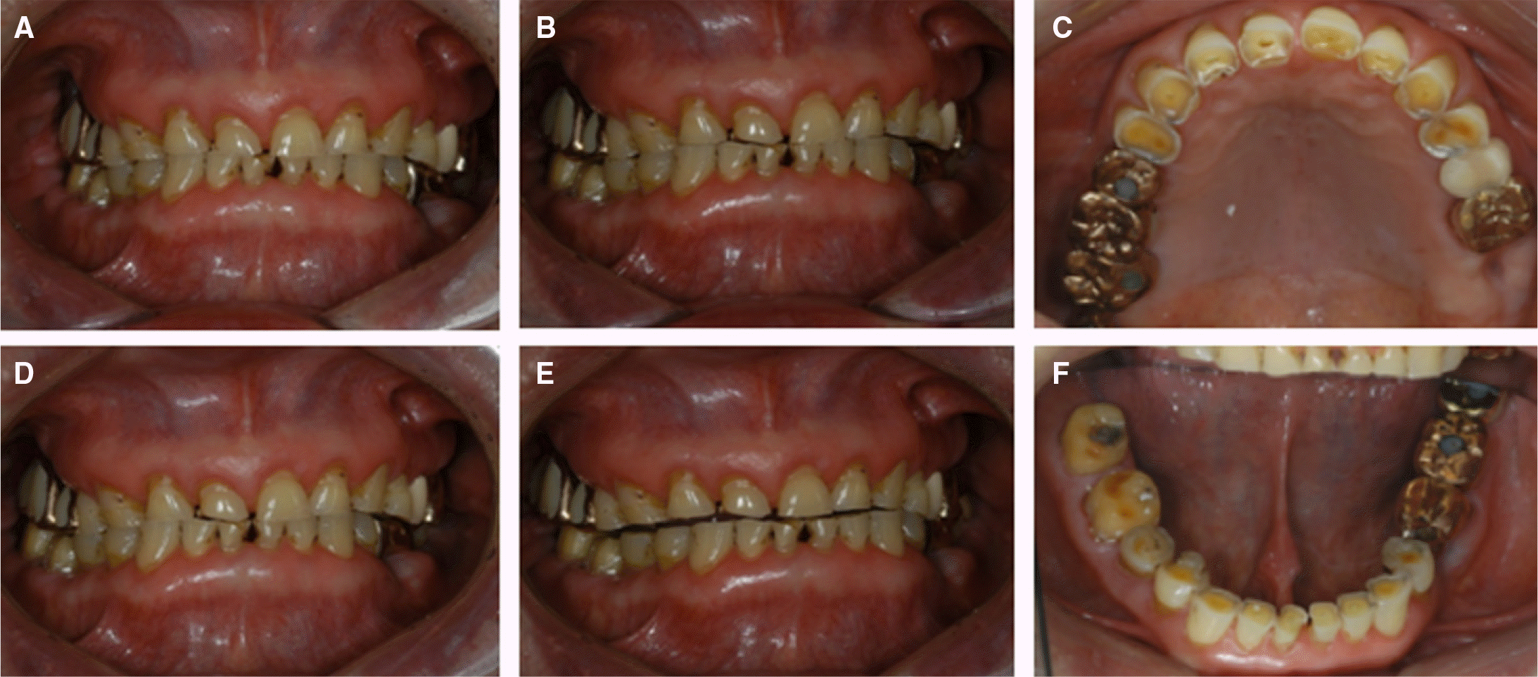
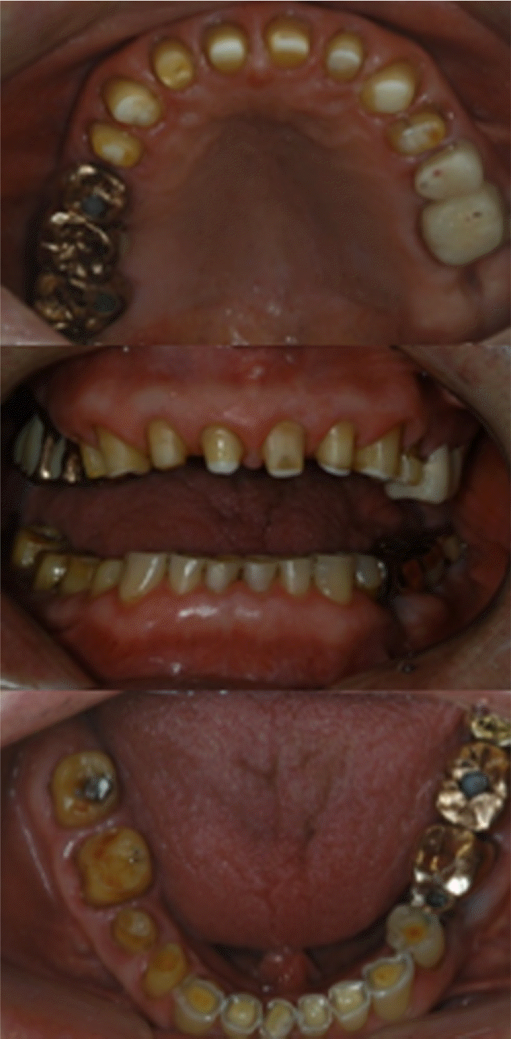
 | Fig. 1.Pretreatment condition. (A) Centric movement, (B) Protrosive movement, (C) Maxillary occlusal, (D) Right movement, (E) Left movement, (F) Mandibular occlusal. |
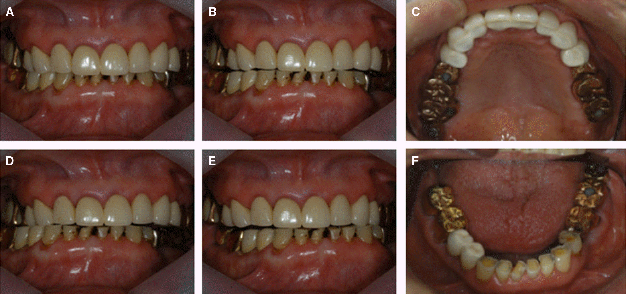
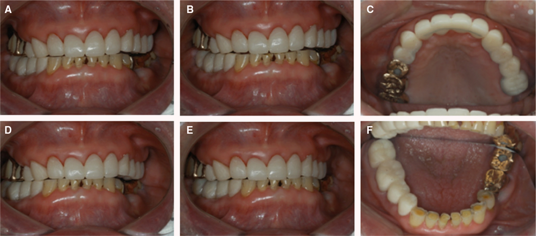 | Fig. 4.Provisional stage (AlikeTM, GC America inc., Alsip, IL, USA) (A) Centric movement, (B) Protrosive movement, (C) Maxillary occlusal view, (D) Right movement, (E) Left movement, (F) Mandibular occlusal view. |
 XML Download