Abstract
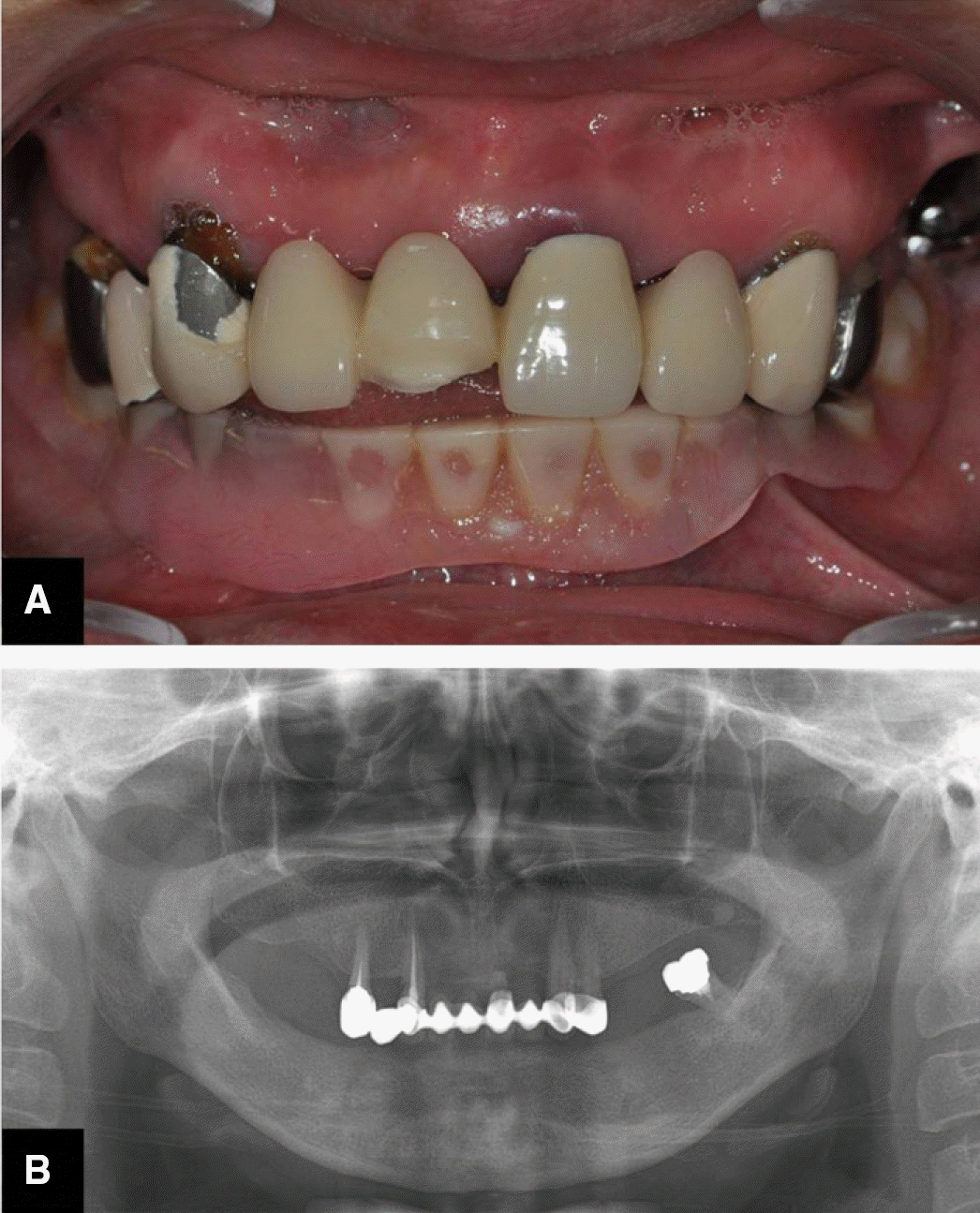
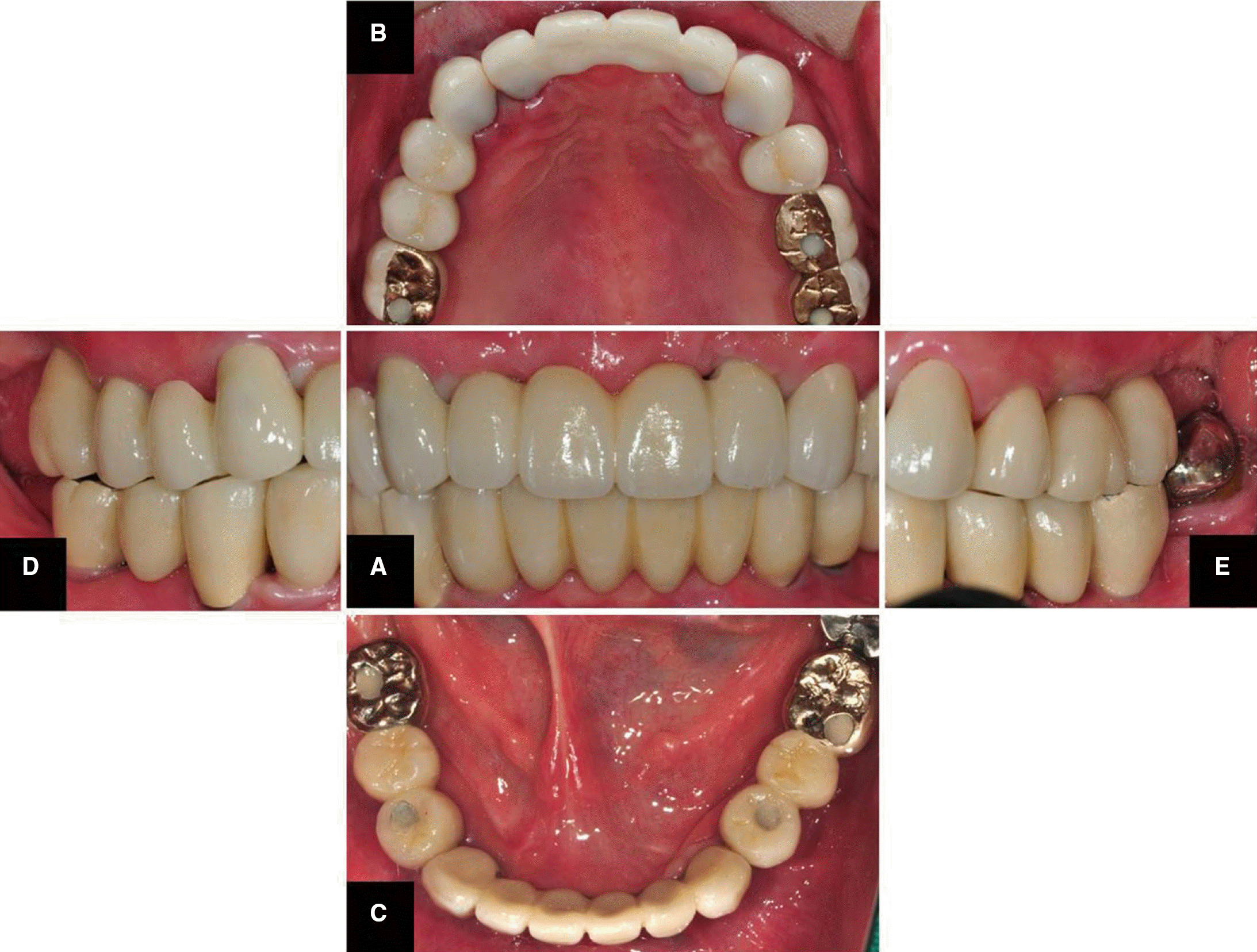
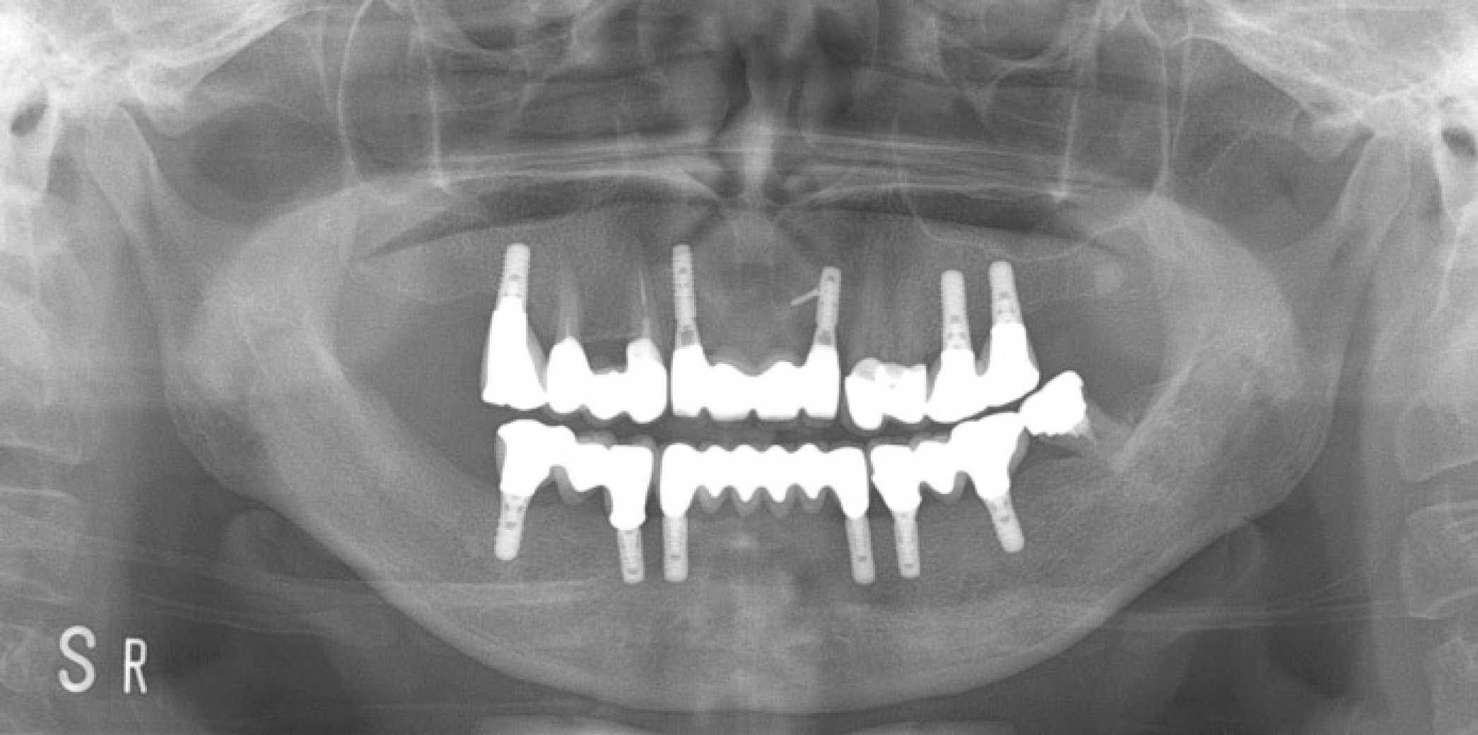
This report describes the prosthetic treatment of a patient with multiple missing teeth. Installation of five fixtures on maxilla with sinus lift and six fixtures on mandible with ramal bone graft were performed. With implant supported all-ceramic with zirconia core using CAD/CAM technology and porcelain-fused-to-gold prosthesis, treatment with positive outcome which satisfies both functional and esthetical aspect was obtained.
REFERENCES
1. Adell R, Lekholm U, Rockler B, Bra�nemark PI. A 15-year study of osseointegrated implants in the treatment of the edentulous jaw. Int J Oral Surg. 1981; 10:387–416.

2. van Steenberghe D, Lekholm U, Bolender C, Folmer T, Henry P, Herrmann I, Higuchi K, Laney W, Linden U, Astrand P. Applicability of osseointegrated oral implants in the rehabilitation of partial edentulism: a prospective multicenter study on 558 fixtures. Int J Oral Maxillofac Implants. 1990; 5:272–81.
3. Pjetursson BE, Karoussis I, Bu¨rgin W, Bra¨gger U, Lang NP. Patients' satisfaction following implant therapy. A 10-year prospective cohort study. Clin Oral Implants Res. 2005; 16:185–93.

4. Budtz-Jo¨rgensen E. Restoration of the partially edentulous mouth-a comparison of overdentures, removable partial dentures, fixed partial dentures and implant treatment. J Dent. 1996; 24:237–44.
5. Raigrodski AJ. Contemporary materials and technologies for all-ceramic fixed partial dentures: a review of the literature. J Prosthet Dent. 2004; 92:557–62.

6. Conrad HJ, Seong WJ, Pesun IJ. Current ceramic materials and systems with clinical recommendations: a systematic review. J Prosthet Dent. 2007; 98:389–404.

7. Pjetursson BE, Tan K, Lang NP, Bra¨gger U, Egger M, Zwahlen M. A systematic review of the survival and complication rates of fixed partial dentures (FPDs) after an observation period of at least 5 years. Clin Oral Implants Res. 2004; 15:625–42.
8. Bozini T, Petridis H, Garefis K, Garefis P. A meta-analysis of prosthodontic complication rates of implant-supported fixed dental prostheses in edentulous patients after an observation period of at least 5 years. Int J Oral Maxillofac Implants. 2011; 26:304–18.
Table 1.
Implant placement data


 PDF
PDF ePub
ePub Citation
 Print
Print


 XML Download