

 PDF
PDF ePub
ePub Citation
 Print
Print


Abstract
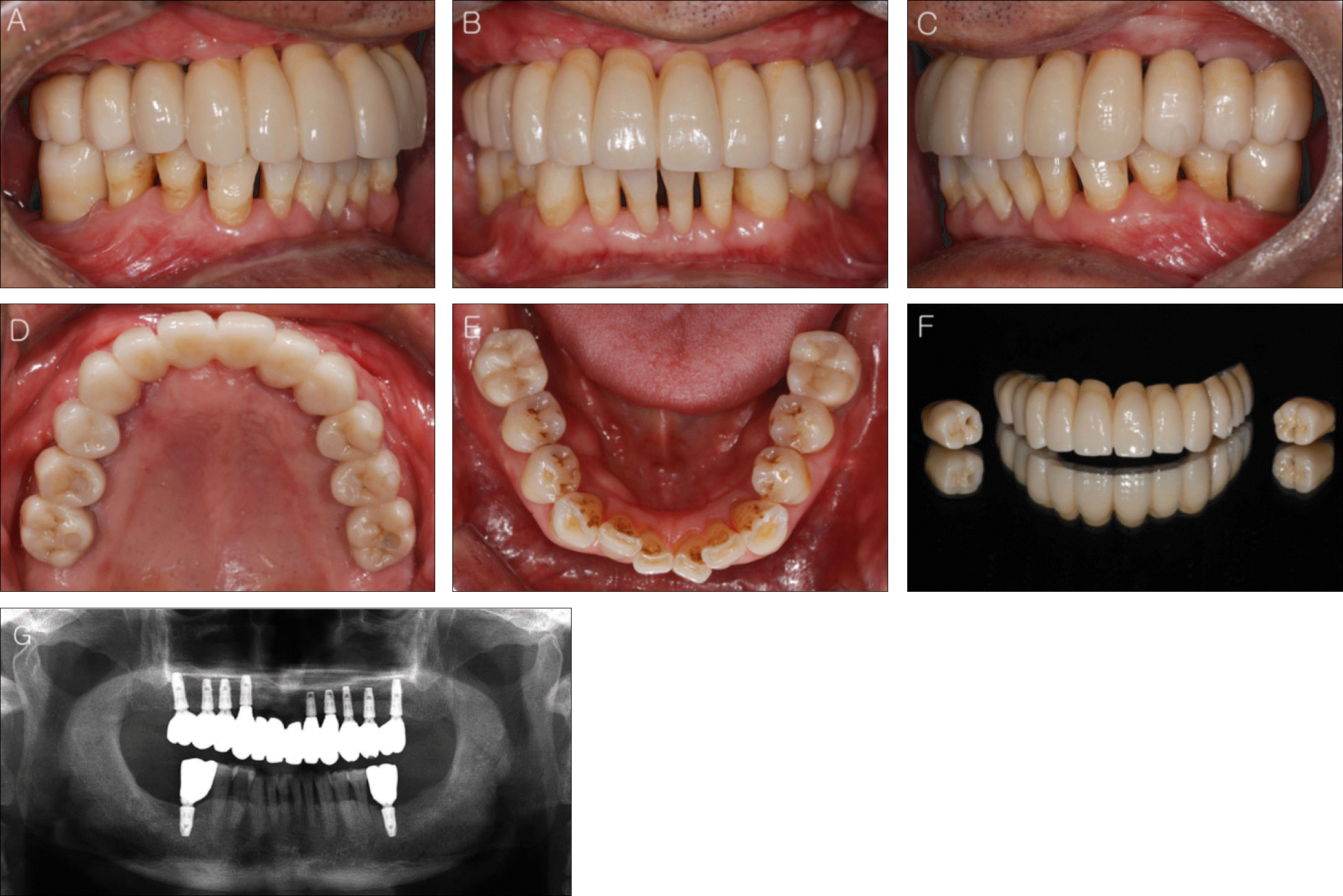
When an implant-supported maxillary full-arch fixed prosthesis is planned for patients with the horizontal and vertical bone resorption induced by periodontal disease, it is necessary to consider the masticatory function, esthetics and phonetics when placing implants. For this reason, thorough clinical and radiological diagnosis is necessary. Extensive bone and soft tissue grafting may be required as well. Since there is no clear guideline for proper number of implants, segment or splinting of substructure and method of retaining prosthesis, these should be considered during diagnostic process. This clinical report describes a patient who has experienced several tooth extractions and periodontal treatment due to severe periodontitis on maxilla and mandible. With bone and soft tissue graft before dental implant placement, the patient have satisfactory result in esthetic and functional aspect with the implant-supported maxillary full-arch fixed prosthesis opposing mandibular natural dentition.
REFERENCES
1. Bra�nemark PI, Svensson B, van Steenberghe D. Ten-year survival rates of fixed prostheses on four or six implants ad modum Bra�nemark in full edentulism. Clin Oral Implants Res. 1995; 6:227–31.
2. Buser D, Ingimarsson S, Dula K, Lussi A, Hirt HP, Belser UC. Long-term stability of osseointegrated implants in augmented bone: a 5-year prospective study in partially edentulous patients. Int J Periodontics Restorative Dent. 2002; 22:109–17.
3. Chiapasco M, Zaniboni M, Boisco M. Augmentation procedures for the rehabilitation of deficient edentulous ridges with oral implants. Clin Oral Implants Res. 2006; 17:136–59.

4. Ha¨mmerle CH, Jung RE, Feloutzis A. A systematic review of the survival of implants in bone sites augmented with barrier mem-branes (guided bone regeneration) in partially edentulous patients. J Clin Periodontol. 2002; 29:226–31.
5. Listgarten MA, Lang NP, Schroeder HE, Schroeder A. Periodontal tissues and their counterparts around endosseous implants. Clin Oral Implants Res. 1991; 2:1–19.
6. Schroeder A, van der Zypen E, Stich H, Sutter F. The reactions of bone, connective tissue, and epithelium to endosteal implants with titanium-sprayed surfaces. J Maxillofac Surg. 1981; 9:15–25.
7. Bachhav VC, Aras MA. Zirconia-based fixed partial dentures: a clinical review. Quintessence Int. 2011; 42:173–82.
8. Misch CE. Contemporary Implant Dentistry. 3rd ed. St. Louis: CV Mosby;2009. p. 367–88.
9. Koo S, Dibart S, Weber HP. Ridge-splitting technique with si-multaneous implant placement. Compend Contin Educ Dent. 2008; 29:106–10.
10. Bra�nemark PI, Hansson BO, Adell R, Breine U, Lindstro¨m J, Halle′n O, Ohman A. Osseointegrated implants in the treatment of the edentulous jaw. Experience from a 10-year period. Scand J Plast Reconstr Surg Suppl. 1977; 16:1–132.
11. Stanford CM. Application of oral implants to the general dental practice. J Am Dent Assoc. 2005; 136:1092–100.
12. Misch CE. Contemporary Implant Dentistry. 2nd ed.St. Louis: CV Mosby;1999. p. 534.
13. Lindhe J. Clinical Periodontology and Implant Dentistry. 5th ed.Blackwell;2008. 1170:p. 1175–6.
14. Taylor TD. Prosthodontic problems and limitations associated with osseointegration. J Prosthet Dent. 1998; 79:74–8.
15. Zarb GA, Schmitt A. The longitudinal clinical effectiveness of osseointegrated dental implants: the Toronto study. Part III: Problems and complications encountered. J Prosthet Dent. 1990; 64:185–94.
16. Goodacre CJ, Kan JY, Rungcharassaeng K. Clinical complications of osseointegrated implants. J Prosthet Dent. 1999; 81:537–52.
17. Pauletto N, Lahiffe BJ, Walton JN. Complications associated with excess cement around crowns on osseointegrated implants: a clinical report. Int J Oral Maxillofac Implants. 1999; 14:865–8.
18. Aparicio C. A new method for achieving passive fit of an interim restoration supported by Bra�nemark implants: a technical note. Int J Oral Maxillofac Implants. 1995; 10:614–8.
19. Preiskel HW, Tsolka P. The DIA anatomic abutment system and telescopic prostheses: a clinical report. Int J Oral Maxillofac Implants. 1997; 12:628–33.
20. Kallus T, Bessing C. Loose gold screws frequently occur in full-arch fixed prostheses supported by osseointegrated implants after 5 years. Int J Oral Maxillofac Implants. 1994; 9:169–78.
21. Salvi GE, Zitzmann NU. The effects of anti-infective preventive measures on the occurrence of biologic implant complications and implant loss: a systematic review. Int J Oral Maxillofac Implants. 2014; 29:292–307.
Fig. 2.
Two months after extractions in maxilla and mandible. (A) Upper occlusal view, (B) Lower occlusal view.
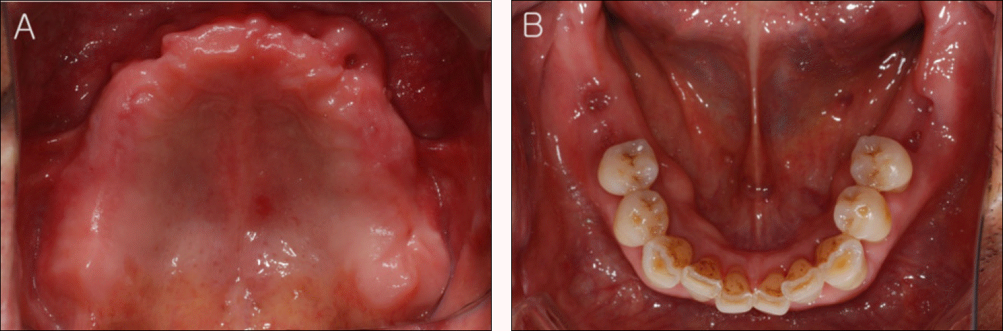
Fig. 3.
Panoramic view was taken after implant placement in maxilla and mandible. There was not any special findings.
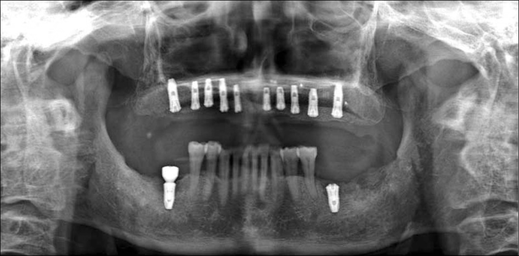
Fig. 4.
Intraoral photographs show healing abutment connection on each implants at second surgery stage. (A) Upper occlusal view, (B) Lower occlusal view.
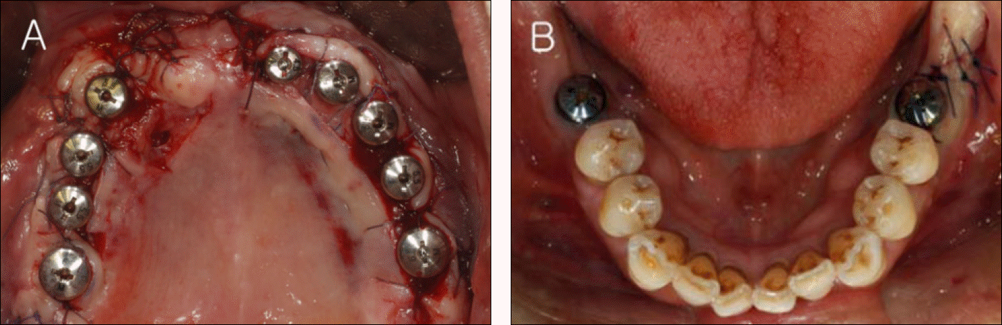
Fig. 7.
Try-in of customized abutment was performed. (A) Upper occlusal view, (B) Lower occlusal view.
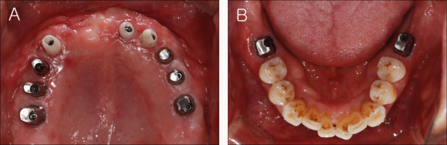
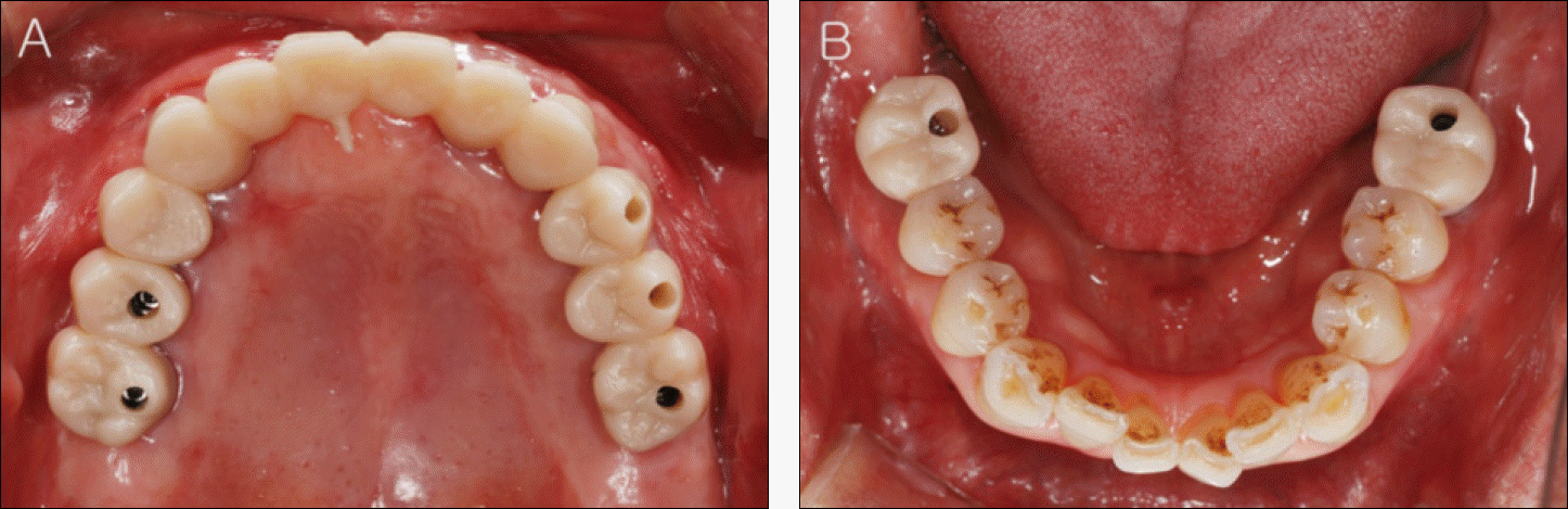
Fig. 8.
Try-in of zirconia framework was performed. (A) Upper occlusal view, (B) Lower occlusal view.
 XML Download