Abstract
Loss of continuity of the mandible destroys the balance and symmetry of mandibular function, leading to altered mandibular movements and deviation of the residual fragment towards the resected side. Apart from deviation, other dysfunctions include difficulty in swallowing, speech, mandibular movements, mastication, and respiration are accompanied. In general, surgical reconstruction is considered first then proceeds to the prosthetic restorations. However, patients with systemic disease such as BRONJ (Bisphosphonate related osteonecrosis of the Jaw), surgical reconstruction may be limited. Thus, the prosthetic restoration remains as the only resort. Numerous prosthetic methods are employed to minimize deviation and to improve masticatory efficiency, function and esthetics. If a removable partial denture is the selected treatment modality, maximum stability of the partial denture base may be accomplished with a functional impression procedure by means of eliminating lateral and horizontal forces caused by the functional movements of the lips, cheeks and tongue. Also, Twin occlusion is used to obtain a favorable occlusal relationship and check support for esthetics. The purpose of this case report is to demon-strate how neutral zone impression technique and twin occlusion scheme were applied to restore a hemimandiblectomy patient with BRONJ syndrome to achieve satisfactory results in functional and esthetic aspects.
REFERENCES
1. Cantor R, Curtis TA. Prosthetic management of edentulous mandibulectomy patients. I. Anatomic, physiologic, and psychologic considerations. J Prosthet Dent. 1971; 25:446–57.
2. Beumer J, Curtis TA, Marunick MT. Maxillofacial rehabilitation: Prosthodontic and surgical considerations. 2nd ed.St. Louis: Ishiyaku Euro America;1996.
3. Beumer J, Curtis T, Firtell D. Maxillofacial Rehabilitation: Prosthodontic and Surgical Considerations. St. Louis: CV Mosby;1979. p. 90–169.
4. Swoope CC. Prosthetic management of resected edentulous mandibles. J Prosthet Dent. 1969; 21:197–202.

5. Rosenthal LE. The edentulous patient with jaw defects. D Clin N Am. 1994; 8:773–9.
6. Shukla P, Hegde C, Rampal N, Pawah S, Gupta A, Shukla M. Modified technique of resection denture prosthesis fabrication for a patient with segmental mandibulectomy-a case report. Eur J Prosthodont Restor Dent. 2011; 19:175–8.
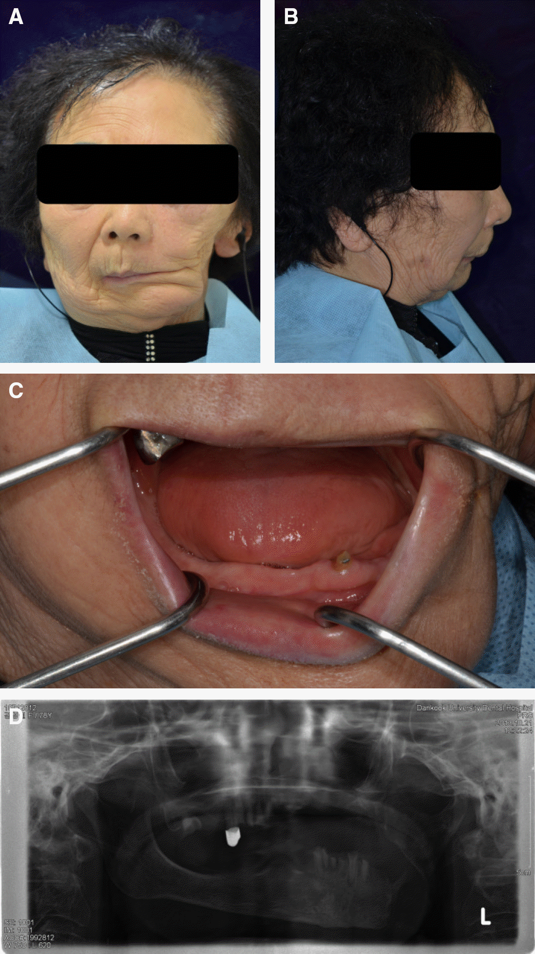
Fig. 1.
Initial photographs. (A) Frontal view, (B) Lateral view, (C) Opening position, (D) Panoramic view.

Fig. 3.
(A) Maxillary temporary denture, (B) Mandibular temporary denture, (C) Intraoral photograph after the placement of temporary denture.

Fig. 4.
Modified mandibular temporary denture. (A) Occlusal surface of temporary denture. (B) Tissue surface of temporary denture. (C) Intraoral photograph after the placement of modified temporary denture.

Fig. 5.
(A) Teeth preparation for attachment & coping, (B) Final impression, (C) Intraoral photograph of placement of attachment & coping.

Fig. 7.
(A) Upper wax rim with two rows on the unaffected side, (B) Resin base with wire loops, (C) Adaptation of wire loops in accordance with obtained vertical dimension.

Fig. 8.
(A) Recording neutral zone with tissue conditioner, (B) Putty index surrounding neutral zone impression.


 PDF
PDF ePub
ePub Citation
 Print
Print


 XML Download