

 PDF
PDF ePub
ePub Citation
 Print
Print


Abstract
Inter-dental arch discrepancy between maxilla and mandible could cause three dimensional occlusal problems, and collapse of occlusal plane, multiple teeth loss and decrease of masticatory efficiency could be observed in patient having unstable occlusal contact. Patient showing posterior bite collapse, unstable occlusal contact and improper anterior guidance should be treated to recover stable centric occlusion, occlusal contact, and anterior guidance in conjunction between prosthodontics and orthodontic treatment. This clinical report describes the favorable results of orthodontic and prosthodontics rehabilitation of patient with above mentioned problems.
Go to : 
REFERENCES
1. Moyers RE. Handbook of orthodontics. Year Book Medical Publishers Inc. Chicago, London, Boca Raton;. 1988. 392–96.
2. Tamamura N, Kuroda S, Sugawara Y, Takano-Yamamoto T, Yamashiro T. Use of palatal miniscrew anchorage and lingual mul-ti-bracket appliances to enhance efficiency of molar scissors-bite correction. Angle Orthod. 2009; 79:577–84.

3. Chugh VK, Sharma VP, Tandon P, Singh GP. Brodie bite with an extracted mandibular first molar in a young adult: a case report. Am J Orthod Dentofacial Orthop. 2010; 137:694–700.
4. Yun SW, Lim WH, Chong DR, Chun YS. Scissors-bite correction on second molar with a dragon helix appliance. Am J Orthod Dentofacial Orthop. 2007; 132:842–7.
5. Tomonari H, Kubota T, Yagi T, Kuninori T, Kitashima F, Uehara S, Miyawaki S. Posterior scissors-bite: masticatory jaw movement and muscle activity. J Oral Rehabil. 2014; 41:257–65.
6. Shifman A, Laufer BZ, Chweidan H. Posterior bite collapse-re-visited. J Oral Rehabil. 1998; 25:376–85.
7. Amsterdam M. Periodontal prosthesis. Twenty-five years in retrospect. Alpha Omegan. 1974; 67:8–52.
8. Ramfjord SP, Ash MM. Occlusion. WB Saunders Co;Philadelphia: 1966. p. 115.
9. The glossary of prosthodontic terms. J Prosthet Dent. 2005; 94:10–92.
10. Dawson PE. Functional occlusion: From TMJ to smile design. Mosby;. Elsevier Health Sciences;2006. p. 453–77.
11. Ishihara Y, Kuroda S, Sugawara Y, Kurosaka H, Takano-Yamamoto T, Yamashiro T. Long-term stability of implant-an-chored orthodontics in an adult patient with a Class II Division 2 malocclusion and a unilateral molar scissors-bite. Am J Orthod Dentofacial Orthop. 2014; 145:S100–13.
12. Weiland FJ, Bantleon HP, Droschl H. Evaluation of continuous arch and segmented arch leveling techniques in adult patients-a clinical study. Am J Orthod Dentofacial Orthop. 1996; 110:647–52.
13. Nanda R. Correction of deep overbite in adults. Dent Clin North Am. 1997; 41:67–87.
14. Wennströ m JL, Stokland BL, Nyman S, Thilander B. Periodontal tissue response to orthodontic movement of teeth with in-frabony pockets. Am J Orthod Dentofacial Orthop. 1993; 103:313–9.
15. McFadden WM, Engstrom C, Engstrom H, Anholm JM. A study of the relationship between incisor intrusion and root shortening. Am J Orthod Dentofacial Orthop. 1989; 96:390–6.
16. Pancherz H, Anehus-Pancherz M. Muscle activity in class II, division 1 malocclusions treated by bite jumping with the Herbst appliance. An electromyographic study. Am J Orthod. 1980; 78:321–9.
17. Kwak YY, Jang I, Choi DS, Cha BK. Functional evaluation of orthopedic and orthodontic treatment in a patient with unilateral posterior crossbite and facial asymmetry. Korean J Orthod. 2014; 44:143–53.
18. Kawakami S, Kumazaki Y, Manda Y, Oki K, Minagi S. Specific diurnal EMG activity pattern observed in occlusal collapse patients: relationship between diurnal bruxism and tooth loss pro-gression. PLoS One. 2014; 9:e101882.
Go to :
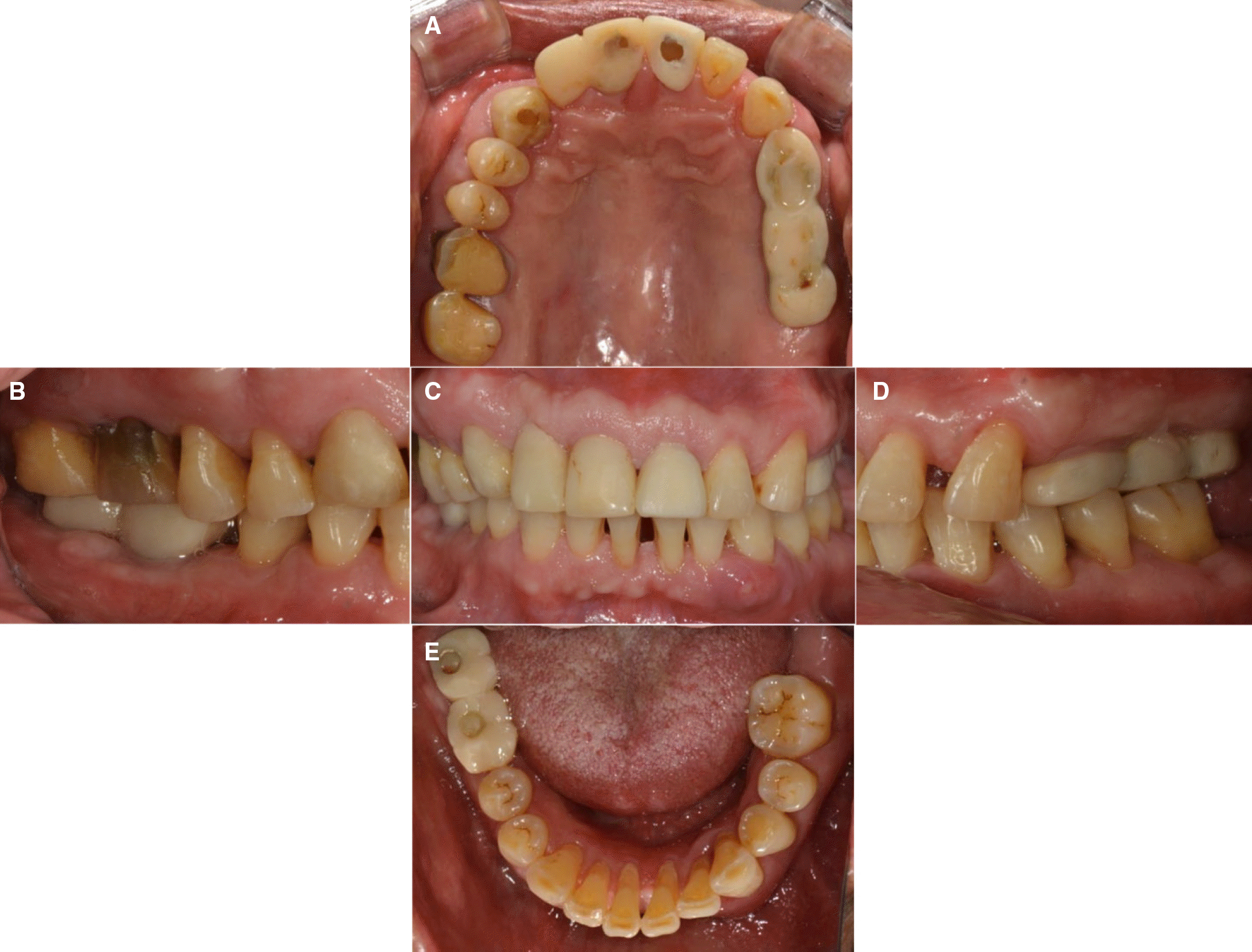
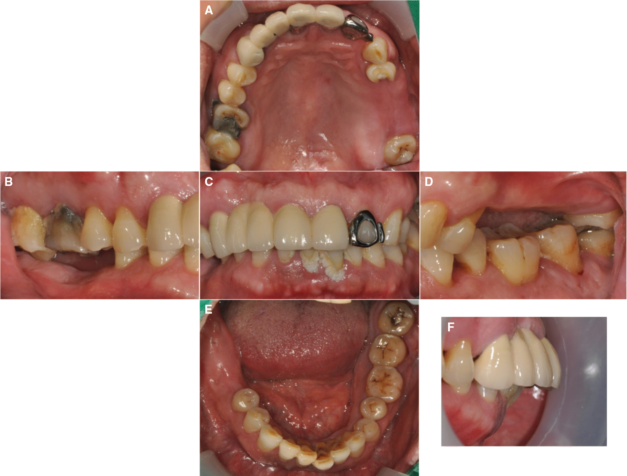 | Fig. 1.Intraoral view at first visit (A) Maxillary occlusal view, (B) Right lateral view, (C) Frontal view, (D) Left lateral view, (E) Mandibular occlusal view, (F) Anterior lateral view. |
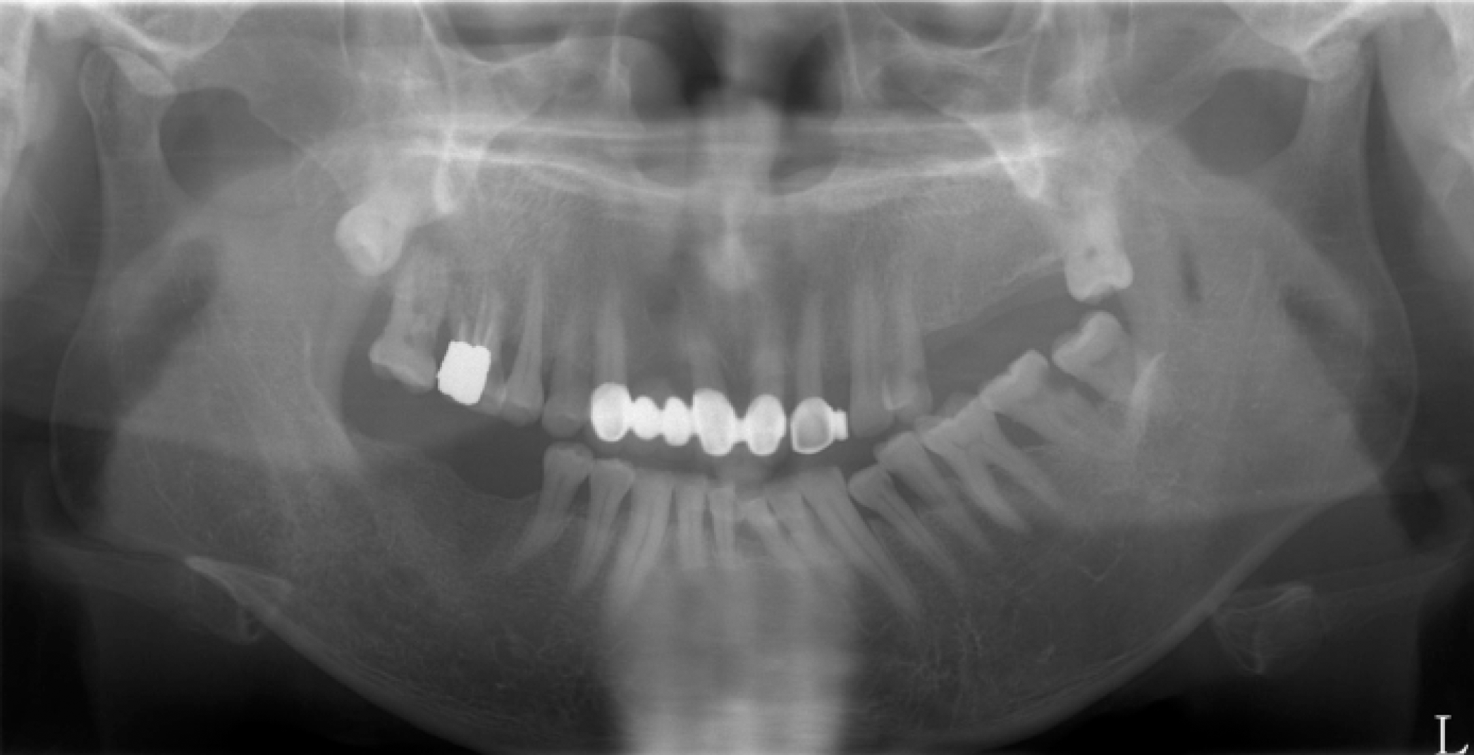
 | Fig. 3.Transcranial radiograph (A) Initial stage, (B) Provisional restoration stage, (C) Definitive restoration stage. |
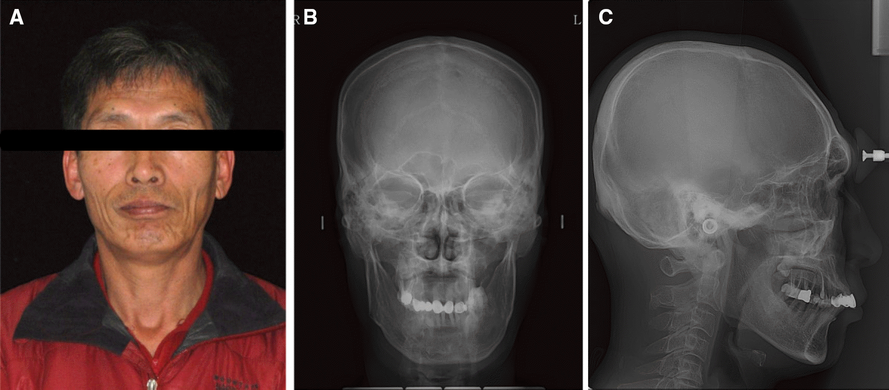 | Fig. 4.(A) Extraoral view at first visit, (B) Posteroanterior cephalometric radiograph, (C) Lateral cephalometric radiograph. |
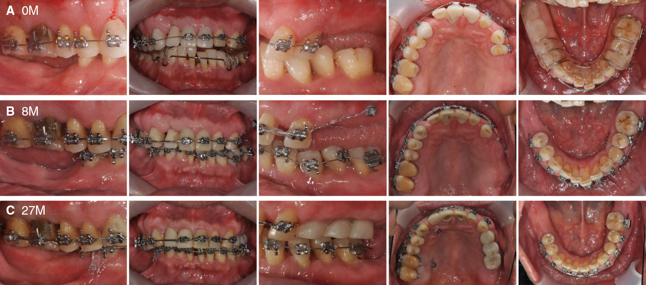 | Fig. 5.The progress of orthodontic treatment (A) Initial leveling of the upper teeth, (B) Retraction of maxillary incisors with chain elastic and initial leveling of the lower teeth, (C) Intrusion of #13 (not seen in the picture) and #44, 45 using the miniscrews to correct scissors bite. |
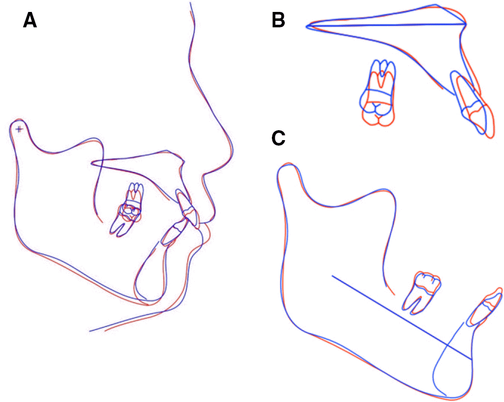 | Fig. 6.Cephalometric superimposition of initial stage (red) and post-orthodontic stage (blue), (A) Overall superimposition, (B) Maxillary superimposition, (C) Mandibular superimposition. |
 | Fig. 7.Intraoral views at post-orthodontic state. (A) Maxillary occlusal view, (B) Right lateral view, (C) Frontal view, (D) Left lateral view, (E) Mandibular occlusal view. |
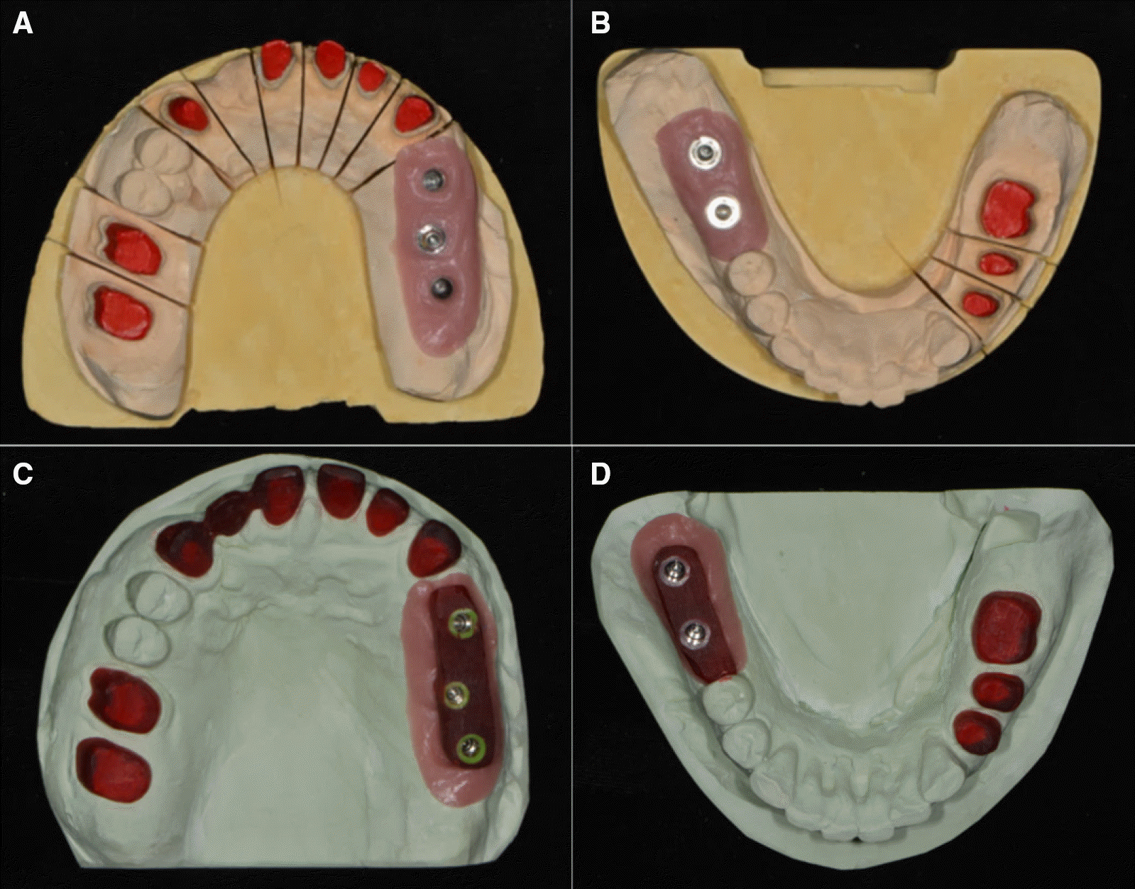
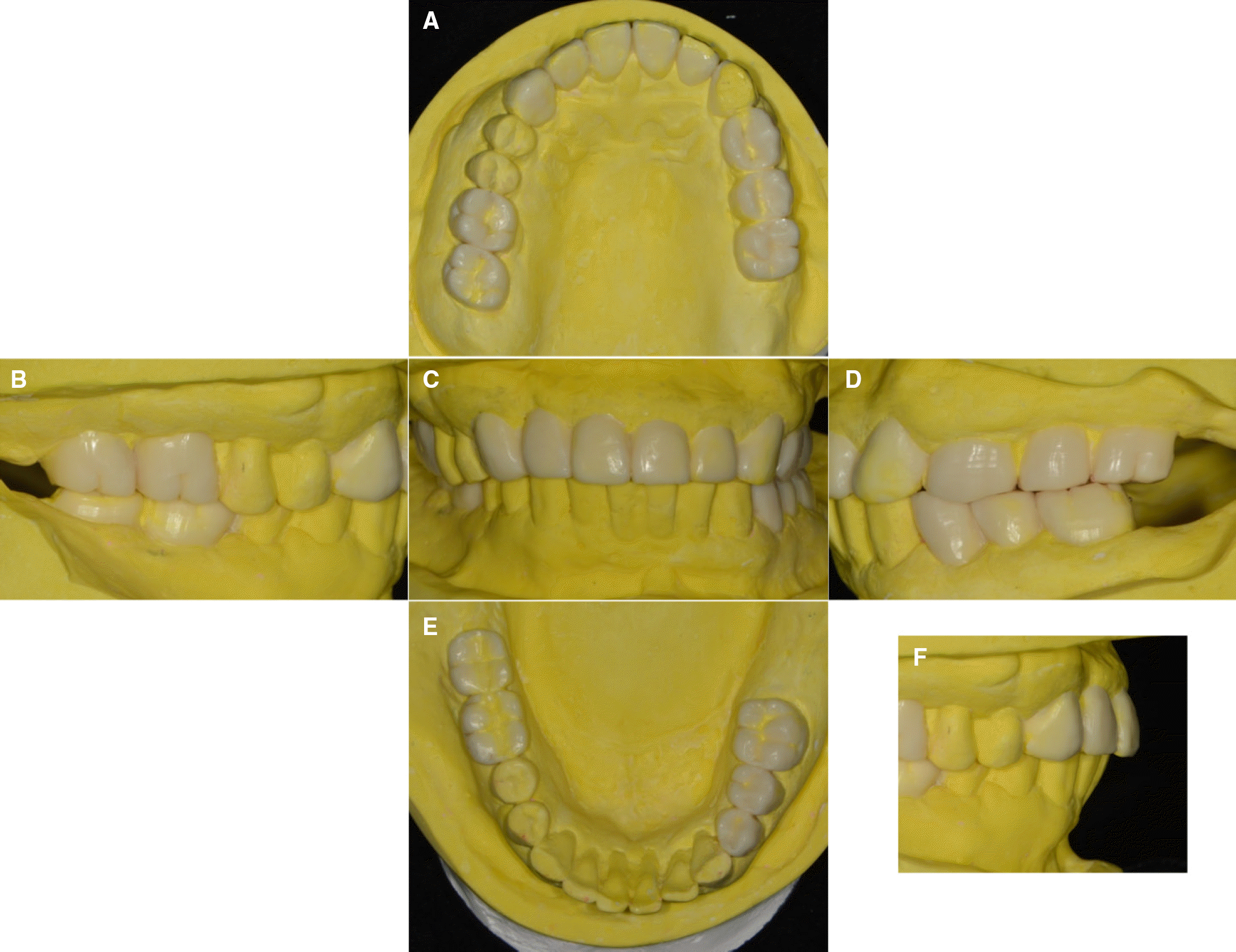 | Fig. 8.Diagnostic wax pattern. (A) Maxillary occlusal view, (B) Right lateral view, (C) Frontal view, (D) Left lateral view, (E) Mandibular occlusal view, (F) Anterior lateral view. |
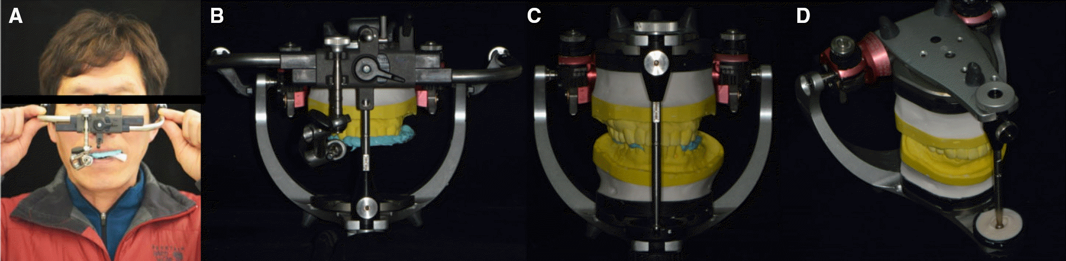 | Fig. 9.(A) Facebow transfer, (B), (C) Mounting procedure to semi-adjustable articulator, (D) Customized anterior guidance table. |
 | Fig. 10.(A) Working cast with a separate die (maxilla), (B) Working cast with a separate die (mandible), (C) Resin coping and pattern resin (maxilla), (D) Resin coping and pattern resin (mandible). |
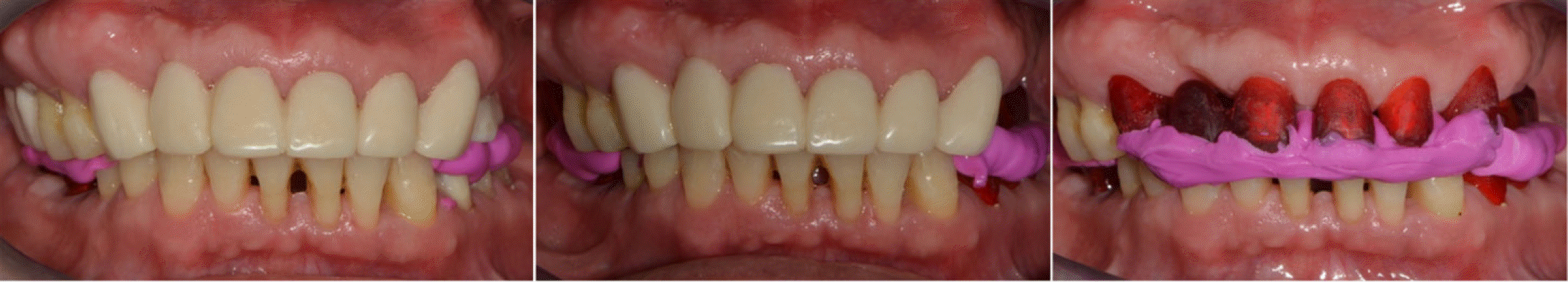 | Fig. 11.Interocclusal relationship registrations using provision restorations and resin coping. |
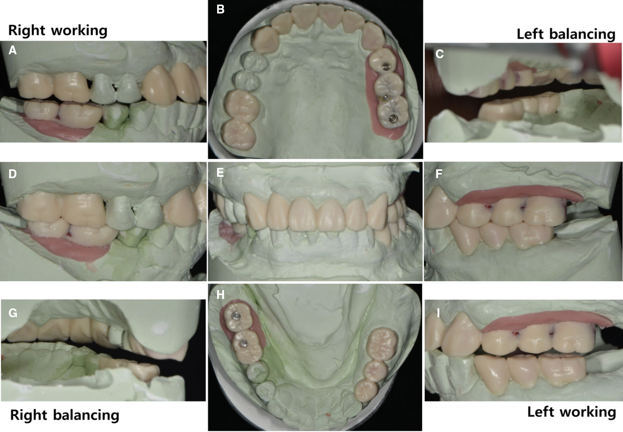 | Fig. 12.Wax pattern for definitive restoration. (A) Working side during right lateral excursion, (B) Maxillary occlusal view, (C) Non-working side during right lateral excursion, (D) Right lateral view at centric occlusion, (E) Frontal view at centric occlusion, (F) Left lateral view at centric occlusion, (G) Non-working side during left lateral excursion, (H) Mandibular occlusal view, (I) Working side during left lateral excursion. |
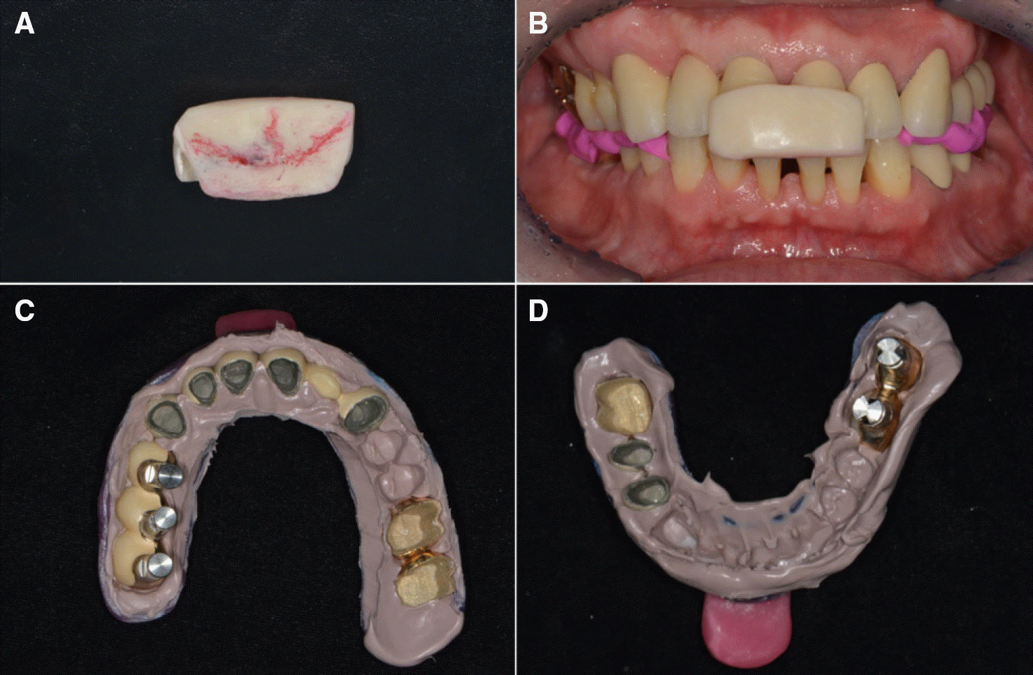 | Fig. 13.Clinical remounting using anterior programming device and pick-up impression. (A) Anterior programming device, (B) Interocclusal relation recording, (C) Pick-up impression for maxillary prosthesis, (D) Pick-up impression for mandibular prosthesis. |
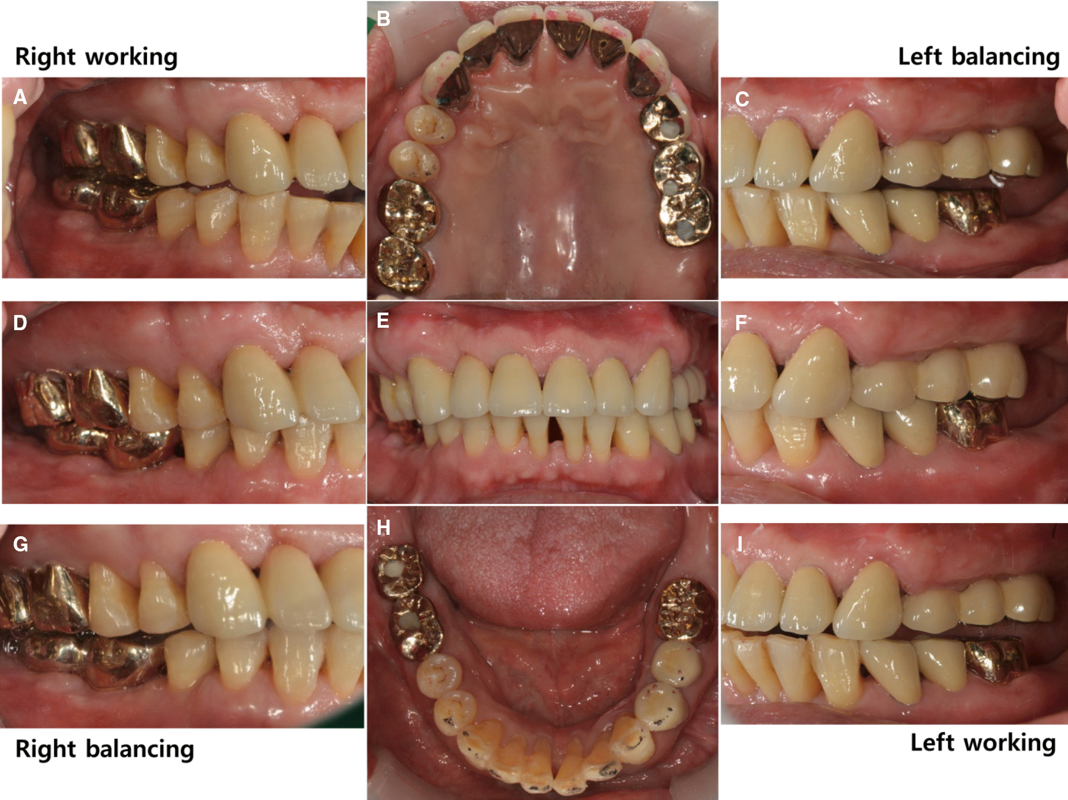 | Fig. 14.Definitive prosthesis. (A) Working side during right lateral excursion, (B) Maxillary occlusal view, (C) Non-working side during right lateral excursion, (D) Left lateral view at centric occlusion, (E) Frontal view at centric occlusion, (F) Right lateral view at centric occlusion, (G) Non-working side during left lateral excursion, (H) Mandibular occlusal view, (I) Working side during left lateral excursion. |
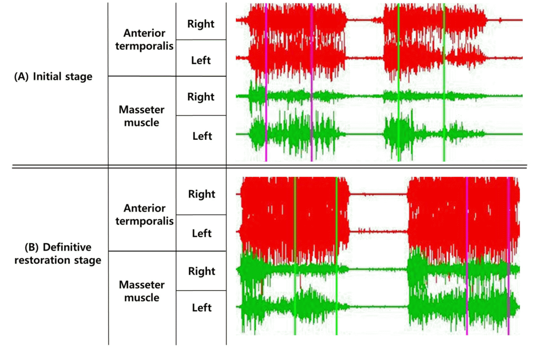 | Fig. 17.Electromyographic recordings of the anterior temporal muscle and masseter muscle, (A) Initial stage, (B) Definitive restoration stage. |
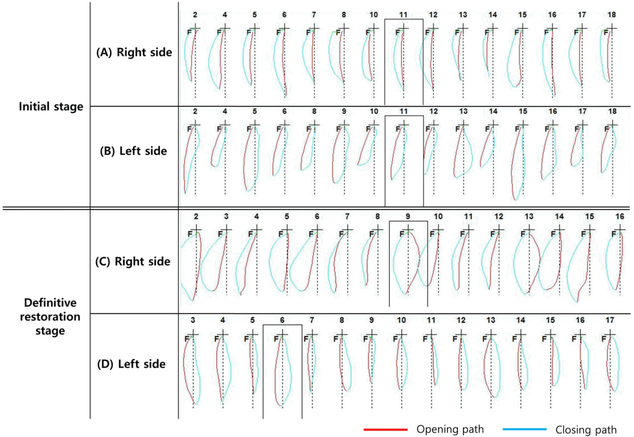 | Fig. 18.Chewing pattern analysis (A) Initial stage of right side (scissor-bite state; chopping chewing pattern), (B) Initial stage of left side, (C) Definitive restoration stage of right side (grinding chewing pattern), (D) Definitive restoration stage of left side. |
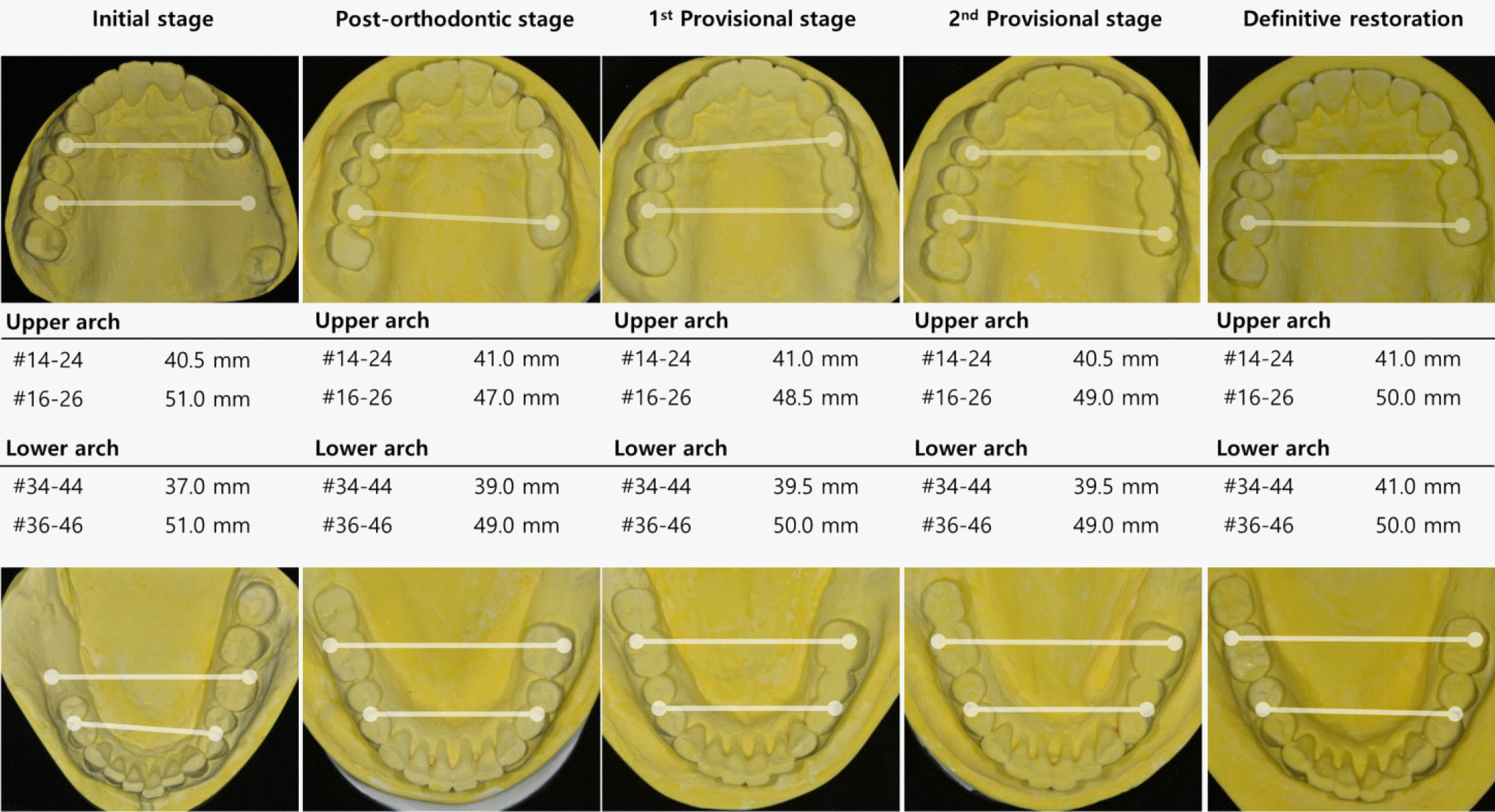
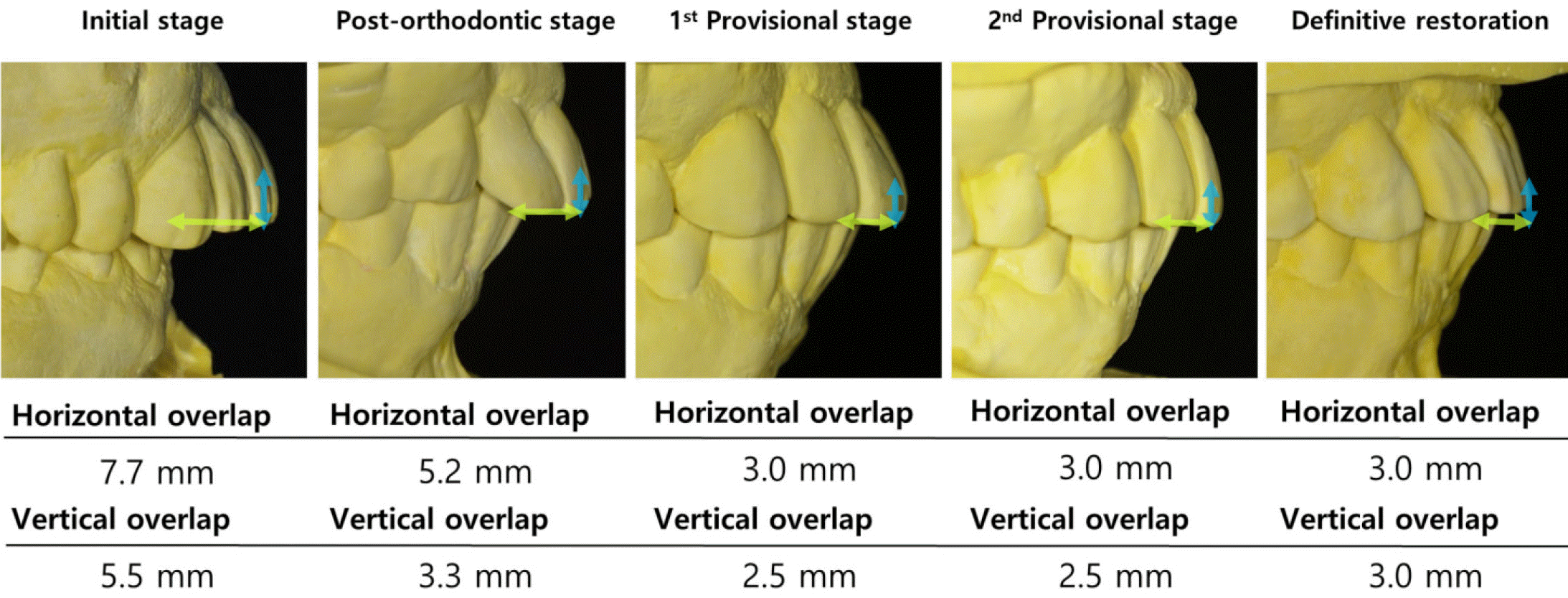
Table 1.
Cephalometric measurements of pretreatment and posttreatment
 XML Download