

 PDF
PDF ePub
ePub Citation
 Print
Print


Abstract
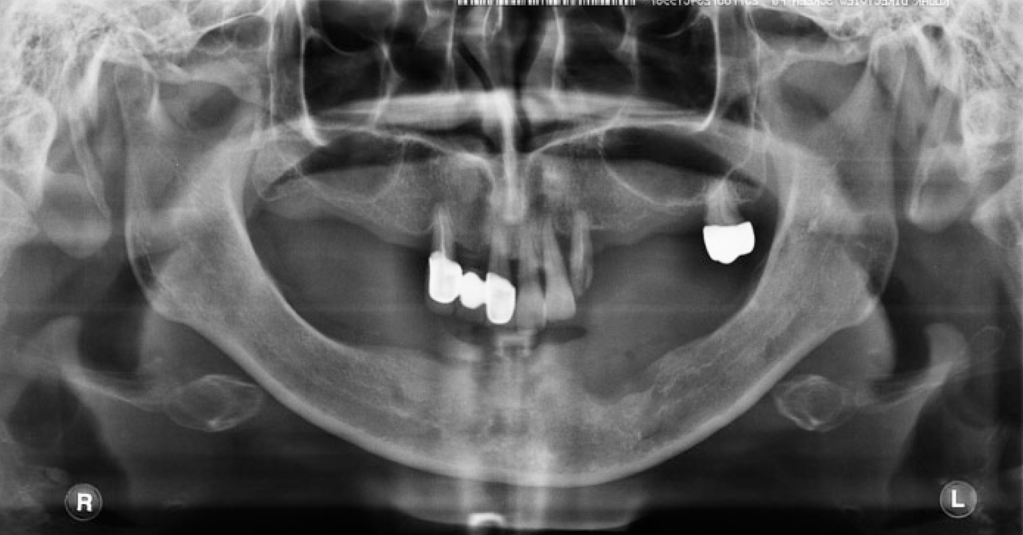
Long-term use of ill-fitting dentures may cause irregular movements of the mandible. In such cases, satisfactory outcomes both in terms of function and esthetics could be achieved by accurate registration of mandibular movement and centric relation when fabricating new dentures. In this case, treatment dentures with flat occlusal tables were used to reg-ister centric relation and mandibular movements of a patient with erratic mandibular movements.
Go to : 
REFERENCES
1. Gunne HS, Bergman B, Enbom L, Hö gströ m J. Masticatory efficiency of complete denture patients. A clinical examination of potential changes at the transition from old to new denture. Acta Odontol Scand. 1982; 40:289–97.
2. Sakurai Y. Treatment denture. Pract Prosthodont. 1990; 23:584–628.
3. Inada M, Yamazaki T, Shinozuka O, Sekiguchi G, Tamamori Y, Ohyama T. Complete denture treatments for a cerebral palsy patient by using a treatment denture. A case report. J Med Dent Sci. 2002; 49:171–7.
4. Sasaki K, Watanabe M. Function of periodontal ligament related to occlusion. Dent Outlook Extra Issue. 1992. 43–54.
5. Ishikawa T. Response properties of single sensory units innervating human temporomandibular joint. Kokubyo Gakkai Zasshi. 1989; 56:528–39.

6. Morimoto T, Takebe H, Sakan I, Kawamura Y. Reflex activation of extrinsic tongue muscles by jaw closing muscle proprio-ceptors. Jpn J Physiol. 1978; 28:461–71.
7. Morimoto T, Nagashima M, Yoshikawa K. Physiological mech-anism controlling biting force and chewing force. Dent Outlook Extra Issue. 1992. 81–93.
8. Abe J. Clinical mandibular position of edentulous patient. The utility and clinical methods of treatment denture. Nippon Dent Rev. 2001; 61:109–16.
9. Nishimura T. The correction of horizontal occlusal relationship in edentulous patients. J Jpn Prosthodont Soc. 1972; 16:420–42.
10. Ohguri T, Kawano F, Ichikawa T, Matsumoto N. Influence of occlusal scheme on the pressure distribution under a complete denture. Int J Prosthodont. 1999; 12:353–8.
Go to :
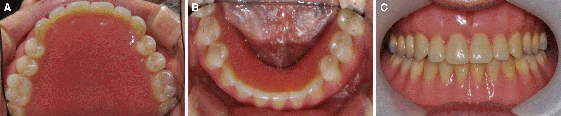
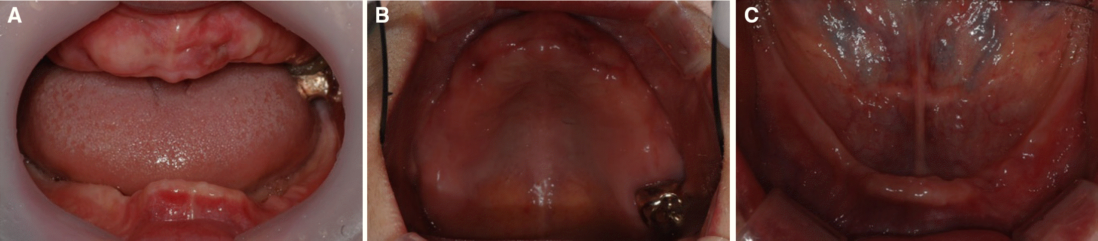 | Fig. 2.Initial intraoral photographs. Severe alveolar ridge atrophy observed on mandible. (A) Frontal view, (B) Maxillary occlusal view, (C) Mandibular occlusal view. |
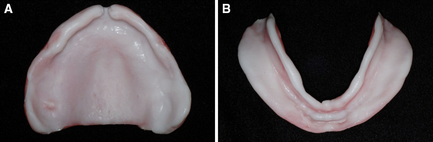
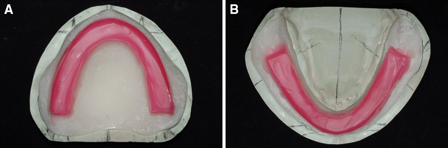 | Fig. 3.Resin recording base fabricated on master casts. (A) Maxillary master cast, (B) Mandibular master cast. |
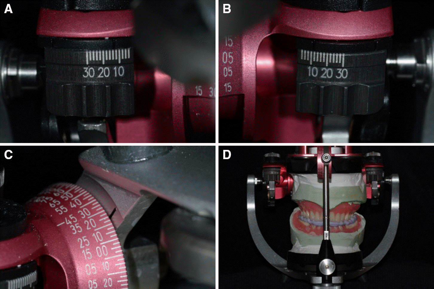
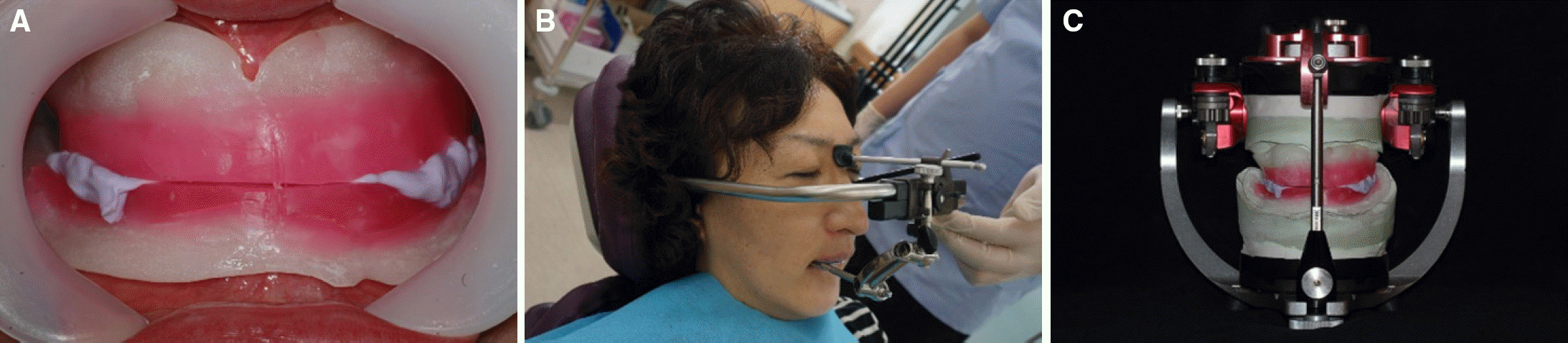 | Fig. 4.CR and VD registration. (A) Frontal view, (B) Facebow transfer, (C) Master cast mounted in the articulator. |
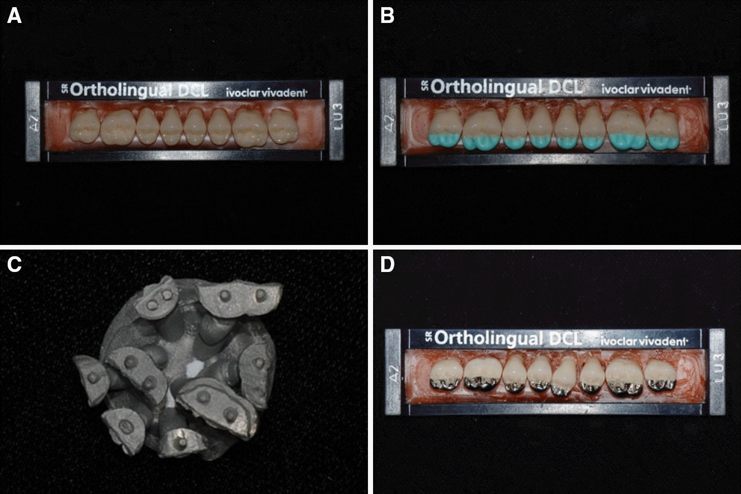 | Fig. 5.Modification of artificial teeth. (A) Artificial teeth, (B) Grinding and wax up of functional cusps, (C) Casting with Ni-Cr alloy, (D) Artificial teeth with Ni-Cr alloy functional cups. |
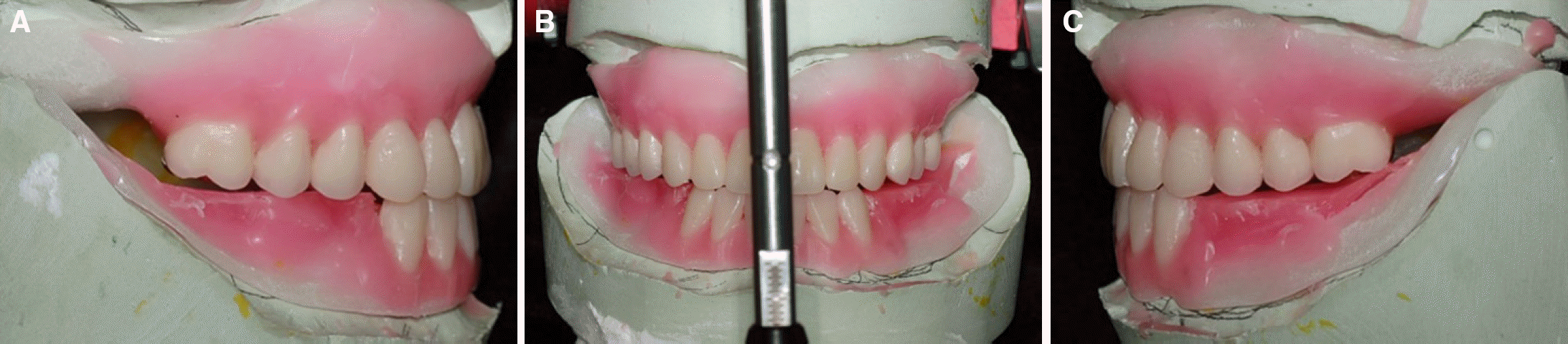 | Fig. 6.Arranging artificial teeth for treatment denture. (A) Lateral view (right), (B) Frontal view, (C) Lateral view (left). |
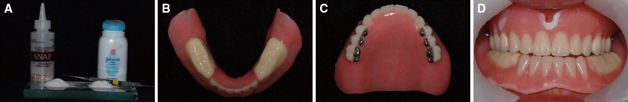 | Fig. 7.Fabrication of treatment dentures. (A) Self cure resin (SNAP, Parke Biomaterials, Edgewood, USA) and baby powder (powder (Johnson’ s baby powder, Johnson & Johnson baby products company, USA), (B) A Flat occlusal tables of mandibular denture, (C) A maxillary treatment denture, (D) Delivery of treatment dentures. |
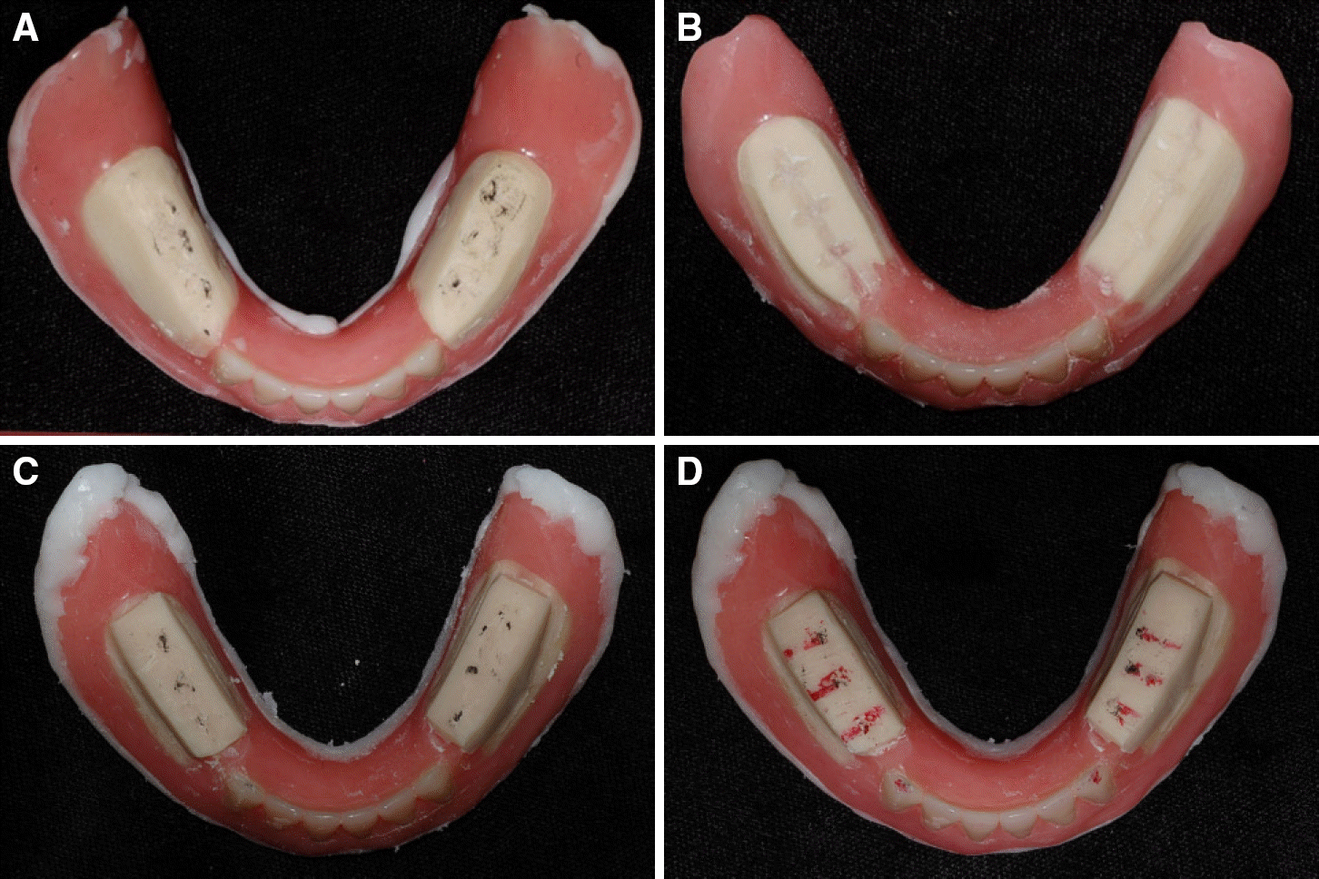 | Fig. 8.(A) Indentation marks on the flat occlusal table of the mandibular denture, (B) Removal of premature contact on the flat occlusal table of the mandibular denture.(C) Indentation marks 5 weeks after delivery. (D) Indentation marks during excursion movements 5 weeks after delivery. |
 | Fig. 9.Functional impressions taken with the treatment dentures and tissue conditioner. (A) Maxillary treatment denture, (B) Mandibular treatment denture. |
 XML Download