

 PDF
PDF ePub
ePub Citation
 Print
Print


Abstract
Purpose
The purpose of present study is to examine the correlation between the accuracy of abutment preparation and the marginal adaptation of metal coping. With this view, this study compared the correlations regard to the three different manufacturing methods of selective laser sintering technique, milling and casting.
Materials and methods
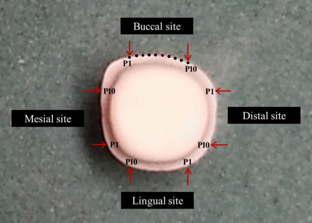
Two master models were made in a different way. First model with deep chamfer margin was prepared directly by a general clinician and the second model was designed by 3-D designing software program with the same abutment preparation principle and produced by computer aided manufacturing. 12 Co-Cr alloy copings were produced respectively with three different method; SLS system, CAD/CAM milling and conventional lost wax technique from each master model. The total 72 copings fully sit on the master model were stereoscopically evaluated at 40 points along the entire circumferential margin.
Results
Significant differences in the absolute marginal discrepancies of Co-Cr copings from SLS system (P=.0231) and casting method (P<.0001) were shown between hand preparation model and computer designed model. However, no significant difference was found between the two model groups from milling method (P=.9962).
Conclusion
Within the limitation of this study, the effect of the accuracy of abutment preparation on the marginal adaptation of Co-Cr coping is statistically significant in SLS system and casting group. The copings produced by SLS system exhibited the low-est marginal discrepancies among all groups, and the marginal gap of this method group was influenced by the accuracy of the abutment preparation.
Go to : 
REFERENCES
1. Anusavice KJ. Phillips' science of dental materials. 11th ed.Philadelphia: W.B. Saunders;2003. p. 621–4.
2. Wataha JC. Alloys for prosthodontic restorations. J Prosthet Dent. 2002; 87:351–63.

3. Viennot S, Dalard F, Lissac M, Grosgogeat B. Corrosion resistance of cobalt-chromium and palladium-silver alloys used in fixed prosthetic restorations. Eur J Oral Sci. 2005; 113:90–5.
4. Rosenstiel SF, Land MF, Fujimoto J. Contemporary fixed prosthodontics. 4th ed.St. Louis: Mosby;2006. 599:p. 606–8.
5. Takaichi A, Suyalatu , Nakamoto T, Joko N, Nomura N, Tsutsumi Y, Migita S, Doi H, Kurosu S, Chiba A, Wakabayashi N, Igarashi Y, Hanawa T. Microstructures and mechanical properties of Co-29Cr-6Mo alloy fabricated by selective laser melting process for dental applications. J Mech Behav Biomed Mater. 2013; 21:67–76.
6. Akova T, Ucar Y, Tukay A, Balkaya MC, Brantley WA. Comparison of the bond strength of laser-sintered and cast base metal dental alloys to porcelain. Dent Mater. 2008; 24:1400–4.
7. Ucar Y, Akova T, Akyil MS, Brantley WA. Internal fit evaluation of crowns prepared using a new dental crown fabrication technique: laser-sintered Co-Cr crowns. J Prosthet Dent. 2009; 102:253–9.
8. Fasbinder DJ. Clinical performance of chairside CAD/CAM restorations. J Am Dent Assoc. 2006; 137:22–31.
9. Miyazaki T, Hotta Y, Kunii J, Kuriyama S, Tamaki Y. A review of dental CAD/CAM: current status and future perspectives from 20 years of experience. Dent Mater J. 2009; 28:44–56.
10. Suleiman SH, Vult von Steyern P. Fracture strength of porcelain fused to metal crowns made of cast, milled or laser-sintered cobalt-chromium. Acta Odontol Scand. 2013; 71:1280–9.
11. White SN, Sorensen JA, Kang SK, Caputo AA. Microleakage of new crown and fixed partial denture luting agents. J Prosthet Dent. 1992; 67:156–61.
12. Lakhani SA, Ercoli C, Moss ME, Graser GN, Tallents RH. Influence of cold working and thermal treatment on the fit of implant-supported metal-ceramic fixed partial dentures. J Prosthet Dent. 2002; 88:159–69.
13. Pierce LH, Goodkind RJ. A status report of possible risks of base metal alloys and their components. J Prosthet Dent. 1989; 62:234–8.
14. Wassell RW, Walls AW, Steele JG. Crowns and extra-coronal restorations: materials selection. Br Dent J. 2002; 192:199–202. 205–11.
15. Quante K, Ludwig K, Kern M. Marginal and internal fit of metal-ceramic crowns fabricated with a new laser melting technology. Dent Mater. 2008; 24:1311–5.
16. Kim KB, Kim WC, Kim HY, Kim JH. An evaluation of marginal fit of three-unit fixed dental prostheses fabricated by direct metal laser sintering system. Dent Mater. 2013; 29:91–6.
17. Kim KB, Kim JH, Kim WC, Kim HY, Kim JH. Evaluation of the marginal and internal gap of metal-ceramic crown fabricated with a selective laser sintering technology: two- and three-di-mensional replica techniques. J Adv Prosthodont. 2013; 5:179–86.
18. Sundar MK, Chikmagalur SB, Pasha F. Marginal fit and microleakage of cast and metal laser sintered copings-an in vitro study. J Prosthodont Res. 2014; 58:252–8.
19. Bhaskaran E, Azhagarasan NS, Miglani S, Ilango T, Krishna GP, Gajapathi B. Comparative evaluation of marginal and internal gap of Co-Cr copings fabricated from conventional wax pattern, 3D printed resin pattern and DMLS tech: an in vitro study. J Indian Prosthodont Soc. 2013; 13:189–95.
20. Castillo-Oyague R, Lynch CD, Turrio′n AS, Lo′pez-Lozano JF, Torres-Lagares D, Sua′rez-Garc l′a MJ. Misfit and microleakage of implant-supported crown copings obtained by laser sintering and casting techniques, luted with glass-ionomer, resin cements and acrylic/urethane-based agents. J Dent. 2013; 41:90–6.
21. Oyague RC, Sa′nchez-Turrio′n A, Lo′pez-Lozano JF, Montero J, Albaladejo A, Sua′rez-Garc l′a MJ. Evaluation of fit of cementretained implant-supported 3-unit structures fabricated with direct metal laser sintering and vacuum casting techniques. Odontology. 2012; 100:249–53.
22. Akova T, Ucar Y, Tukay A, Balkaya MC, Brantley WA. Comparison of the bond strength of laser-sintered and cast base metal dental alloys to porcelain. Dent Mater. 2008; 24:1400–4.
23. Al Jabbari YS, Koutsoukis T, Barmpagadaki X, Zinelis S. Metallurgical and interfacial characterization of PFM Co-Cr dental alloys fabricated via casting, milling or selective laser melting. Dent Mater. 2014; 30:79–88.
24. Xin XZ, Chen J, Xiang N, Wei B. Surface properties and corrosion behavior of Co-Cr alloy fabricated with selective laser melting technique. Cell Biochem Biophys. 2013; 67:983–90.
25. Holmes JR, Bayne SC, Holland GA, Sulik WD. Considerations in measurement of marginal fit. J Prosthet Dent. 1989; 62:405–8.
26. Sua′rez MJ, Gonza′lez de Villaumbrosia P, Prad l′es G, Lozano JF. Comparison of the marginal fit of Procera AllCeram crowns with two finish lines. Int J Prosthodont. 2003; 16:229–32.
27. Molin M, Karlsson S. The fit of gold inlays and three ceramic in-lay systems. A clinical and in vitro study. Acta Odontol Scand. 1993; 51:201–6.
28. Rahme HY, Tehini GE, Adib SM, Ardo AS, Rifai KT. In vitro evaluation of the “replica technique”in the measurement of the fit of Procera crowns. J Contemp Dent Pract. 2008; 9:25–32.
29. Ortorp A, Jonsson D, Mouhsen A, Vult von Steyern P. The fit of cobalt-chromium three-unit fixed dental prostheses fabricated with four different techniques: a comparative in vitro study. Dent Mater. 2011; 27:356–63.
Go to :
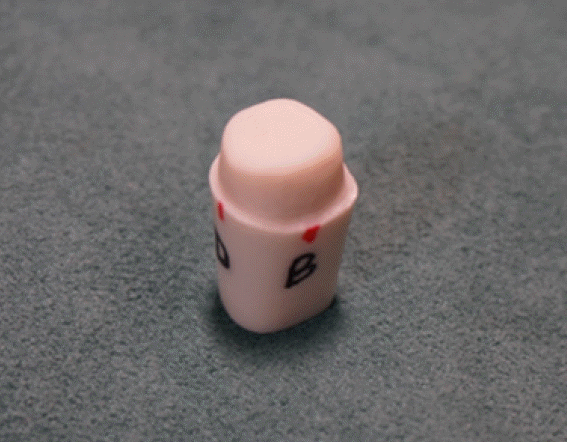 | Fig. 1.Resin tooth master model with hand perparation (1.2 mm deep chamfer margin preparation was made on a mandibular right first molar). |
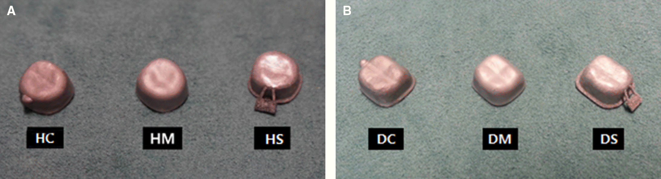 | Fig. 3.Copings produced by three different fabricating method; selective laser sintering, milling and casting. (A) HS, HM and HC from the hand preparation model, (B) DS, DM and DC from the computer designed model. |
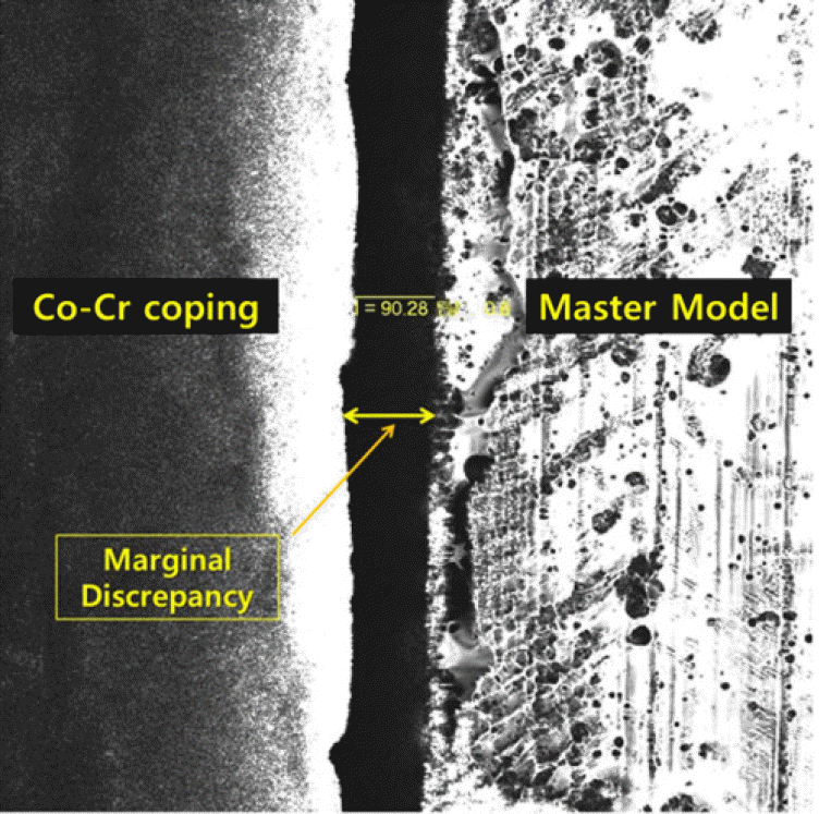 | Fig. 5.Measurement of absolute marginal discrepancy by the confocal laser scanning microscope at ×150 magnification. |
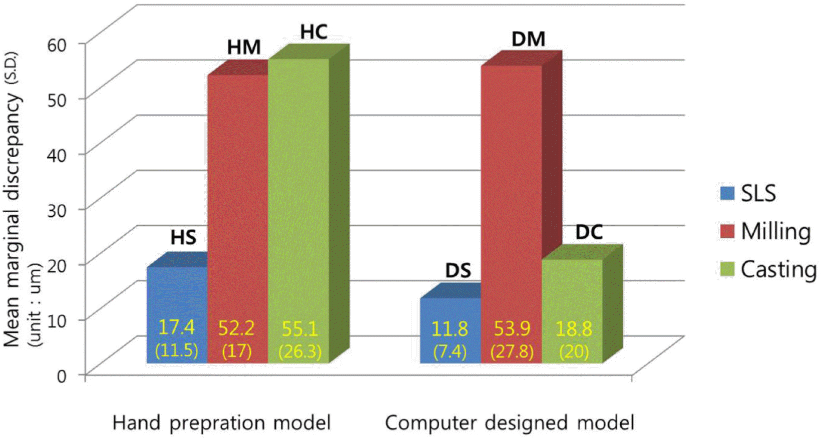 | Fig. 6.Total mean (standard deviations) of absolute marginal discrepancy for metal copings from two master models. Statistically significant differences between hand preparation model and computer designed model in SLS group (P=.0231) and casting group (P<.0001). |
Table 1.
Mean (SD) value of absolute marginal discrepancy for four site of the metal copings with the results of the Wilcoxon / Kruskal-Wallis Tests (unit: ㎛)
 XML Download