Abstract
The treatment of areas demanding esthetic requirements, such as maxillary anterior teeth, should take into account the achievement of a healthy, harmonious to the surrounding tissue, and an attractive smile line. In this case report, smile line, soft tissue and hard tissue morphology, and the anatomy and proportion of the tooth, must be considered. In patients with unesthetic maxillary anterior ratio due to inadequate gingival contour and diastema caused by peg lateralis, it would be challenging to achieve an esthetic restoration by orthodontic treatment alone. In such case, after orthodontic treatment, dento-gingivo-facial compositional diagnosis and analysis, followed by gingivectomy and prosthodontic restoration is needed to improve the interdental mesial/distal, width/length ratio to achieve a satisfactory esthetic result. In addition, when improving the tooth proportion of peg lateralis by prosthodontic treatment, Porcelain laminate veneer (PLV), which results in less tooth structure loss, reproduction of similar shade to that of the proximal tooth and high transparency, is recommended. This case report demonstrates esthetic improvements by prosthodontic restoration through systematic diagnosis and treatment procedure in patients with unesthetic maxillary anterior proportion after orthodontic treatment due to peg lateralis by means of two female patients aged twenty years old.
REFERENCES
1. Pinto RC, Chambrone L, Colombini BL, Ishikiriama SK, Britto IM, Romito GA. Minimally invasive esthetic therapy: a case report describing the advantages of a multidisciplinary approach. Quintessence Int. 2013; 44:385–91.
2. Lampreia M, Perez J. Aesthetic porcelain laminate veneer restoration following orthodontic treatment: sequential technique. Pract Proced Aesthet Dent. 2008; 20:545–7.
3. Ittipuriphat I, Leevailoj C. Anterior space management: inter-disciplinary concepts. J Esthet Restor Dent. 2013; 25:16–30.

4. Calamia JR, Calamia CS. Porcelain laminate veneers: reasons for 25 years of success. Dent Clin North Am. 2007; 51:399–417.

5. Beier US, Kapferer I, Burtscher D, Dumfahrt H. Clinical performance of porcelain laminate veneers for up to 20 years. Int J Prosthodont. 2012; 25:79–85.
6. da Cunha LF, Pedroche LO, Gonzaga CC, Furuse AY. Esthetic, occlusal, and periodontal rehabilitation of anterior teeth with min-imum thickness porcelain laminate veneers. J Prosthet Dent. 2014; 112:1315–8.

7. Mistry S. Principles of smile demystified design. J Cosmet Dent. 2012; 28:116–24.
8. Gresnigt M, Ozcan M. Esthetic rehabilitation of anterior teeth with porcelain laminates and sectional veneers. J Can Dent Assoc. 2011; 77:b143.
9. Ahmad I. Geometric considerations in anterior dental aesthetics: restorative principles. Pract Periodontics Aesthet Dent. 1998; 10:813–22.
14. Pini NP, Aguiar FH, Lima DA, Lovadino JR, Terada RS, Pascotto RC. Advances in dental veneers: materials, applications, and techniques. Clin Cosmet Investig Dent. 2012; 4:9–16.
15. Giordano R, McLaren EA. Ceramics overview: classification by microstructure and processing methods. Compend Contin Educ Dent. 2010; 31:682–4. 686, 688.
16. Turgut S, Bagis B. Colour stability of laminate veneers: an in vitro study. J Dent. 2011; 39:e57–64.

17. Reshad M, Cascione D, Magne P. Diagnostic mock-ups as an objective tool for predictable outcomes with porcelain laminate veneers in esthetically demanding patients: a clinical report. J Prosthet Dent. 2008; 99:333–9.

18. Lopes LG, Vaz MM, de Magalhaes AP, Cardoso PC, de Souza JB, de Torres EM. Shade evaluation of ceramic laminates according to different try-in materials. Gen Dent. 2014; 62:32–5.
19. Xing W, Jiang T, Ma X, Liang S, Wang Z, Sa Y, Wang Y. Evaluation of the esthetic effect of resin cements and try-in pastes on ceromer veneers. J Dent. 2010; 38:e87–94.

20. Kampouropoulos D, Gaintantzopoulou M, Papazoglou E, Kakaboura A. Colour matching of composite resin cements with their corresponding try-in pastes. Eur J Prosthodont Restor Dent. 2014; 22:84–8.
Fig. 1.
Initial extraoral photographs in frontal view. (A) Facial midline with the midline of central incisors, (B) Horizontal parameters relating the inter-pupillary line to the incisal edge position, (C) Full-facial analysis.
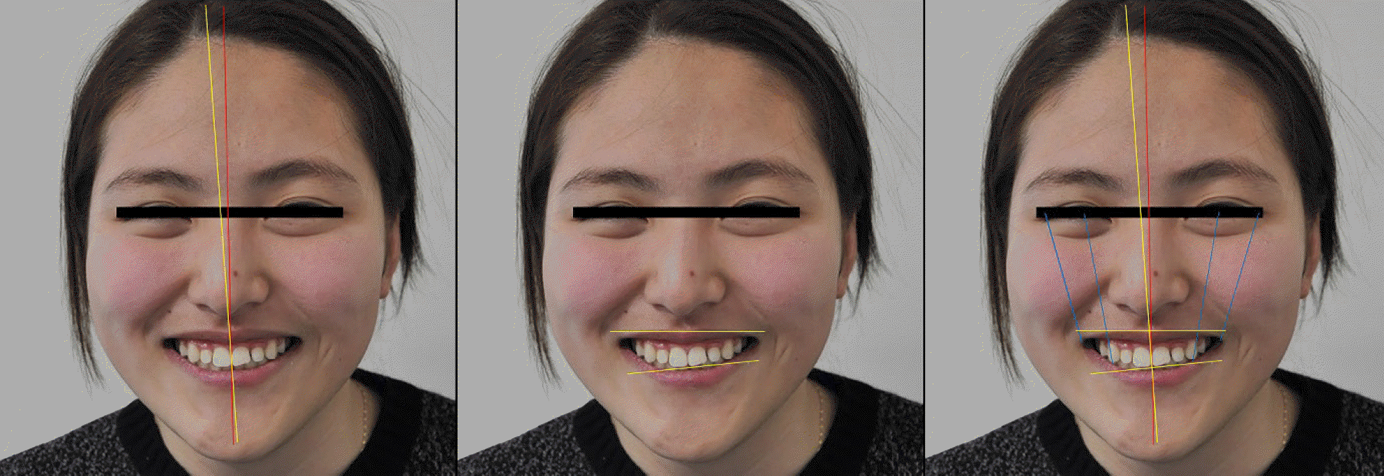
Fig. 2.
Initial extraoral photographs in lateral view. (A) Rickett' s E-plane indicating the distance to the upper lip less than 4 mm, (B) Nasio-labial angle less than 90®.
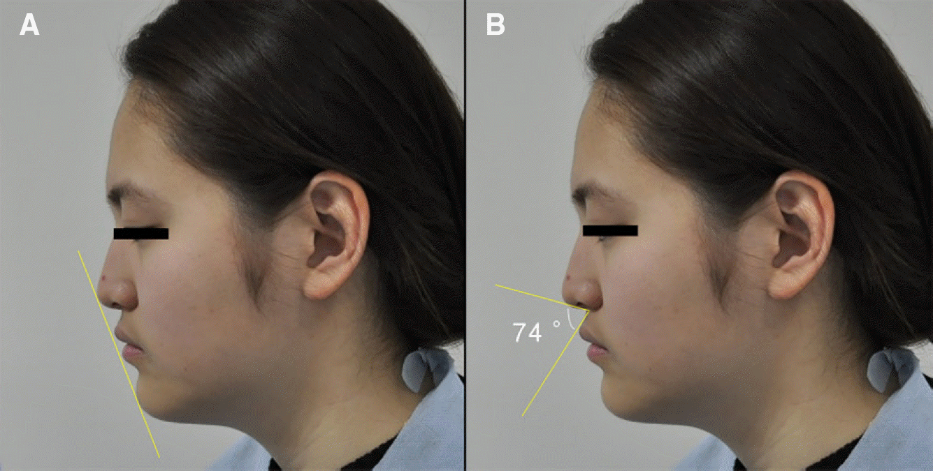
Fig. 3.
Initial intraoral photographs in centric occlusion. (A) Right lateral view, (B) Frontal view, (C) Left lateral view.

Fig. 4.
Initial intraoral photographs. (A) Right lateral view, (B) Maxillary anterior view, (C) Left lateral view.

Fig. 5.
Analysis of smile line at initial visit. (A) Rest state view, (B) Slight smile view, (C) Maximum smile view.

Fig. 6.
Analysis of anterior smile line.7 (A) Calculating the length of the teeth using the width/length ratio of 78%, (B) Designing the gingival esthetic line based upon the esthetic parameters, (C) Determining of the position of the central incisors using facial midline, (D) Creating midline symmetry, (E, F) Determinig of the size of the lateral incisors and canines using principles of tooth proportions, the "golden proportion".

Fig. 7.
Semi-articulator mounting procedures. (A) Facebow transfer (B) Mounting the diagnostic model to an articulator in frontal view (C) Mounting the diagnostic model to an articulator in lateral view (D) Diagnostic model.

Fig. 8.
Model Analysis. (A) Analysis of diagnostic model (B) Diagnostic waxup in diagnostic model (Application of ideal teeth width/length ratio 78% and golden proportion 62%).

Fig. 9.
Gingivectomy using template. (A) Template (Duran®, SCHEU-DENTAL GmbH, Iserlohn, Germany) fabrication, (B) Template application, (C) After gingivectomy.

Fig. 10.
Follow-up after one month. (A) Right lateral view, (B) Frontal view, (C) Left lateral view.

Fig. 12.
Mock-up fabrication and abutment preparation. (A) Spot etching, (B) Mock-up fabircation, (C) Abutment preparation, (D) Removal of resin excess.

Fig. 13.
Final impression taking. (A) Cord packing, (B) Final impression, (C) Master cast fabrication.

Fig. 14.
Laminate fabrication. (A) Final Wax-up, (B) IPS e.max® Press HT ingot (Shade A1, Ivoclar vivadent, Schaan, Leichtenstein), (C) Laminate try-in at master cast.

Fig. 15.
Final setting by Variolink® N (Yellow shade, Ivoclar vivadent, Schaan, Leichtenstein). (A) Right lateral view, (B) Semi-right lateral view, (C) Frontal view, (D) Semi-left lateral view, (E) Left lateral view.

Fig. 16.
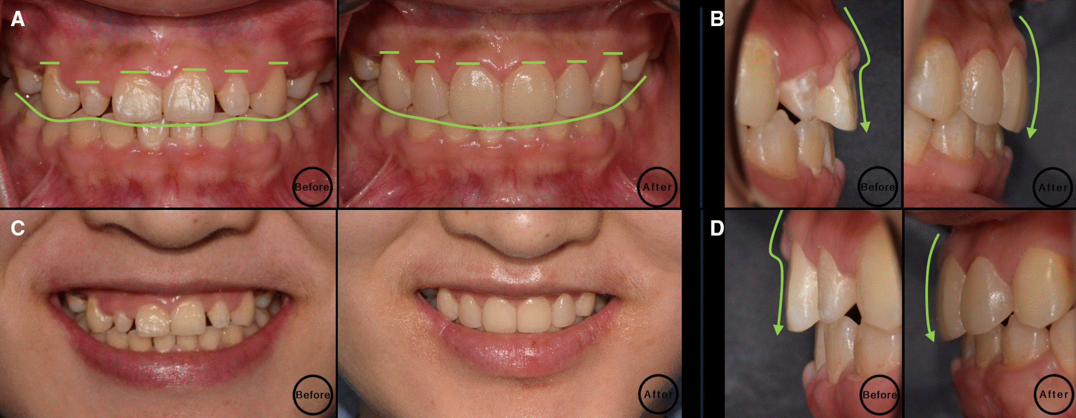
Comparison of before and after treatment. (A) Frontal view, (B) Right lateral view, (C) Maxillary anterior view, (D) Left lateral view.
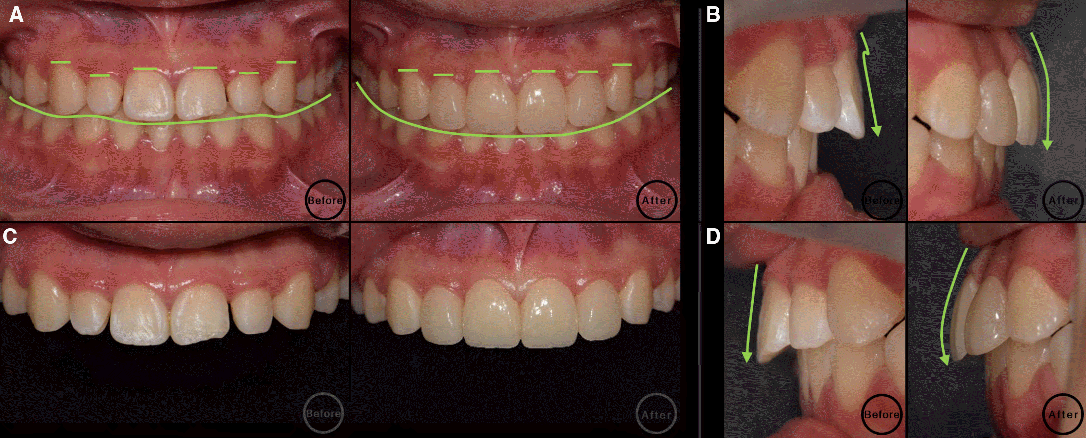
Fig. 17.
Smile line improvement. (A) Dentofacial view, (B) Frontal view, (C) Semi-lateral view, (D) Lateral view.

Fig. 18.
Initial extraoral photographs in frontal view. (A) Facial midline with the midline of central incisors, (B) Horizontal parameters relating the inter-pupillary line to the incisal edge position, (C) Full-facial analysis.

Fig. 19.
Initial extraoral photographs in lateral view. (A) Rickett' s E-plane indicating the distance to the upper lip less than 4 mm, (B) Nasio-labial angle less than 90®.
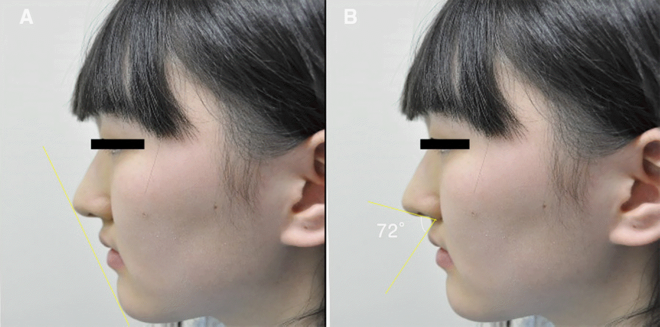
Fig. 20.
Initial intraoral photographs in centric occlusion. (A) Right lateral view, (B) Frontal view, (C) Left lateral view.

Fig. 21.
Initial intraoral photographs. (A) Right lateral view, (B) Maxillary anterior view, (C) Left lateral view.

Fig. 22.
Analysis of smile line at initial visit. (A) Rest state view, (B) Slight smile view, (C) Maximum smile view.

Fig. 23.
Model Analysis. (A) Analysis of diagnostic model, (B) Diagnostic waxup in diagnostic model (Application of ideal teeth width/length ratio 78% and golden proportion 62%).

Fig. 24.
Gingivectomy using template. (A) Template (Duran®, SCHEU-DENTAL GmbH, Iserlohn, Germany) fabrication, (B) Template application, (C) After gingivectomy.

Fig. 25.
Follow-up after one month. (A) Right lateral view, (B) Frontal view, (C) Left lateral view.

Fig. 26.
Mock-up fabrication and abutment preparation. (A) Spot etching, (B) Mock-up fabircation, (C) Abutment preparation, (D) Removal of resin excess.

Fig. 27.
Final impression taking. (A) Cord packing, (B) Final impression, (C) Master cast fabrication.

Fig. 28.
Laminate fabrication. (A) Final Wax-up, (B) IPS e.max® Press HT ingot (Shade A2, Ivoclar vivadent, Schaan, Leichtenstein), (C) Laminate try-in at master cast.

Fig. 29.
Final setting by Variolink® N (Transparent shade, Ivoclar vivadent, Schaan, Leichtenstein). (A) Right lateral view, (B) Semi-right lateral view, (C) Frontal view,(D) Semi-left lateral view, (E) Left lateral view.


 PDF
PDF ePub
ePub Citation
 Print
Print



 XML Download