

 PDF
PDF ePub
ePub Citation
 Print
Print


Abstract
Developing of digital technique, it is possible to fabricate implant prostheses for edentulous area using intraoral 3-dimentional information throughout implant diagnosis and treatment process. It is being changed that from the method using CAD/CAM, producing prostheses by model scanning after conventional impression and model processing, to the method of fabricating implant provisional restorations and customized abutments by digital impression after connecting digital impression copings (scanbody) and implant fixtures without models. But, this digital method has not been actively used for implant prostheses not yet. Specially, it is short of intraoral digital impression cases for immediate provisional restorations of the maxillary anterior implants. The gingival contour impression of maxillary anterior area is very important for esthetic restorations. Accordingly, in this case report, the using a digital impression coping (scanbody) and digital impression by CEREC Omnicam (Sirona, Bensheim, Germany) or Trios (3shape, Copenhagen, Denmark) were introduced for immediate provisional restorations in 3 cases needed a single implant restoration in maxillary anterior area. The clinical results were satisfactory on the convenience and accuracy of digital impression technique and the good esthetics of final restorations.
Go to : 
REFERENCES
1. Duret F, Blouin JL, Duret B. CAD-CAM in dentistry. J Am Dent Assoc. 1988; 117:715–20.

2. Leinfelder KF, Isenberg BP, Essig ME. A new method for gen-erating ceramic restorations: a CAD-CAM system. J Am Dent Assoc. 1989; 118:703–7.
3. Nayyar N, Yilmaz B, McGlumphy E. Using digitally coded healing abutments and an intraoral scanner to fabricate implant-supported, cement-retained restorations. J Prosthet Dent. 2013; 109:210–5.
4. Begum A, Ahmed R, Islam MS. Digital Impression. City Dent Coll J. 2012; 9:31–4.
5. Yoo JY, Kim YG, Lee BS, Kwon YD, Choi BJ, Kim YR. Immediate implant placement after extraction of retained deciduous teeth and impacted canines: Report of a case. J Korean Assoc Maxillofac Plastic Reconstr Surg. 2009; 31:330–3.
6. De Rouck T, Collys K, Cosyn J. Immediate single-tooth implants in the anterior maxilla: a 1-year case cohort study on hard and soft tissue response. J Clin Periodontol. 2008; 35:649–57.
7. den Hartog L, Raghoebar GM, Stellingsma K, Meijer HJ. Immediate loading and customized restoration of a single implant in the maxillary esthetic zone: a clinical report. J Prosthet Dent. 2009; 102:211–5.
8. Mijiritsky E1, Mardinger O, Mazor Z, Chaushu G. Immediate provisionalization of single-tooth implants in fresh-extraction sites at the maxillary esthetic zone: up to 6 years of follow-up. Implant Dent. 2009; 18:326–33.
9. Hong YS, Park EJ, Kim SJ, Kim MR, Heo SJ, Park JM. Customized abutment and screw-type implant prostheses after cementation based on the digital intraoral impression technique. J Korean Acad Prosthodont. 2012; 50:67–73.
10. Lin WS, Harris BT, Morton D. The use of a scannable impression coping and digital impression technique to fabricate a customized anatomic abutment and zirconia restoration in the esthetic zone. J Prosthet Dent. 2013; 109:187–91.
11. Kim Y, Oh TJ, Misch CE, Wang HL. Occlusal considerations in implant therapy: clinical guidelines with biomechanical rationale. Clin Oral Implants Res. 2005; 16:26–35.
12. Kan JY, Rungcharassaeng K, Lozada J. Immediate placement and provisionalization of maxillary anterior single implants: 1-year prospective study. Int J Oral Maxillofac Implants. 2003; 18:31–9.
13. Neale D, Chee WW. Development of implant soft tissue emergence profile: a technique. J Prosthet Dent. 1994; 71:364–8.
14. Shadid R1, Sadaqa N. A comparison between screw- and cement-retained implant prostheses. A literature review. J Oral Implantol. 2012; 38:298–307.
15. Guichet D. Digitally enhanced dentistry: the power of digital design. J Calif Dent Assoc. 2015; 43:135–41.
Go to :
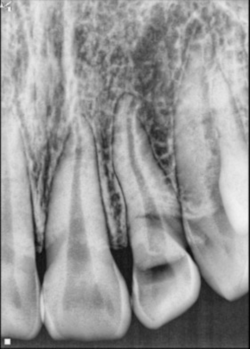
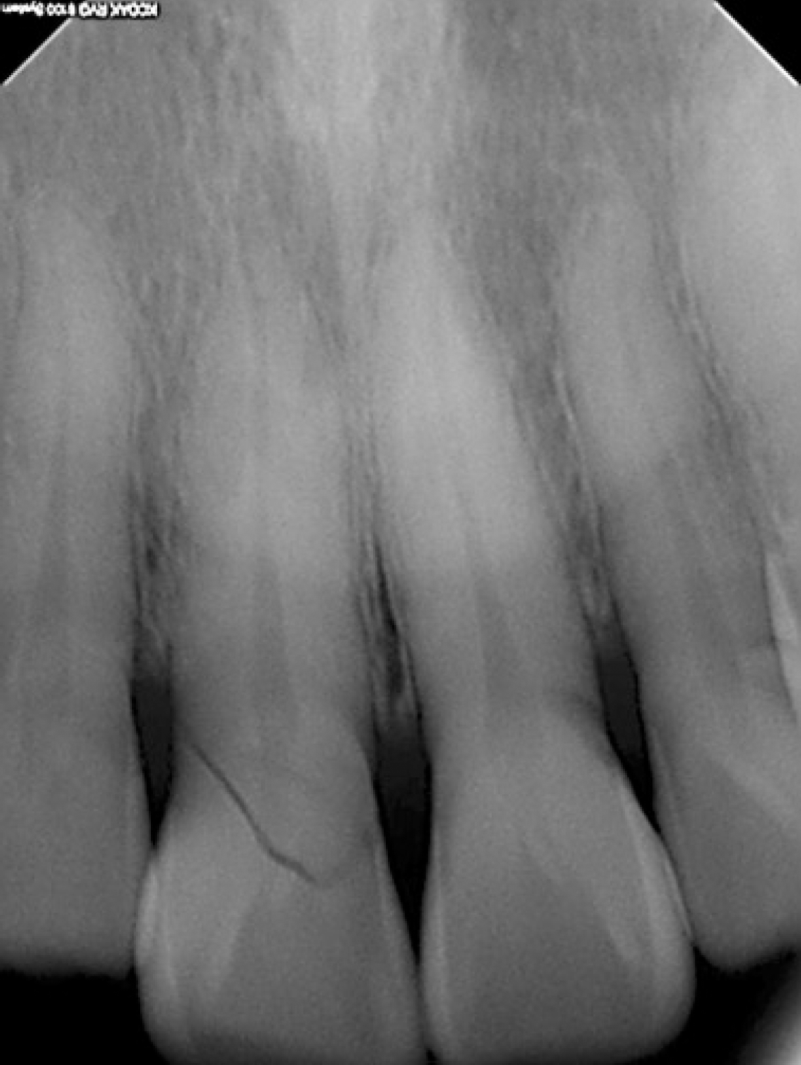
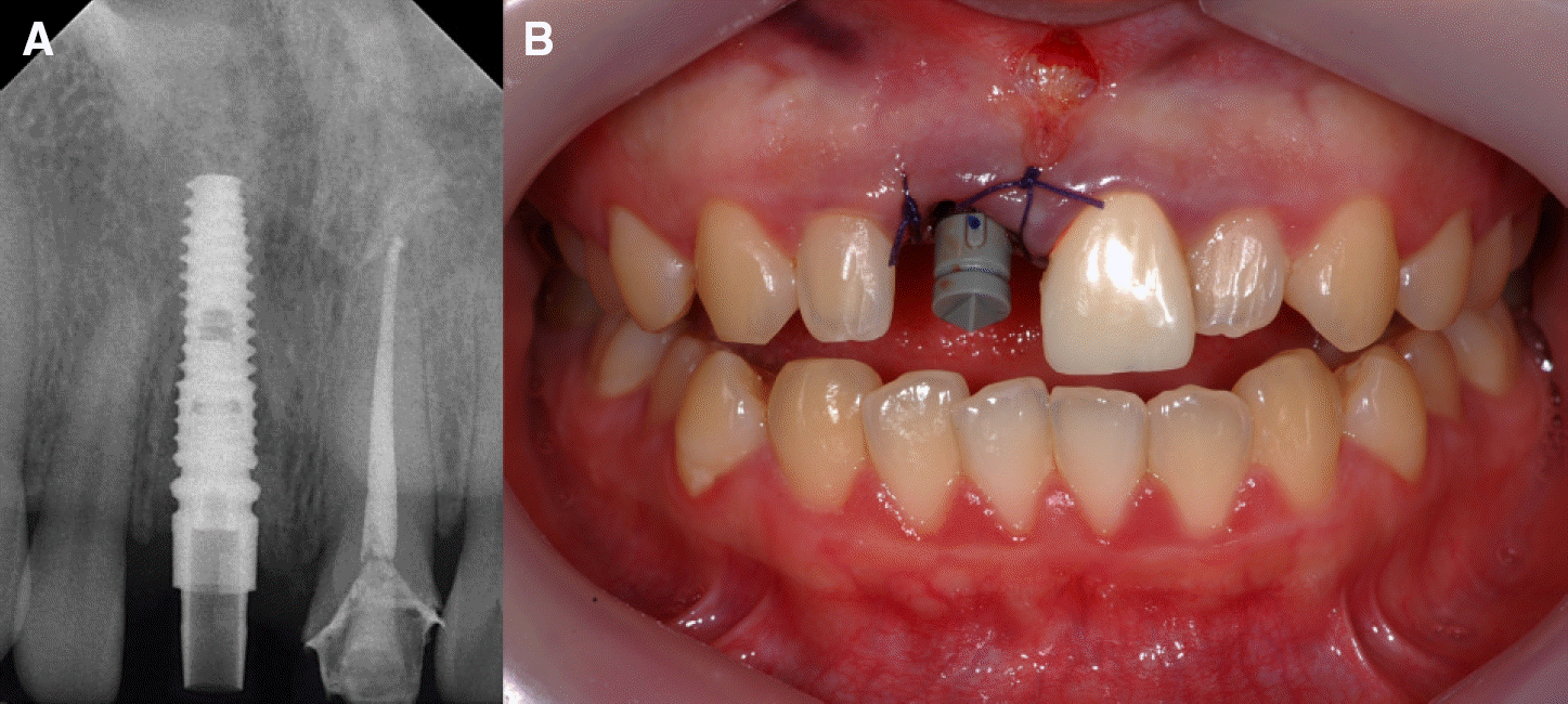 | Fig. 2.(A) Periapical radiograph of scanpost (RaphaBio, Seoul, Korea), (B) Scanbody (Sirona, Bensheim, Germany) adaptation for digital impression. |
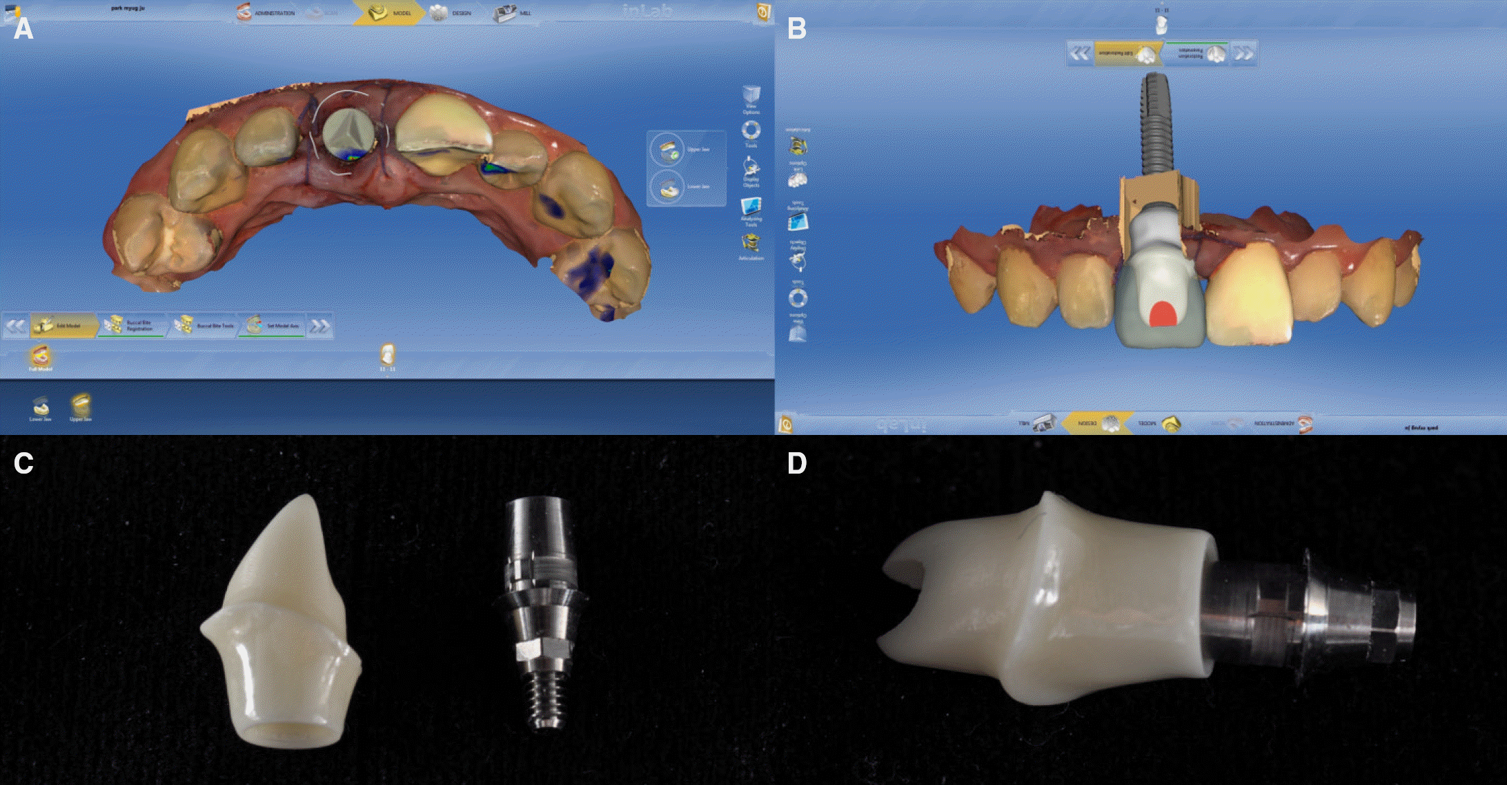 | Fig. 3.(A) Scanned scanbody (grey) and gingiva (pink) by CEREC Omnicam (Sirona, Bensheim, Germany), (B) Design of customized abutment by CEREC software (Sirona, Bensheim, Germany), (C) Zirconia upper abutment part (left) and Ti-base (right), (D) Completed customized zirconia abutment. |
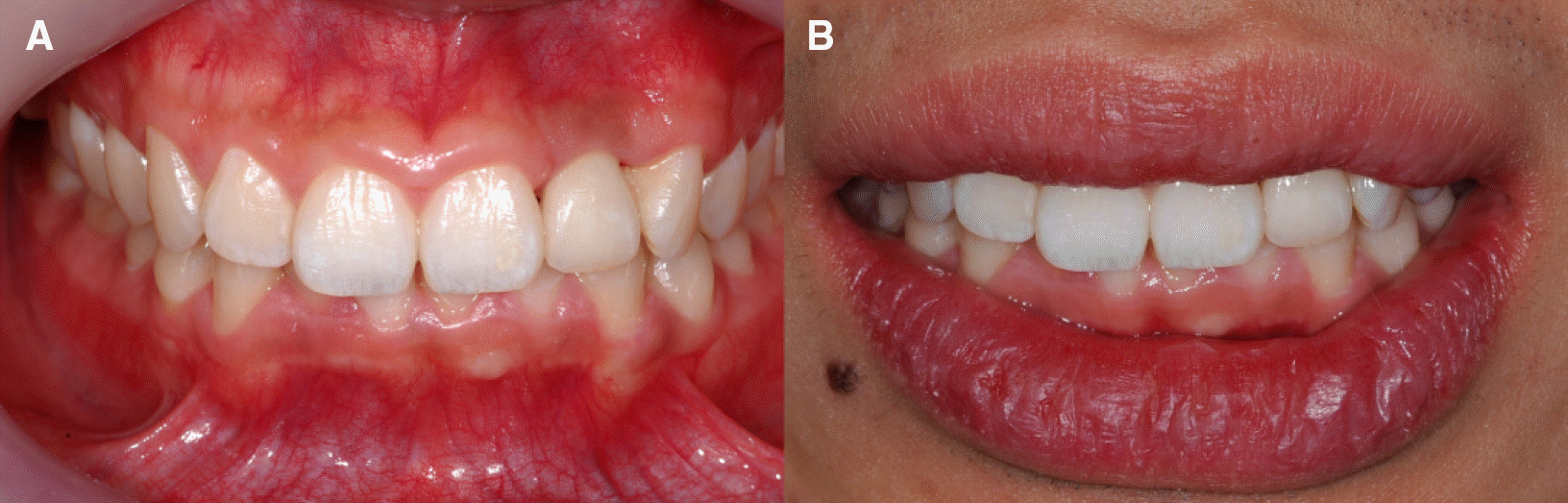
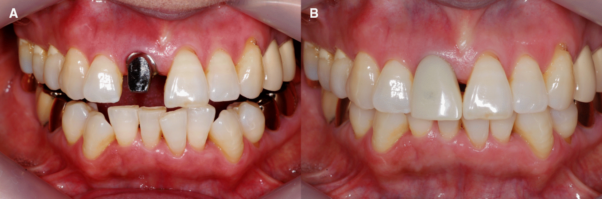
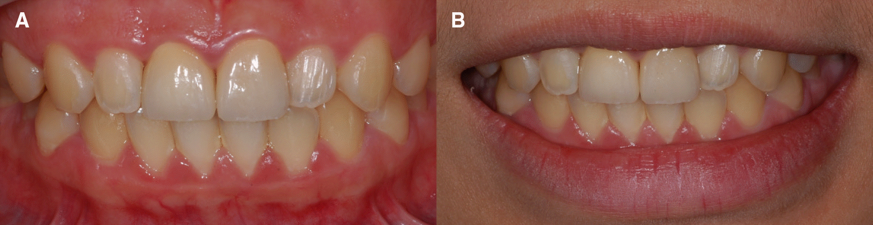 | Fig. 5.(A) Final prosthesis with temporary cementation, (B) Final prosthesis at 3 month’ s check. |
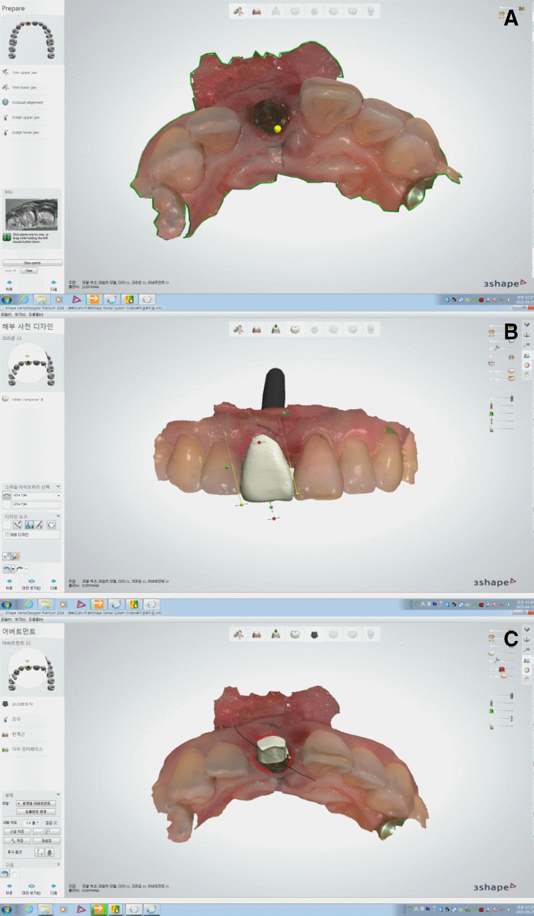
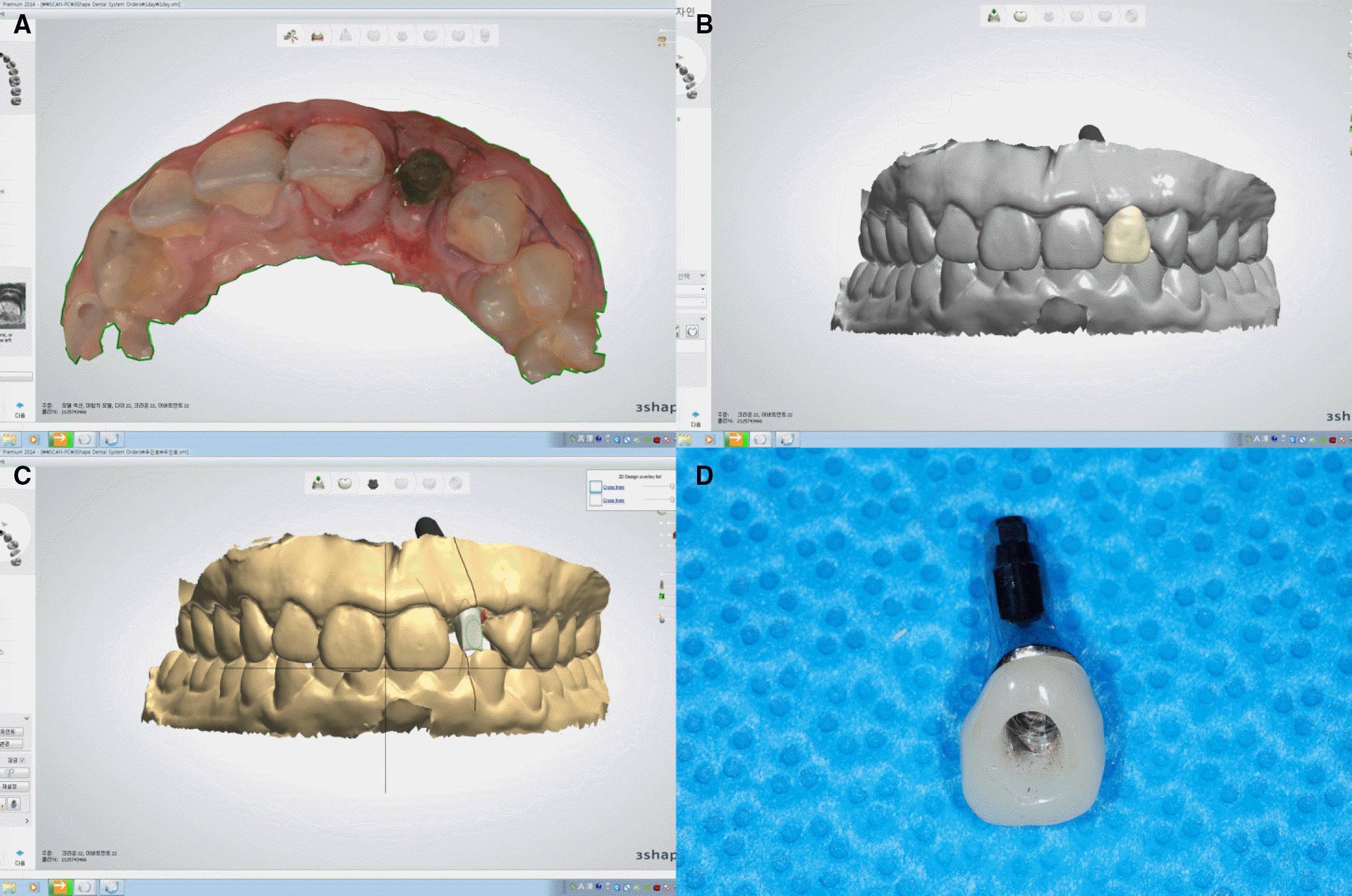 | Fig. 8.(A) Scanned scanbody(black) and gingiva(pink) by Trios (3shape, Copenhagen, Denmark), (B) Digitally diagnostic waxup of final prosthesis by Trios (3shape, Copenhagen, Denmark), (C) Design of customized abutment by 3shape software (3shape, Copenhagen, Denmark), (D) Provisional abutment. |
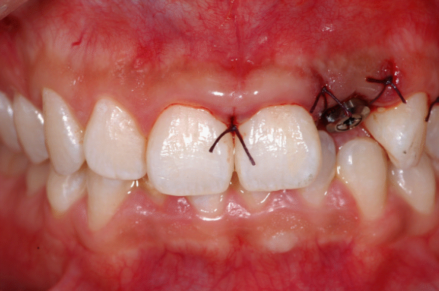
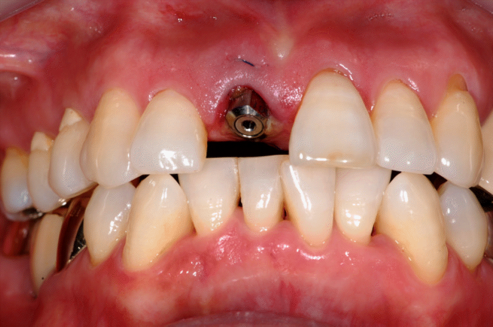
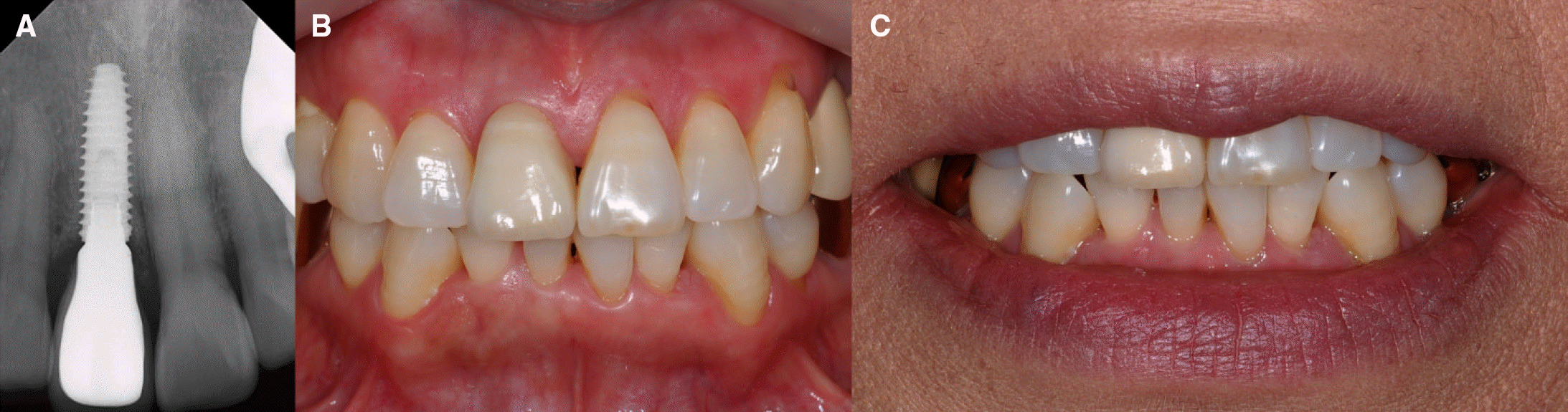
 | Fig. 9.(A) Delivered provisional abutment and temporary crown, (B) After stich out, (C) Delivered customized zirconia abutment, (D) Temporary crown on the abutment. |
 XML Download