Abstract
Loss of teeth may not only imply impaired oral function and loss of alveolar bone but is also often accompanied by reduced self-confidence. This results in a larger problem with the fully edentulous patient. The patient introduced in this study showed multiple missing teeth and mobility of remaining teeth and wanted to have fixed dental prosthesis using implants. Remaining teeth were extracted because of periodontally bad prognosis. This article reports a satisfactory clinical and esthetic outcome of full mouth rehabilitation using implant hybrid prosthesis in fully edentulous patient.
Go to : 
REFERENCES
1. Adell R, Lekholm U, Rockler B, Brå nemark PI. A 15-year study of osseointegrated implants in the treatment of the edentulous jaw. Int J Oral Surg. 1981; 10:387–416.

2. Misch CE. Dental implant prosthetics. 1st ed.Mosby;2004. p. 43–52.
3. Waskewicz GA, Ostrowski JS, Parks VJ. Photoelastic analysis of stress distribution transmitted from a fixed prosthesis at-tached to osseointegrated implants. Int J Oral Maxillofac Implants. 1994; 9:405–11.
4. Assuncao WG, Filho HG, Zaniquelli O. Evaluation of transfer impressions for osseointegrated implants at various angulations. Implant Dent. 2004; 13:358–66.

5. Assif D, Nissan J, Varsano I, Singer A. Accuracy of implant impression splinted techniques: effect of splinting material. Int J Oral Maxillofac Implants. 1999; 14:885–8.
6. Daoudi MF, Setchell DJ, Searson LJ. A laboratory investigation of the accuracy of the repositioning impression coping technique at the implant level for single-tooth implants. Eur J Prosthodont Restor Dent. 2003; 11:23–8.
7. Vigolo P, Majzoub Z, Cordioli G. Evaluation of the accuracy of three techniques used for multiple implant abutment impressions. J Prosthet Dent. 2003; 89:186–92.

8. Kokubo Y, Ohkubo C. Occlusion recording device for dental implant-supported restoration. J Prosthet Dent. 2006; 95:262–3.
9. Hobo S, Ichida E, Garcia LT. Osseointegration and Occlusal Rehabilitation. Chicago; III: Quintessence;1989. p. 159–62. p. 171–3.
10. Loos LG. A fixed prosthodontic technique for mandibular osseointegrated titanium implants. J Prosthet Dent. 1986; 55:232–42.

Go to : 
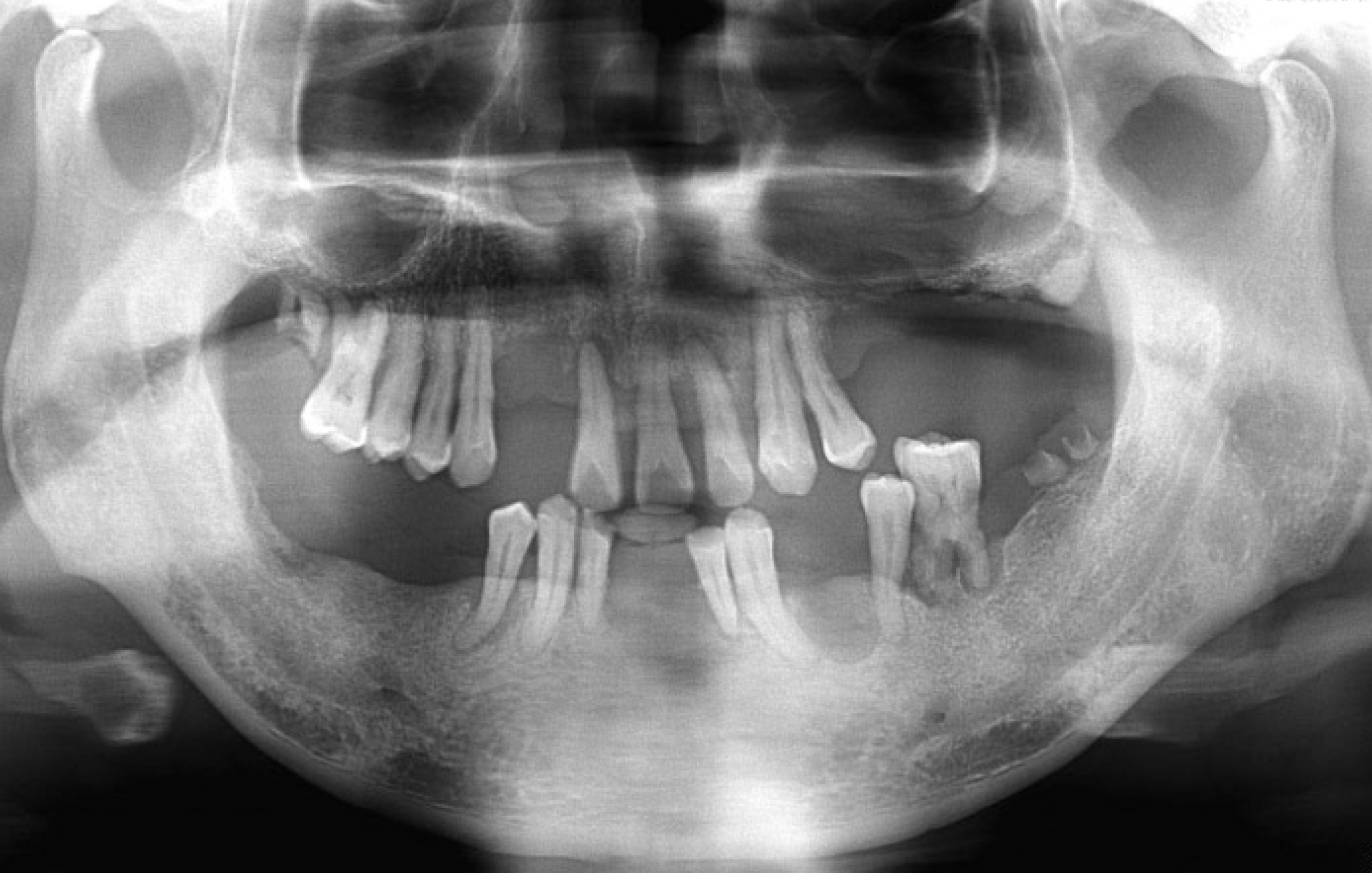
 | Fig. 2.(A) Maxillary occlusal view after implant surgery, (B) Mandibular occlusal view after implant surgery, (C) Panoramic radiograph after implant surgery. |
 | Fig. 3.(A) Upper implant supported record base, (B) Lower implant supported record base, (C) CR recording was carried out with implant supported record base. |
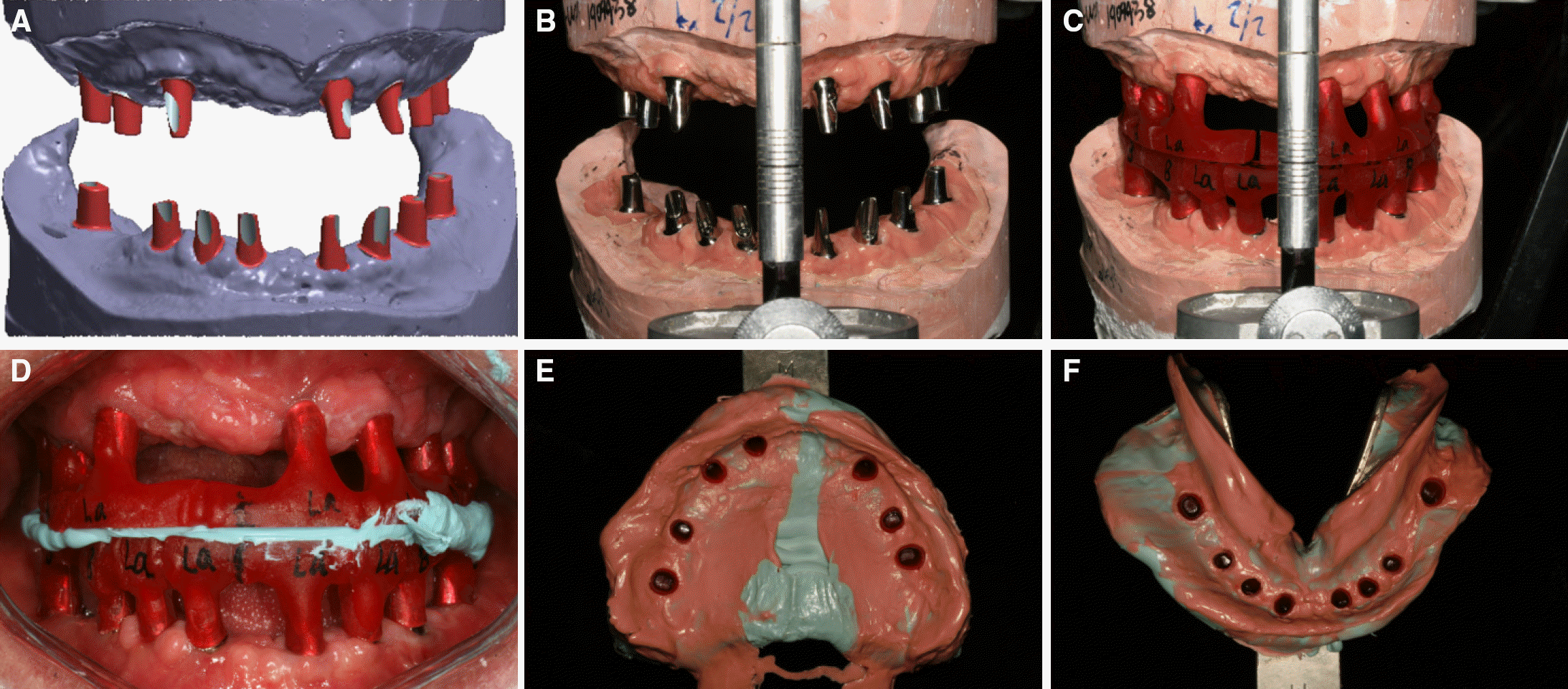
 | Fig. 4.(A) Computer aided design of customized abutment, (B) Customized titanium abutments was connected to working model, (C) Pattern resin block, (D) CR recording was carried out with pattern resin, (E) Final impression of maxillary implants, (F) Final impression of mandibular implants. |
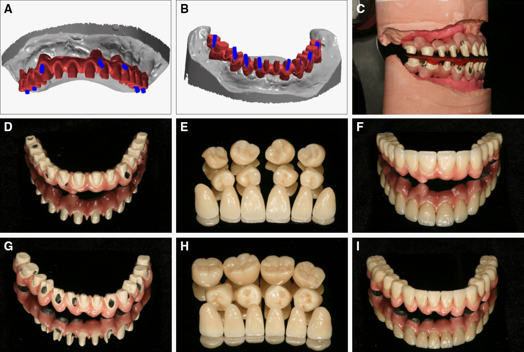
 | Fig. 5.(A) Computer aided design of upper mesostructure, (B) Computer aided design of lower mesostructure, (C) Upper and lower mesostructures were positioned to mounted master cast, (D) Upper mesostructure, (E) Upper zirconia restorations, (F) Upper zirconia restorations were positioned to upper mesostructure, (G) Lower mesostructure, (H) Lower zirconia restorations (I) Lower zirconia restorations were positioned to lower mesostructure. |


 PDF
PDF ePub
ePub Citation
 Print
Print


 XML Download