Abstract
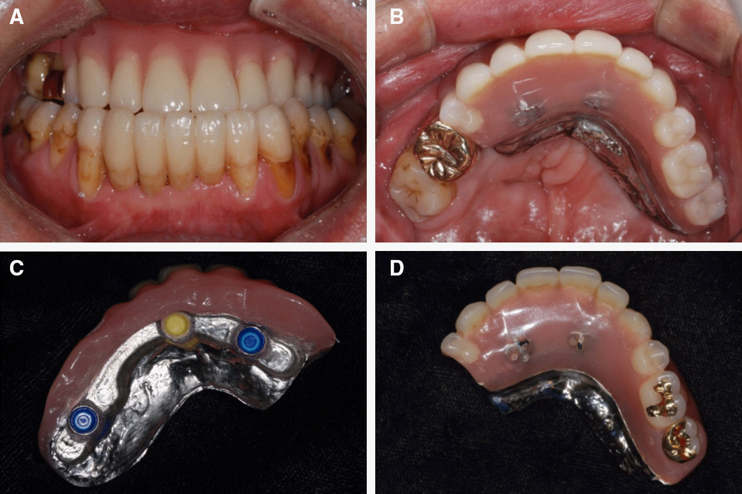
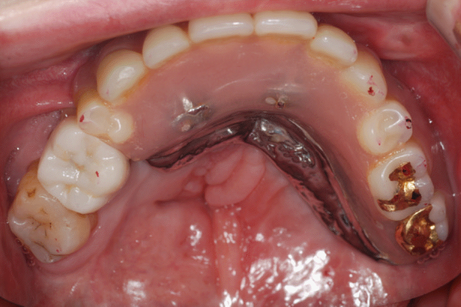
Due to the limitations of conventional removable partial denture prostheses to treat a cleft lip & palate patient who shows scar tissue on upper lip, excessive absorption of the maxillary residual alveolar ridge, and class III malocclusion with narrow palate and undergrowth of the maxilla, 4 implants were placed on the maxillary edentulous region and a maxillary removable implant-supported partial denture was planned using a CAD/CAM milled titanium bar. Unlike metal or gold casting technique which has shrinkage after the molding, CAD/CAM milled titanium bar is highly-precise, economical and lightweight. In practice, however, it is very hard to obtain accurate friction-fit from the milled bar and reduction in retention can occur due to repetitive insertion and removal of the denture. Various auxiliary retention systems (e.g. ERA®, CEKA®, magnetics, Locator® attachment), in order to deal with these problems, can be used to obtain additional retention, cost-effectiveness and ease of replacement. Out of diverse auxiliary attachments, Locator® has characteristics that are dual retentive, minimal in vertical height and convenient of attachment replacement. Drill and tapping method is simple and the replacement of the metal female part of Locator® attachment is convenient. In this case, the Locator® attachment is connected to the milled titanium bar fabricated by CAD/CAM, using the drill and tapping technique. Afterward, screw holes were formed and 3 Locator® attachments were secured with 20 Ncm holding force for additional retention. Following this procedure, satisfactory results were obtained in terms of aesthetic facial form, masticatory function and denture retention, and I hereby report this case.
Go to : 
REFERENCES
1. Baek SH, Yang WS. Clinical study on the anomalies of number and morphology in cleft lip and palate patients'teeth. Korean J Orthod. 2001; 31:51–61.
2. Galindo DF. The implant-supported milled-bar mandibular overdenture. J Prosthodont. 2001; 10:46–51.

3. Drago C, Howell K. Concepts for designing and fabricating metal implant frameworks for hybrid implant prostheses. J Prosthodont. 2012; 21:413–24.

4. Sadowsky SJ, Fitzpatrick B, Curtis DA. Evidence-based crite-ria for differential treatment planning of implant restorations for the maxillary edentulous patient. J Prosthodont. 2014; 22:319–329.

5. Kan JY, Rungcharassaeng K, Bohsali K, Goodacre CJ, Lang BR. Clinical methods for evaluating implant framework fit. J Prosthet Dent. 1999; 81:7–13.

6. Chan MF, Nä rhi TO, de Baat C, Kalk W. Treatment of the atrophic edentulous maxilla with implant-supported overdentures: a review of the literature. Int J Prosthodont. 1998; 11:7–15.
7. Lewis S, Sharma A, Nishimura R. Treatment of edentulous maxillae with osseointegrated implants. J Prosthet Dent. 1992; 68:503–8.

8. Trakas T, Michalakis K, Kang K, Hirayama H. Attachment systems for implant retained overdentures: a literature review. Implant Dent. 2006; 15:24–34.

9. Watson CJ, Tinsley D, Sharma S. Implant complications and failures: the complete overdenture. Dent Update. 2001; 28:234–8. 240.

10. Naert I, Gizani S, Vuylsteke M, Van Steenberghe D. A 5-year prospective randomized clinical trial on the influence of splinted and unsplinted oral implants retaining a mandibular overdenture: prosthetic aspects and patient satisfaction. J Oral Rehabil. 1999; 26:195–202.

11. Wismeijer D, van Waas MA, Kalk W. Factors to consider in se-lecting an occlusal concept for patients with implants in the edentulous mandible. J Prosthet Dent. 1995; 74:380–4.

12. Oh SC, Han JS, Kim MJ. Implant supported overdenture using milled titanium bar with Locator attachment on fully edentulous maxillae: a case report. J Dent Rehab App Sci. 2011; 27:223–30.
13. Kurtzman GM. Lab techniques for use of the locator attachment in bar-overdenture applications. TeamWork. 2008; 1:72–8.
Go to : 
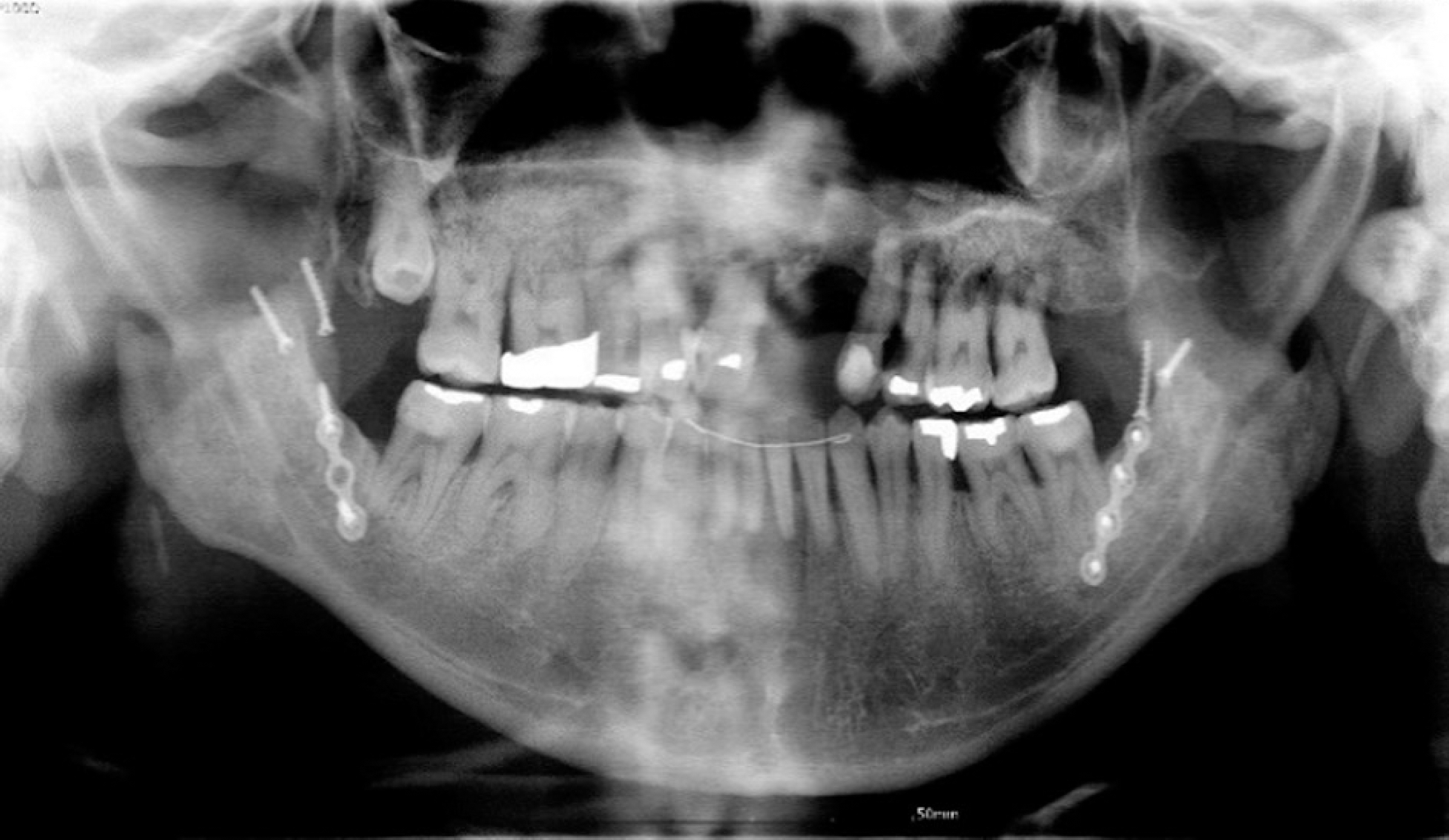 | Fig. 1.Panoramic radiographic view shows residual dentition and alveolar bone loss due to advance periodontal disease. |
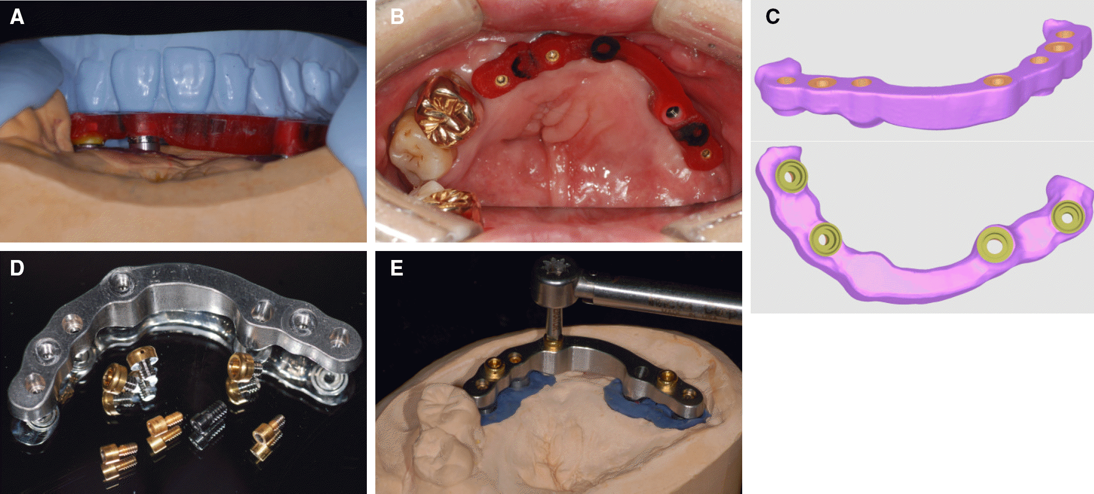 | Fig. 5.(A) Space evaluation for milled bar, (B) Try-in of bar pattern resin, (C) Bar design after scanned pattern resin, (D) Titanium milled bar and Locator® attachment,(E) Female part of Locator® attachment tightening by 20 Ncm torque. |
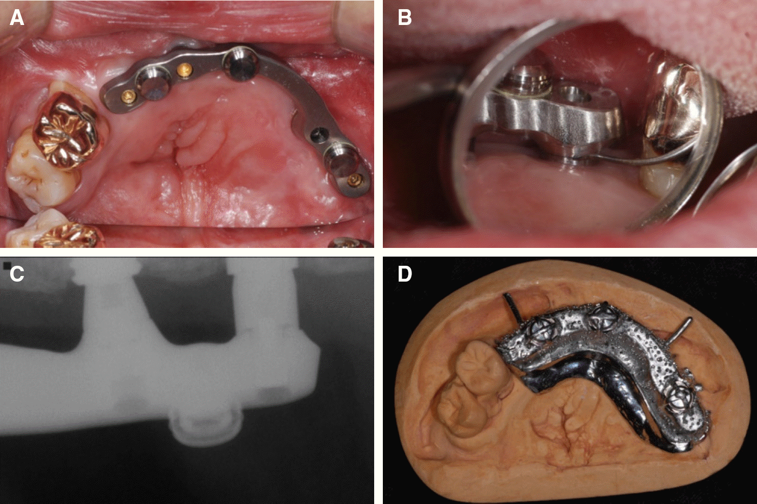 | Fig. 6.(A) Titanium milled bar try-in, (B) Passive fit evaluation: direct vision and tactile sensation, (C) Passive fit evaluation: periapical radiographic image, (D) Fabricated metal framework. |


 PDF
PDF ePub
ePub Citation
 Print
Print



 XML Download