Abstract
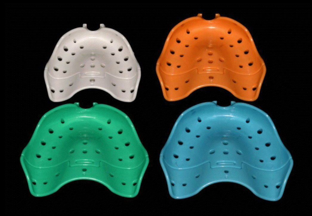
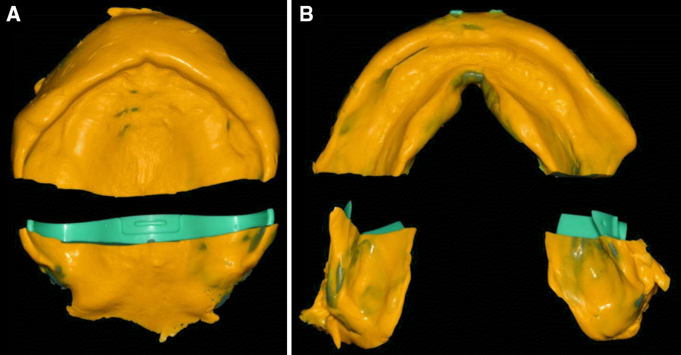
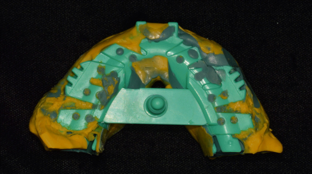
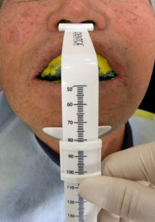
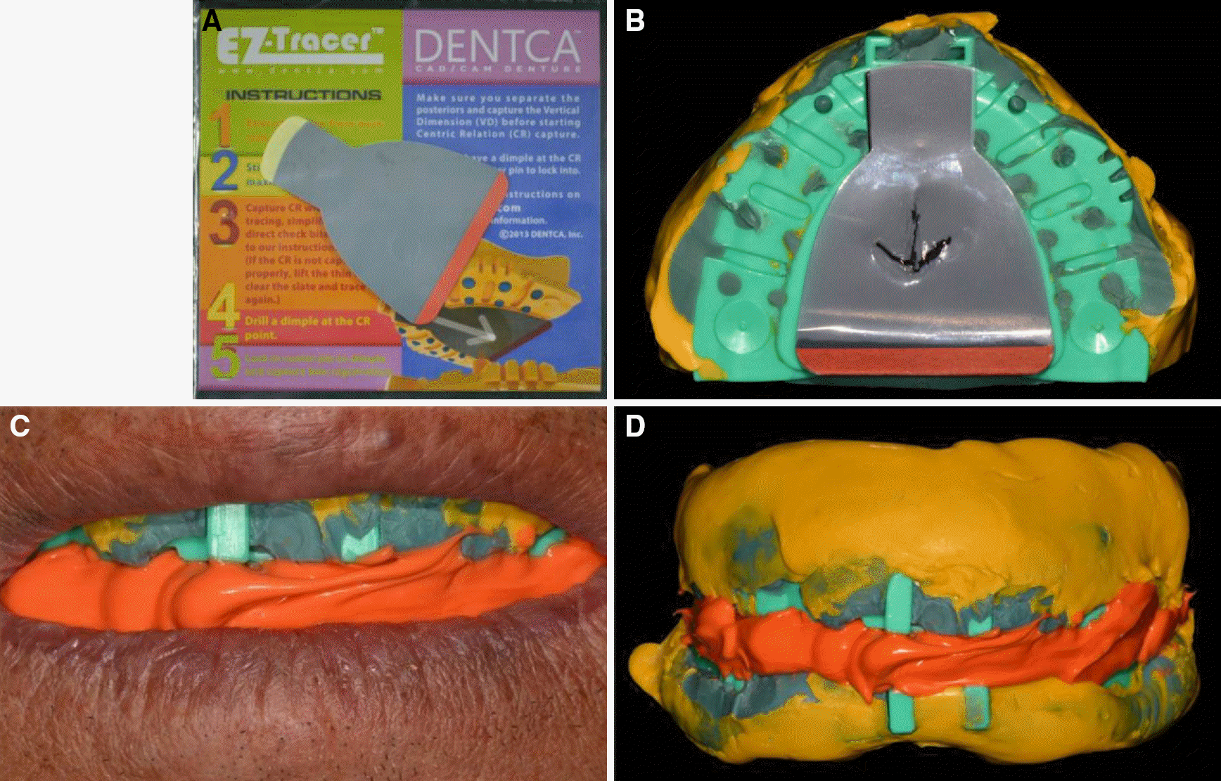
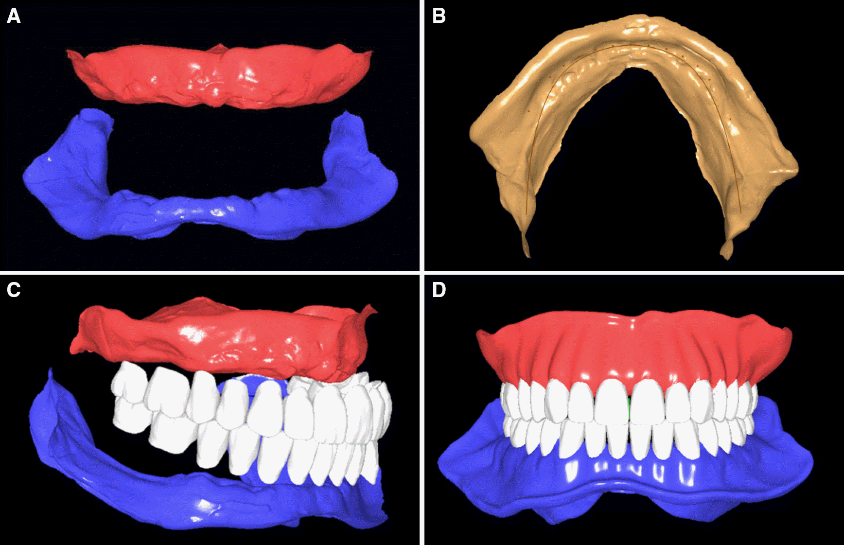
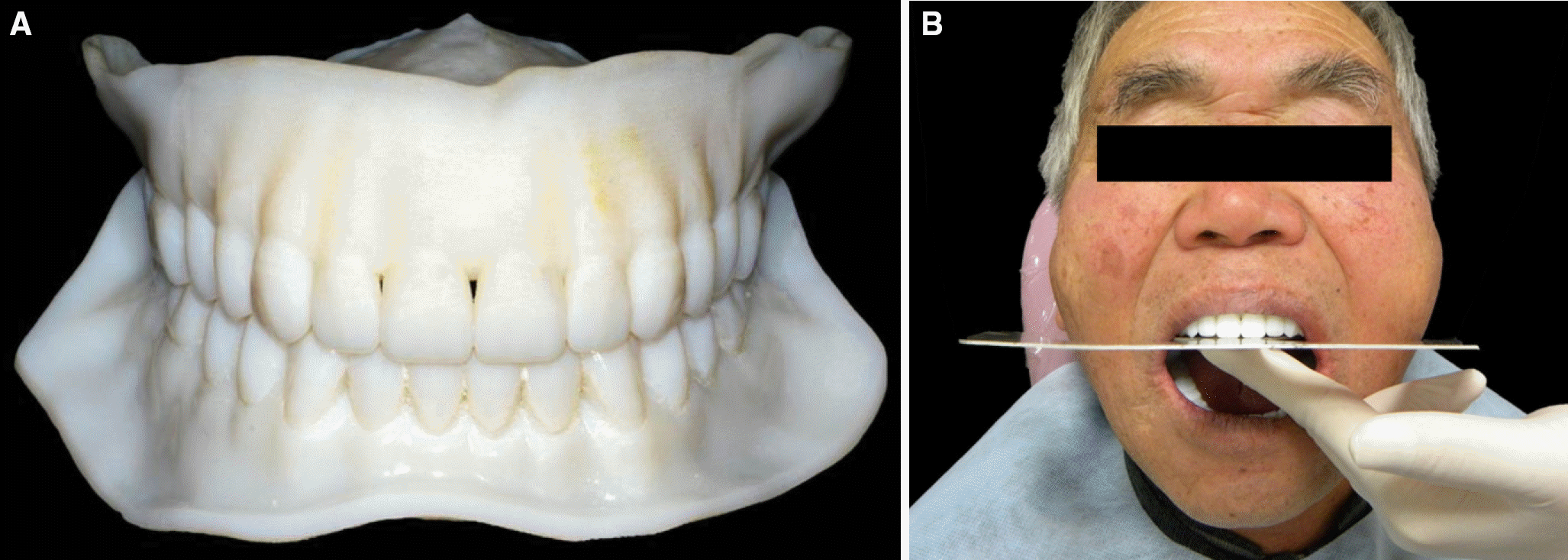
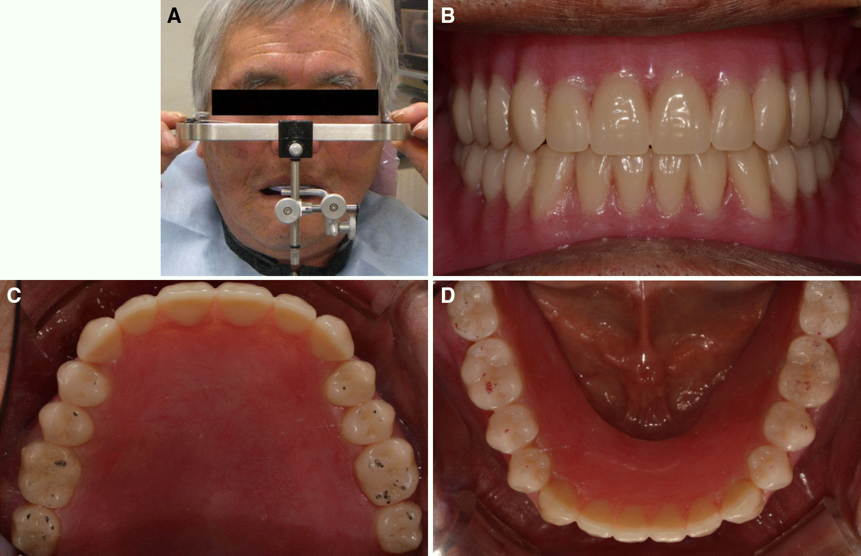
Nowadays, CAD/CAM is broadly used in dentistry for inlays, crowns, implant abutments and its spectrum is expanding to complete dentures. Utilizing CAD/CAM to fabricate complete dentures is expected to decrease chair time and the number of visits, thus decreasing total fabrication time, expenses and errors caused during fabrication processes. One of the systems using CAD/CAM, DENTCA™ CAD/CAM denture (DENTCA Inc. Los Angeles, USA) scans edentulous impressions, designs dentures digitally, fabricates try-in dentures by 3D printing and converts them into final dentures. Patients can wear final dentures after only 2 - 3 visits with satisfying adaptation. This case report introduces a 71-year-old male patient who visited to consult remaking of existing old dentures. Residual teeth with bad prognosis and root remnants were extracted and the patient used reformed existing mandibular denture for 2 months. And then DENTCA system started. One-step border molding was done using conventional tray of adequate size provided by DENTCA system and wash impression was taken. Gothic arch tracing was completed based on the vertical dimension of existing dentures. Both maxillary and mandibular trays were placed to the resultant centric relation and bite registration was taken. Then DENTCA scanned the bite registration, arranged the teeth, completed the festooning and fabricated the try-in dentures by 3D printing. The try-in dentures were positioned, occlusal plane and occlusal relations were evaluated. The try-in dentures were converted to final dentures. To create bilateral balanced occlusion, occlusal adjustment was done after clinical remounting using facebow transfer. The result was satisfactory and it was confirmed by patient and operator.
REFERENCES
1. Miyazaki T, Hotta Y, Kunii J, Kuriyama S, Tamaki Y. A review of dental CAD/CAM: current status and future perspectives from 20 years of experience. Dent Mater J. 2009; 28:44–56.

2. Goodacre CJ, Garbacea A, Naylor WP, Daher T, Marchack CB, Lowry J. CAD/CAM fabricated complete dentures: concepts and clinical methods of obtaining required morphological data. J Prosthet Dent. 2012; 107:34–46.

3. Maeda Y, Minoura M, Tsutsumi S, Okada M, Nokubi T. A CAD/CAM system for removable denture. Part I: Fabrication of complete dentures. Int J Prosthodont. 1994; 7:17–21.
4. Kawahata N, Ono H, Nishi Y, Hamano T, Nagaoka E. Trial of duplication procedure for complete dentures by CAD/CAM. J Oral Rehabil. 1997; 24:540–8.

5. Sun Y, Lu¨ P, Wang Y. Study on CAD&RP for removable complete denture. Comput Methods Programs Biomed. 2009; 93:266–72.
6. Inokoshi M, Kanazawa M, Minakuchi S. Evaluation of a complete denture trial method applying rapid prototyping. Dent Mater J. 2012; 31:40–6.

7. Kattadiyil MT, Goodacre CJ, Baba NZ. CAD/CAM complete dentures: a review of two commercial fabrication systems. J Calif Dent Assoc. 2013; 41:407–16.


 PDF
PDF ePub
ePub Citation
 Print
Print



 XML Download