Abstract
This article describes how to use CBCT and an intraoral scanner in a fully edentulous case that enables the clinician to place implants with flapless guided surgery and to engage prefabricated, customized implant abutments at the time of implant surgery, with only 1 clinical consultation before implant surgery. The patient's existing denture is used to simulate the teeth, the soft tissue and the vertical dimension of occlusion, and jaw relationship in the fully edentulous jaw. It provides clinicians with a fast workflow and improves clinical efficiency.
Go to : 
REFERENCES
1. Derhalli M. The digitalizing of implant dentistry: a clinical evaluation of 15 patients. Compend Contin Educ Dent. 2013; 34:192–6.
2. Di Giacomo GA, Cury PR, de Araujo NS, Sendyk WR, Sendyk CL. Clinical application of stereolithographic surgical guides for implant placement: preliminary results. J Periodontol. 2005; 76:503–7.
3. Ruppin J, Popovic A, Strauss M, Spü ntrup E, Steiner A, Stoll C. Evaluation of the accuracy of three different computer-aided surgery systems in dental implantology: optical tracking vs. stereolithographic splint systems. Clin Oral Implants Res. 2008; 19:709–16.

4. Van Assche N, van Steenberghe D, Guerrero ME, Hirsch E, Schutyser F, Quirynen M, Jacobs R. Accuracy of implant placement based on pre-surgical planning of three-dimensional cone-beam images: a pilot study. J Clin Periodontol. 2007; 34:816–21.

5. van Steenberghe D, Naert I, Andersson M, Brajnovic I, Van Cleynenbreugel J, Suetens P. A custom template and definitive prosthesis allowing immediate implant loading in the maxilla: a clinical report. Int J Oral Maxillofac Implants. 2002; 17:663–70.
6. Hermann S, Carsten T, Stephan I, Gü nter B, Robert S, Hans-Florian Z. Rapid Prototyping models for surgical planning with hard and soft tissue representation. Int Congress Series. 2004; 1268:567–72.
7. Ganz SD. Use of stereolithographic models as diagnostic and restorative aids for predictable immediate loading of implants. Pract Proced Aesthet Dent. 2003; 15:763–71.
8. Hajeer MY, Millett DT, Ayoub AF, Siebert JP. Applications of 3D imaging in orthodontics: part II. J Orthod. 2004; 31:154–62.
Go to : 
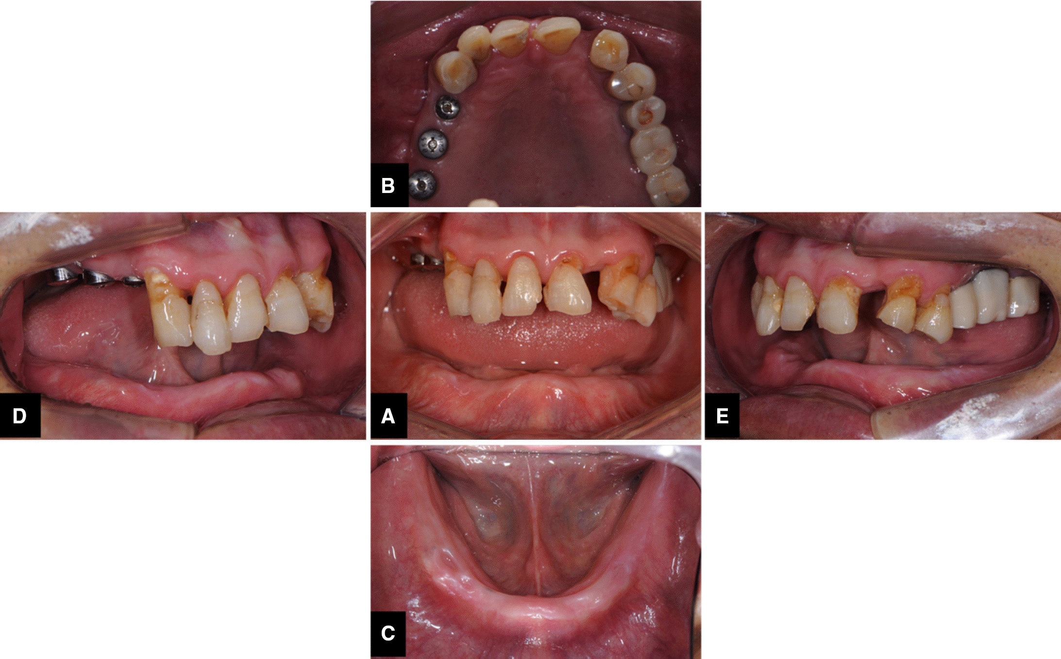 | Fig. 1.Pretreatment condition. (A) Frontal view, (B) Maxillary occlusal, (C) Mandibular occlusal, (D) Right posterior, (E) Left posterior. |
 | Fig. 3.Scanning of the denture, including the denture base, teeth and markers. (A) Scanning the denture base, (B) Scanning the marker and edge of denture base, (C) Scanning the denture teeth and marker. |
 | Fig. 4.Patient wearing the denture with markers. (A) Patient wearing the denture and taking CBCT, (B) 3 markers bonded to the denture. |
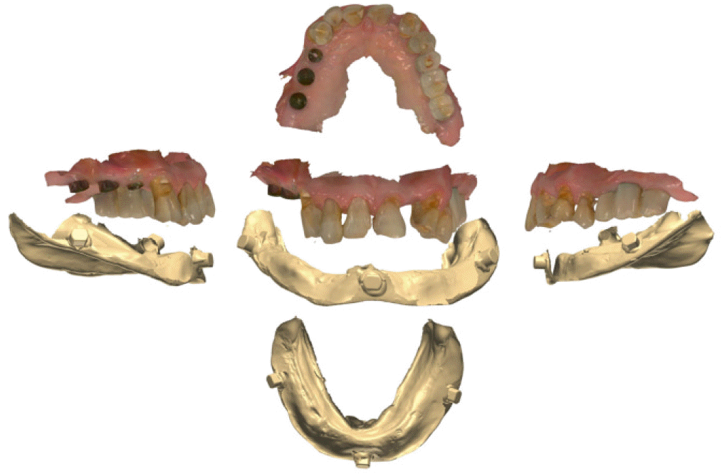
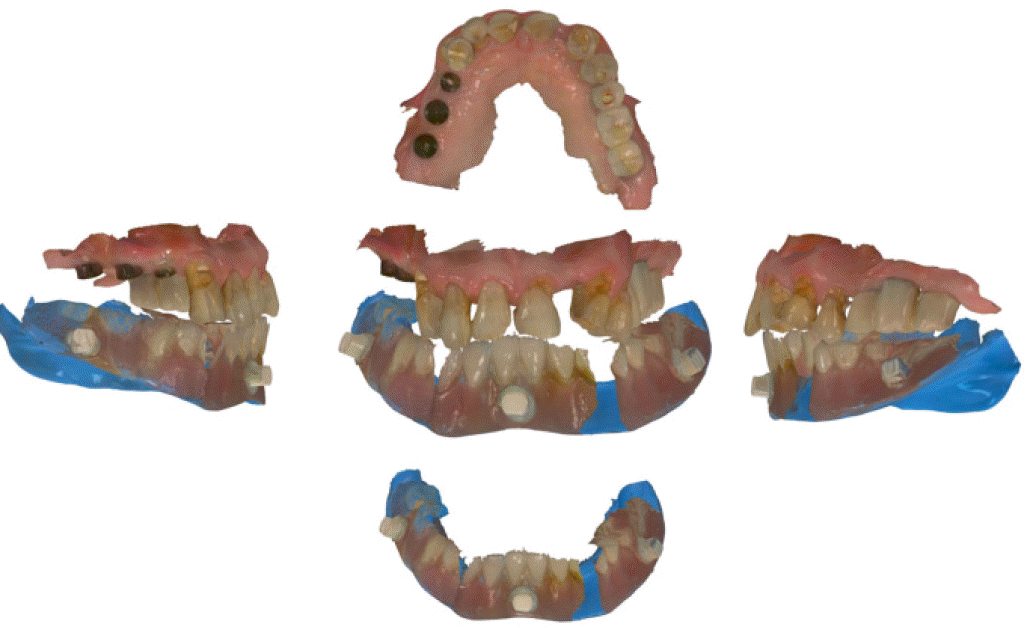 | Fig. 5.Digital images of the denture and the opposing teeth with interocclusal relationship. |
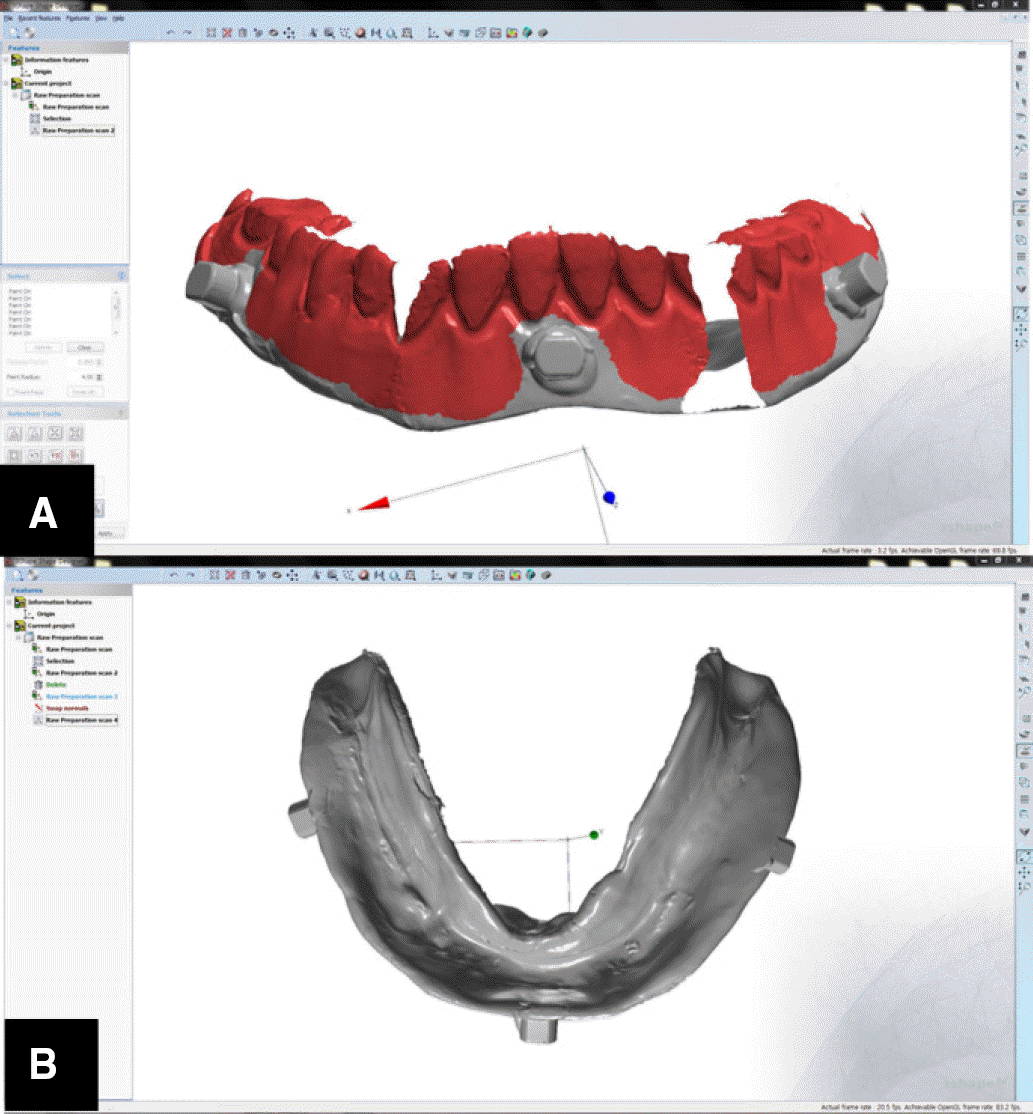 | Fig. 6.Virtual process of deleting aspects of the denture image and inverted image of the denture base. (A) Deleting aspects of denture image by Shape Designer, (B) The inverted image of denture base. |
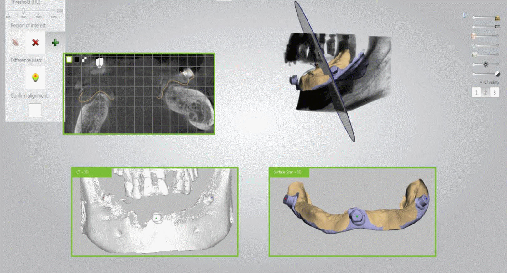 | Fig. 8.Image fusion of the inverted images from the intraoral scanner with images from the CBCT scan (Image matching of the inverted scan image and CBCT image). |
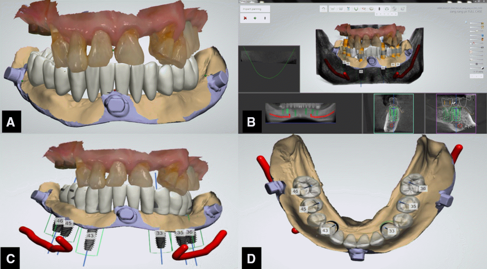 | Fig. 9.Implant planning. (A)Virtual tooth arrangement, (B) Implant planning, (C) After the implant planning, (D) Check implant position in the occlusial view. |
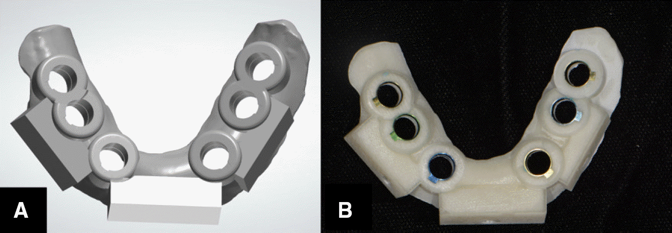 | Fig. 10.Surgical guide designed and printed. (A) The surgical guide designed by computer software, (B) The surgical guide fabricated by 3D printer. |
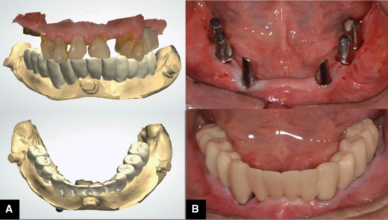
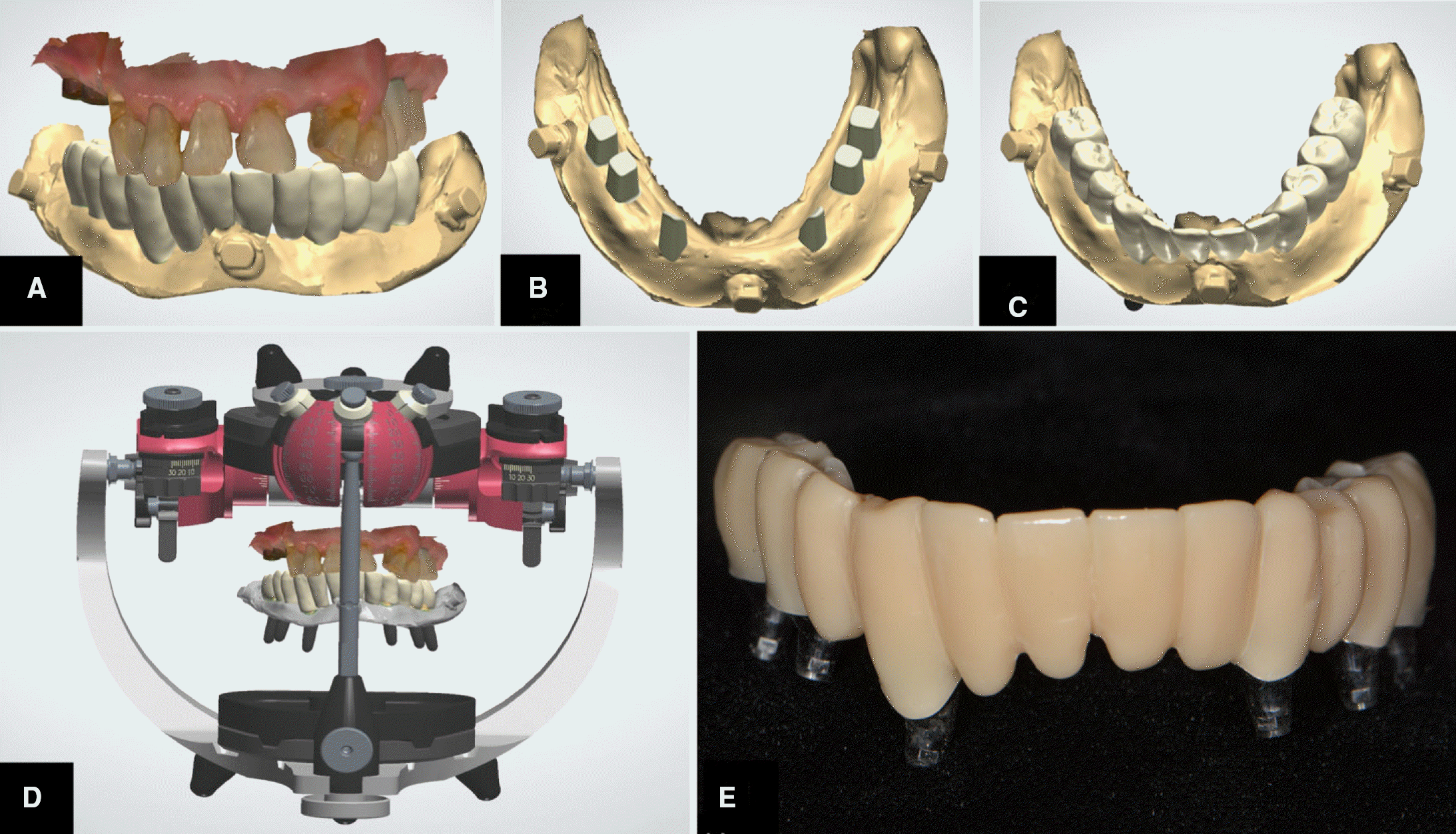 | Fig. 11.The designed provisional restoration in the frontal view (A) Customized abutments designed by computer software (B) Check the abutment and provisional restoration in occlusal view (C) Check occlusion with vitual articulator (D) Fabricated abutments and provisional restoration (E). |
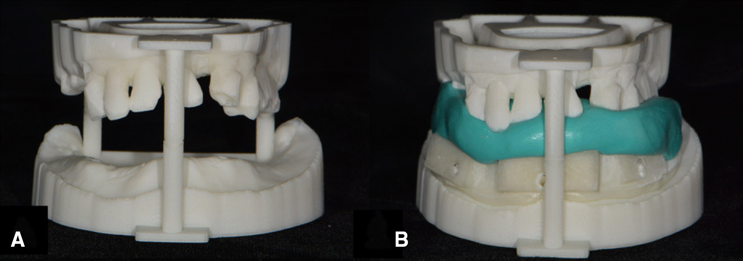 | Fig. 12.Stereolithographic model (A) and Interocclusal record made using the stereolithographic model (B). |


 PDF
PDF ePub
ePub Citation
 Print
Print



 XML Download