

 PDF
PDF ePub
ePub Citation
 Print
Print


Abstract
Purpose
Most of the former studies about the occlusal contact patterns during the mandibular movement focused on foreigner. The purpose of this study is analyzing the occlusal contacts of young Koreans by using T-Scan system.
Materials and methods
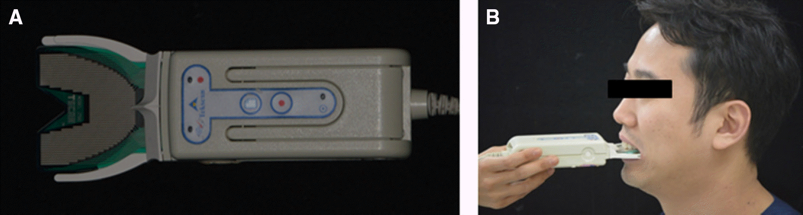
The sample size was 87 and the occlusal contacts of each right and left lateral movements were measured from the maximum intercuspation to the 3mm excursive position for three times respectively. All of the occlusal contacts were double checked through the thin metal foil. The results were categorized as two; 1) considering occlusal contact patterns on working side only, 2) considering occlusal contact patterns on working and nonworking sides.
Results
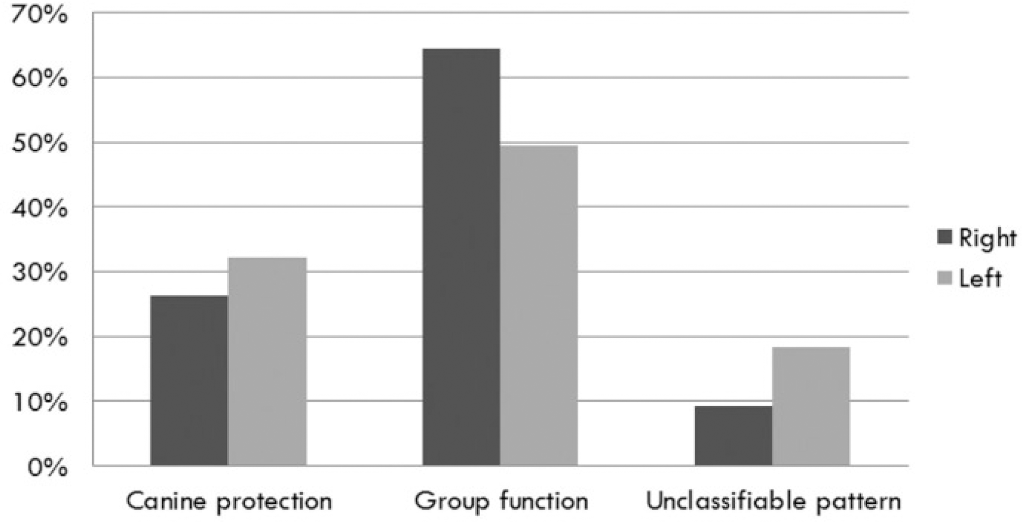
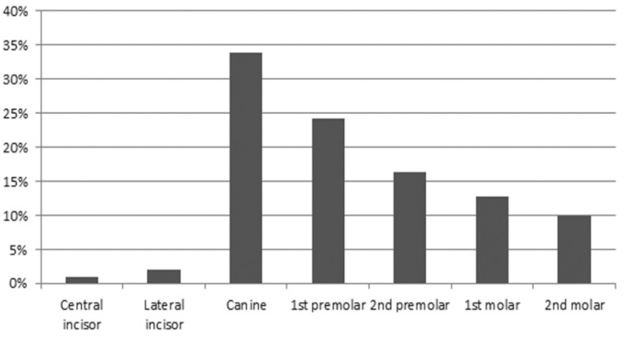
The results showed that the nonworking side occlusal contacts play major roles in the lateral mandibular movement. In both cases of considering with and without the nonworking side occlusal contacts, the group function was the most prevalent. In the working side, the contacts were the most frequent in canine and the frequency of contacts was decreased as the distance was increased from canine to molar. In the nonworking side, the contacts were the most frequent in second molar. And the gender factor was statistically significant (α =.05), as females have more nonworking side occlusal contacts in this study.
Go to : 
REFERENCES
1. Thornton LJ. Anterior guidance: group function/canine guidance. A literature review. J Prosthet Dent. 1990; 64:479–82.

2. Ogawa T, Ogimoto T, Koyano K. Pattern of occlusal contacts in lateral positions: canine protection and group function validity in classifying guidance patterns. J Prosthet Dent. 1998; 80:67–74.
3. The glossary of prosthodontic terms. J Prosthet Dent. 2005; 94:10–92.
4. Schuyler CH. Factors of occlusion applicable to restorative dentistry. J Prosthet Dent. 1953; 3:772–82.
5. Weinberg LA. A cinematic study of centric and eccentric occlusions. J Prosth Dent. 1964; 14:290–3.
6. Goldstein GR. The relationship of canine-protected occlusion to a periodontal index. J Prosthet Dent. 1979; 41:277–83.
7. Madone G, Ingervall B. Stability of results and function of the masticatory system in patients treated with the Herren type of ac-tivator. Eur J Orthod. 1984; 6:92–106.
8. Agerberg G, Sandströ m R. Frequency of occlusal interferences: a clinical study in teenagers and young adults. J Prosthet Dent. 1988; 59:212–7.
9. Saraç oğlu A, Ozpinar B. In vivo and in vitro evaluation of occlusal indicator sensitivity. J Prosthet Dent. 2002; 88:522–6.
10. Maness WL, Benjamin M, Podoloff R, Bobick A, Golden RF. Computerized occlusal analysis: a new technology. Quintessence Int. 1987; 18:287–92.
11. Baba K, Tsukiyama Y, Clark GT. Reliability, validity, and util-ity of various occlusal measurement methods and techniques. J Prosthet Dent. 2000; 83:83–9.
12. Harvey WL, Hatch RA, Osborne JW. Computerized occlusal analysis: an evaluation of the sensors. J Prosthet Dent. 1991; 65:89–92.
13. Olivieri F, Kang KH, Hirayama H, Maness WL. New method for analyzing complete denture occlusion using the center of force concept: a clinical report. J Prosthet Dent. 1998; 80:519–23.
14. Pokorny PH, Wiens JP, Litvak H. Occlusion for fixed prosthodontics: a historical perspective of the gnathological influence. J Prosthet Dent. 2008; 99:299–313.
15. Mann AW, Pankey LD. Oral rehabilitation: Part I. Use of the P-M instrument in treatment planning and in restoring the lower posterior teeth. J Prosthet Dent. 1960; 10:135–50.
16. D’ Amico A. Functional occlusion of the natural teeth of man. J Prosthet Dent. 1961; 11:899–915.
17. Nagao M. Comparative studies on the curve of Spee in mammals, with a discussion of its relation to the form of the fossa mandibularis. J Dent Res. 1919; 1:159–202.
18. Ingervall B, Hä hner R, Kessi S. Pattern of tooth contacts in eccentric mandibular positions in young adults. J Prosthet Dent. 1991; 66:169–76.
19. Takai A, Nakano M, Bando E, Hewlett ER. Evaluation of three occlusal examination methods used to record tooth contacts in lateral excursive movements. J Prosthet Dent. 1993; 70:500–5.
20. Scaife RR Jr. Holt JE. Natural occurrence of cuspid guidance. J Prosthet Dent. 1969; 22:225–9.
21. Ingervall B. Tooth contacts on the functional and nonfunction-al side in children and young adults. Arch Oral Biol. 1972; 17:191–200.
22. Ogawa T, Koyano K, Suetsugu T. The relationship between inclination of the occlusal plane and jaw closing path. J Prosthet Dent. 1996; 76:576–80.
23. Anderson GC, Schulte JK, Aeppli DM. Reliability of the evaluation of occlusal contacts in the intercuspal position. J Prosthet Dent. 1993; 70:320–3.
24. Rinchuse DJ, Sassouni V. An evaluation of functional occlusal interferences in orthodontically treated and untreated subjects. Angle Orthod. 1983; 53:122–30.
25. Yaffe A, Ehrlich J. The functional range of tooth contact in lateral gliding movements. J Prosthet Dent. 1987; 57:730–3.
26. Sadowsky C, BeGole EA. Long-term status of temporomandibular joint function and functional occlusion after orthodontic treatment. Am J Orthod. 1980; 78:201–12.
27. de Laat A, van Steenberghe D. Occlusal relationships and temporomandibular joint dysfunction. Part I: Epidemiologic findings. J Prosthet Dent. 1985; 54:835–42.
28. Ingervall B, Meyer D, Stettler B. Tooth contacts in eccentric mandibular positions and facial morphology. J Prosthet Dent. 1992; 67:317–22.
29. Ogawa T, Koyano K, Suetsugu T. The influence of anterior guidance and condylar guidance on mandibular protrusive movement. J Oral Rehabil. 1997; 24:303–9.
30. Koyano K, Ogawa T, Suetsugu T. The influence of canine guidance and condylar guidance on mandibular lateral movement. J Oral Rehabil. 1997; 24:802–7.
31. Mohlin B, Kopp S. A clinical study on the relationship between malocclusions, occlusal interferences and mandibular pain and dysfunction. Swed Dent J. 1978; 2:105–12.
32. Troeltzsch M, Troeltzsch M, Cronin RJ, Brodine AH, Frankenberger R, Messlinger K. Prevalence and association of headaches, temporomandibular joint disorders, and occlusal interferences. J Prosthet Dent. 2011; 105:410–7.
33. Brandini DA, Trevisan CL, Panzarini SR, Pedrini D. Clinical evaluation of the association between noncarious cervical lesions and occlusal forces. J Prosthet Dent. 2012; 108:298–303.
34. Droukas B, Lindee C, Carlsson GE. Occlusion and mandibular dysfunction: a clinical study of patients referred for functional disturbances of the masticatory system. J Prosthet Dent. 1985; 53:402–6.
35. Kahn J, Tallents RH, Katzberg RW, Ross ME, Murphy WC. Prevalence of dental occlusal variables and intraarticular temporomandibular disorders: molar relationship, lateral guidance, and nonworking side contacts. J Prosthet Dent. 1999; 82:410–5.
Go to :
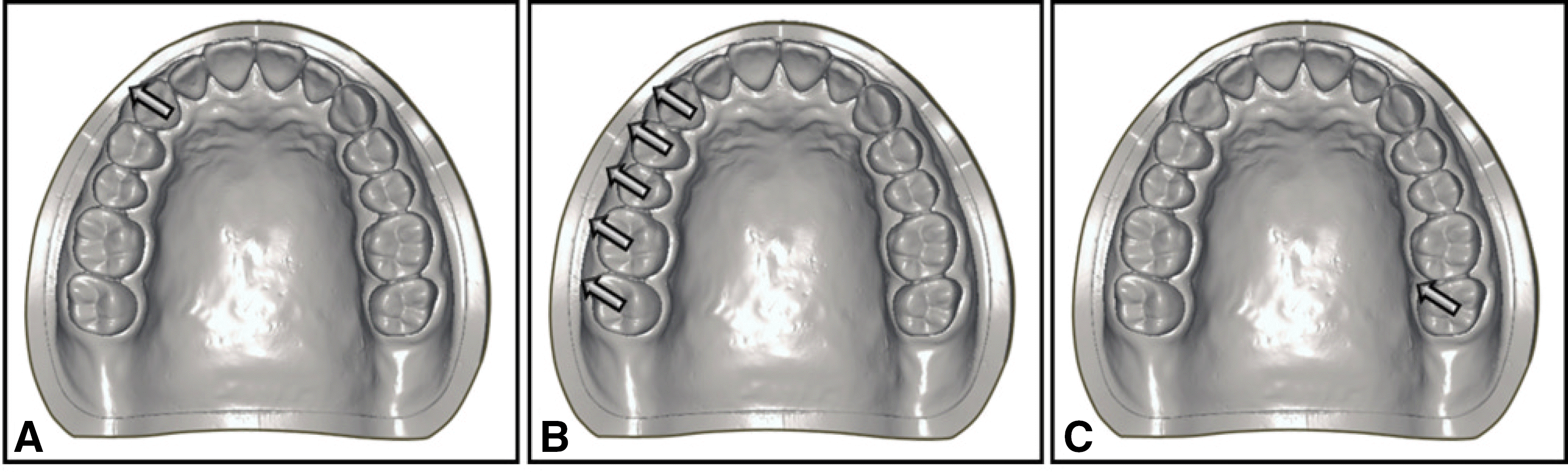 | Fig. 2.Classification of occlusal contact patterns in the system 1 (considering occlusal contact patterns on working side only). (A) Canine protection,(B) Group function, (C) Unclassifiable pattern. |
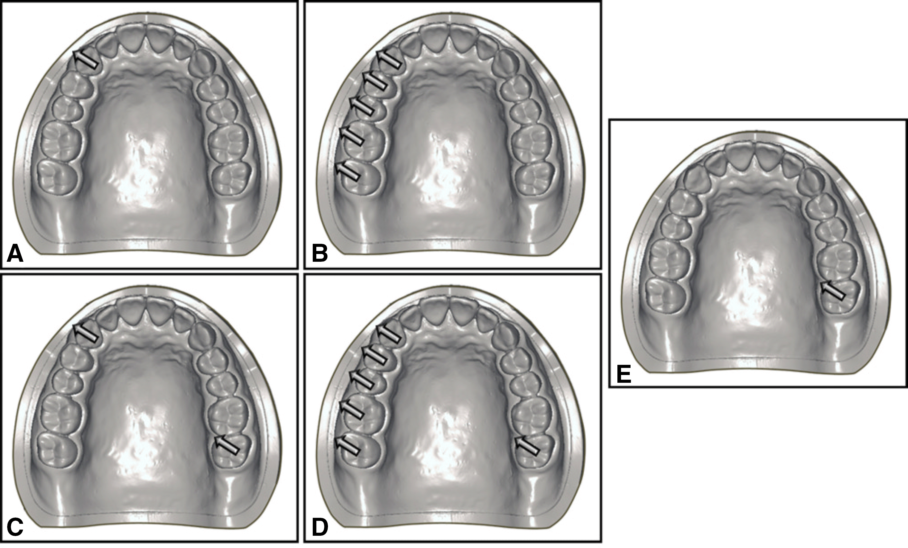 | Fig. 3.Classification of occlusal contact patterns in the system 2 (considering occlusal contact patterns on working and nonworking sides). (A) Canine protection (without nonworking side contacts), (B) Group function (without non-working side contacts), (C, D) With working & nonworking side occlusal contacts, (E) Unclassifiable pattern. |
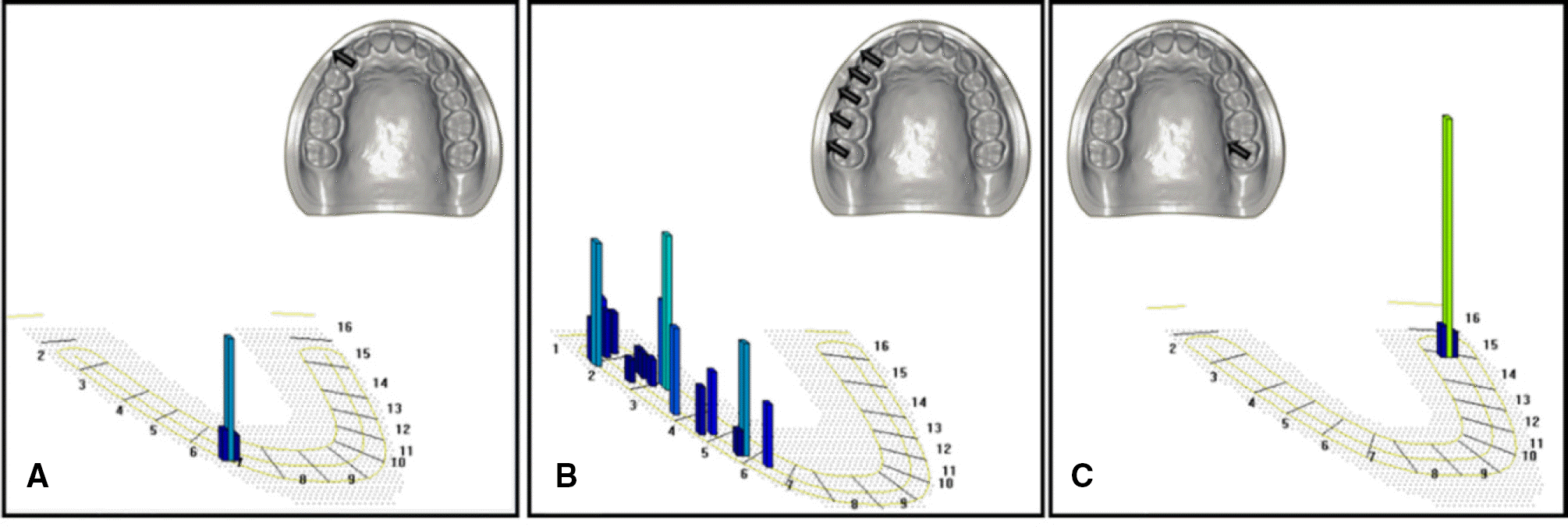 | Fig. 4.Result of T-Scan analysis in the system 1. (A) Canine protection, (B) Group function, (C) Unclassifiable pattern. |
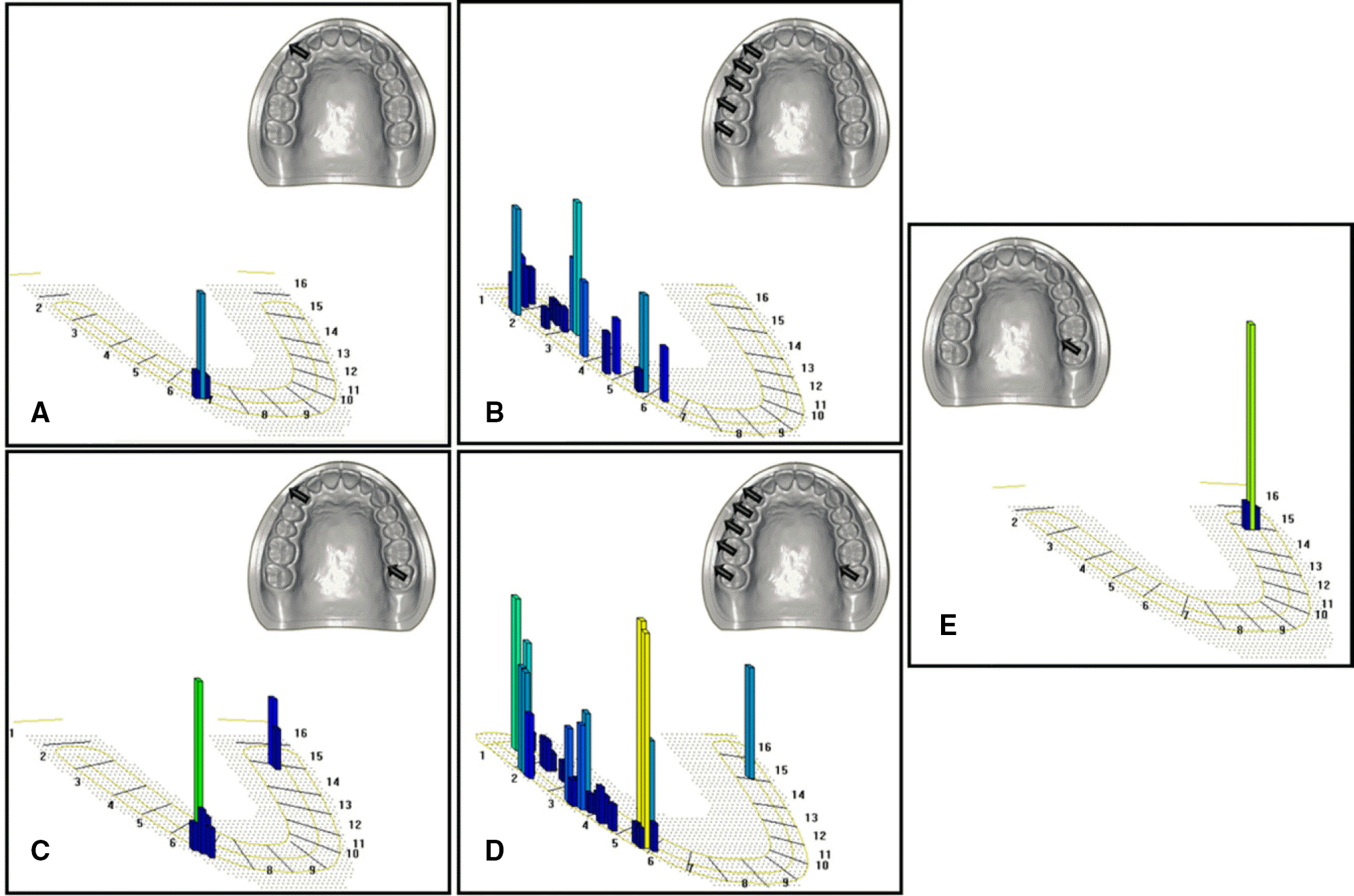 | Fig. 7.Result of T-Scan analysis in the system 2. (A) Canine protection (without nonworking side contacts), (B) Group function (without nonworking side contacts), (C, D) With working & nonworking side occlusal contacts, (E) Unclassifiable pattern. |
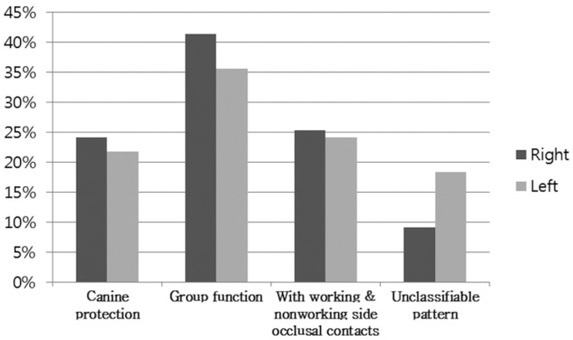
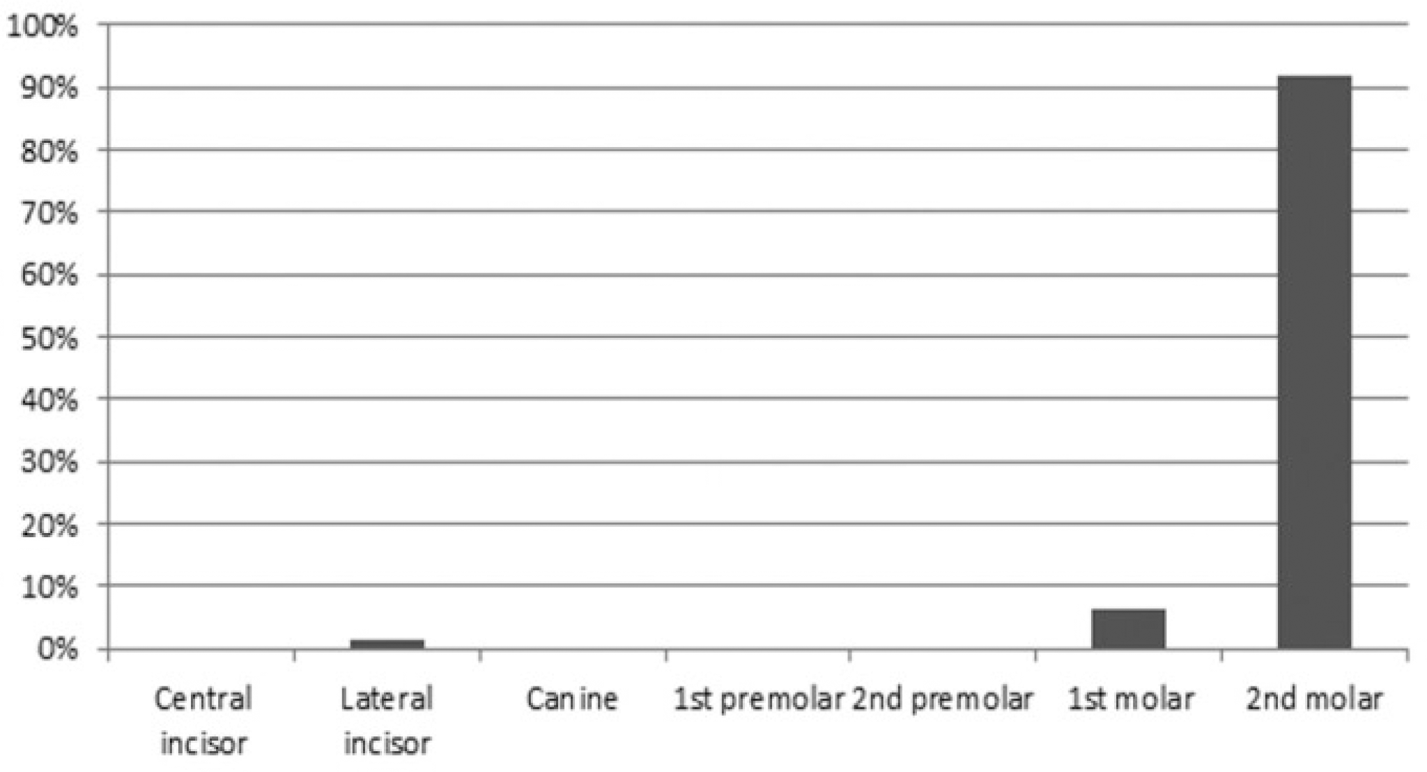
Table 1.
Participants in this study
| Number | Mean age (year) | |
|---|---|---|
| Male | 57 | 30.6 |
| Female | 30 | 28.7 |
| Total | 87 | 29.9 |
Table 2.
Percentage of nonworking side occlusal contacts (n=87)
| Without nonworking side occlusal contacts | 48 (55.2%) | ||
|---|---|---|---|
| With nonworking side occlusal contacts | Working & nonworking side contacts | 35 (40.2%) | 39 (44.8%) |
| Only nonworking side contacts | 4 (4.6%) |
Table 3.
Nonworking side occlusal contact frequency according to the gender
| Female | Male | Total | x2 | |
|---|---|---|---|---|
| With nonworking side contacts | 18 | 21 | 39 | |
| Without nonworking side contacts | 12 | 36 | 48 | 4.262 |
| Total | 30 | 57 | 87 | (df=1) |
 XML Download