Abstract
Purpose
This study was aimed to compare the radiopacity of four kinds of currently available resin based implant cements using digital radiography.
Materials and Methods
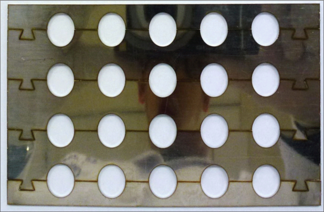
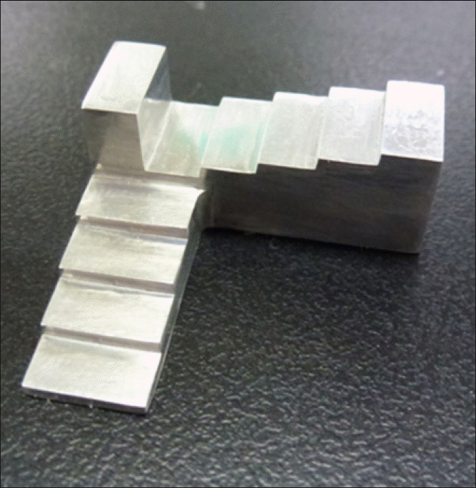
Four resin-based implant cements((Estemp ImplantTM (Spident, Incheon, Korea), Premier®Implant (Premier, Pennsylvania, USA), Cem-ImplantTM (B.J.M lab, Or-yehuda, Israel), InterCemTM (SCI-PHARM, California, USA)) and control group (Elite Cement 100TM (GC, Tokyo, Japan)) were mixed and cured according to the manufacturer's instructions on the custom made split-type metal mold. A total of 150 specimens of each cement were prepared and each specimen (purity over 99%) was placed side-by-side with an aluminum step wedge for image taking with Intraoral X-ray unit (Esx, Vatech, Korea) and digital X-ray sensor (EzSensor, Vatech, Korea). For the evaluation of aluminum wedge equivalent thickness (mm Al), ImageJ 1.47 m (Wayne Rasband, National Institutes of Health, USA) and Color inspector 3D ver 2.0 (Interaktive Visualisierung von Farbra¨umen, Berlin, Germany) programs were used.
Result
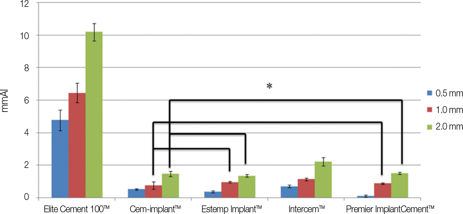
Among the 5 cements, Elite cement 100TM (control group) showed the highest radio-opacity in all thickness. In the experimental group, InterCemTM had the highest radio-opacity followed by Premier® Implant CementTM, Cem-ImplantTMand Estemp ImplantTM. In addition, InterCemTM showed radio-opacity that met the ISO No. 4049 standard in all the tested specimen thickness. Cem-Implant on 0.5 mm thickness showed radiopacity that met the ISO No. 4049 standard.
Conclusion
Among the implant resin-based cements tested in the study, Premier® Implant Cement and Estemp ImplantTM did not show appropriate radio-opacity. Only InterCemTM and Cem-ImplantTM 0.5 mm specimen had the proper radiopacity and met the experiment standard.(J Korean Acad Prosthodont 2014;52:97-104)
Go to : 
REFERENCES
1.De Rouck T., Collys K., Cosyn J. Single-tooth replacement in the anterior maxilla by means of immediate implantation and pro-visionalization: a review. Int J Oral Maxillofac Implants. 2008. 23:897–904.
2.Lee A., Okayasu K., Wang HL. Screwversus cement-retained implant restorations: current concepts. Implant Dent. 2010. 19:8–15.
3.Chee W., Jivraj S. Screw versus cemented implant supported restorations. Br Dent J. 2006. 201:501–7.

4.Chaar MS., Att W., Strub JR. Prosthetic outcome of cement-retained implant-supported fixed dental restorations: a systematic review. J Oral Rehabil. 2011. 38:697–711.

5.Michalakis KX., Hirayama H., Garefis PD. Cement-retained versus screw-retained implant restorations: a critical review. Int J Oral Maxillofac Implants. 2003. 18:719–28.
6.Nissan J., Narobai D., Gross O., Ghelfan O., Chaushu G. Long-term outcome of cemented versus screw-retained implant-supported partial restorations. Int J Oral Maxillofac Implants. 2011. 26:1102–7.
7.Ramp MH., Dixon DL., Ramp LC., Breeding LC., Barber LL. Tensile bond strengths of provisional luting agents used with an implant system. J Prosthet Dent. 1999. 81:510–4.

8.Koka S., Ewoldsen NO., Dana CL., Beatty MW. The effect of cementing agent and technique on the retention of a CeraOne gold cylinder: a pilot study. Implant Dent. 1995. 4:32–5.

9.Clayton GH., Driscoll CF., Hondrum SO. The effect of luting agents on the retention and marginal adaptation of the CeraOne implant system. Int J Oral Maxillofac Implants. 1997. 12:660–5.
10.Akça K., Iplikçioğlu H., Cehreli MC. Comparison of uniaxial resistance forces of cements used with implant-supported crowns. Int J Oral Maxillofac Implants. 2002. 17:536–42.
11.GaRey DJ., Tjan AH., James RA., Caputo AA. Effects of ther-mocycling, load-cycling, and blood contamination on cemented implant abutments. J Prosthet Dent. 1994. 71:124–32.

12.Cho-Yan Lee J., Mattheos N., Nixon KC., Ivanovski S. Residual periodontal pockets are a risk indicator for peri-implantitis in patients treated for periodontitis. Clin Oral Implants Res. 2012. 23:325–33.

13.Shapoff CA., Lahey BJ. Crestal bone loss and the consequences of retained excess cement around dental implants. Compend Contin Educ Dent. 2012. 33:94–6. 98–101.
14.Wilson TG Jr. The positive relationship between excess cement and peri-implant disease: a prospective clinical endoscopic study. J Periodontol. 2009. 80:1388–92.
15.Dumbrigue HB., Abanomi AA., Cheng LL. Techniques to minimize excess luting agent in cement-retained implant restorations. J Prosthet Dent. 2002. 87:112–4.

16.Schwedhelm ER., Lepe X., Aw TC. A crown venting technique for the cementation of implant-supported crowns. J Prosthet Dent. 2003. 89:89–90.

17.Wadhwani C., Piñeyro A. Technique for controlling the cement for an implant crown. J Prosthet Dent. 2009. 102:57–8.

18.Wadhwani C., Rapoport D., La Rosa S., Hess T., Kretschmar S. Radiographic detection and characteristic patterns of residual excess cement associated with cement-retained implant restorations: a clinical report. J Prosthet Dent. 2012. 107:151–7.

19.Pauletto N., Lahiffe BJ., Walton JN. Complications associated with excess cement around crowns on osseointegrated implants: a clinical report. Int J Oral Maxillofac Implants. 1999. 14:865–8.
20.Gapski R., Neugeboren N., Pomeranz AZ., Reissner MW. Endosseous implant failure influenced by crown cementation: a clinical case report. Int J Oral Maxillofac Implants. 2008. 23:943–6.
21.O'Rourke B., Walls AW., Wassell RW. Radiographic detection of overhangs formed by resin composite luting agents. J Dent. 1995. 23:353–7.
22.Soares CJ., Santana FR., Fonseca RB., Martins LR., Neto FH. In vitro analysis of the radiodensity of indirect composites and ceramic inlay systems and its influence on the detection of cement overhangs. Clin Oral Investig. 2007. 11:331–6.

23.Reis JM., Jorge EG., Ribeiro JG., Pinelli LA., Abi-Rached FO., Tanomaru-Filho M. Radiopacity evaluation of contemporary luting cements by digitization of images. ISRN Dent. 2012. 2012:1–5.

24.Pette GA., Ganeles J., Norkin FJ. Radiographic appearance of commonly used cements in implant dentistry. Int J Periodontics Restorative Dent. 2013. 33:61–8.

25.ISO 4049. Dentistry: Polymer-based restorative materials. Geneva: ISO International Organization for Standardization;2009.
26.Wadhwani C., Hess T., Faber T., Pin ̃eyro A., Chen CS. A descriptive study of the radiographic density of implant restorative cements. J Prosthet Dent. 2010. 103:295–302.

27.Agar JR., Cameron SM., Hughbanks JC., Parker MH. Cement removal from restorations luted to titanium abutments with simulated subgingival margins. J Prosthet Dent. 1997. 78:43–7.

28.Altintas SH., Yildirim T., Kayipmaz S., Usumez A. Evaluation of the radiopacity of luting cements by digital radiography. J Prosthodont. 2013. 22:282–6.

29.Begoña Ormaechea M., Millstein P., Hirayama H. Tube angulation effect on radiographic analysis of the implant-abutment interface. Int J Oral Maxillofac Implants. 1999. 14:77–85.
30.Attar N., Tam LE., McComb D. Mechanical and physical properties of contemporary dental luting agents. J Prosthet Dent. 2003. 89:127–34.

31.Callan DP., Cobb CM. Excess cement and peri-implant disease. J Implant Adv Clin Dent. 2009. 1:61–8.
32.Wilson TG Jr. The positive relationship between excess cement and peri-implant disease: a prospective clinical endoscopic study. J Periodontol. 2009. 80:1388–92.
Go to : 
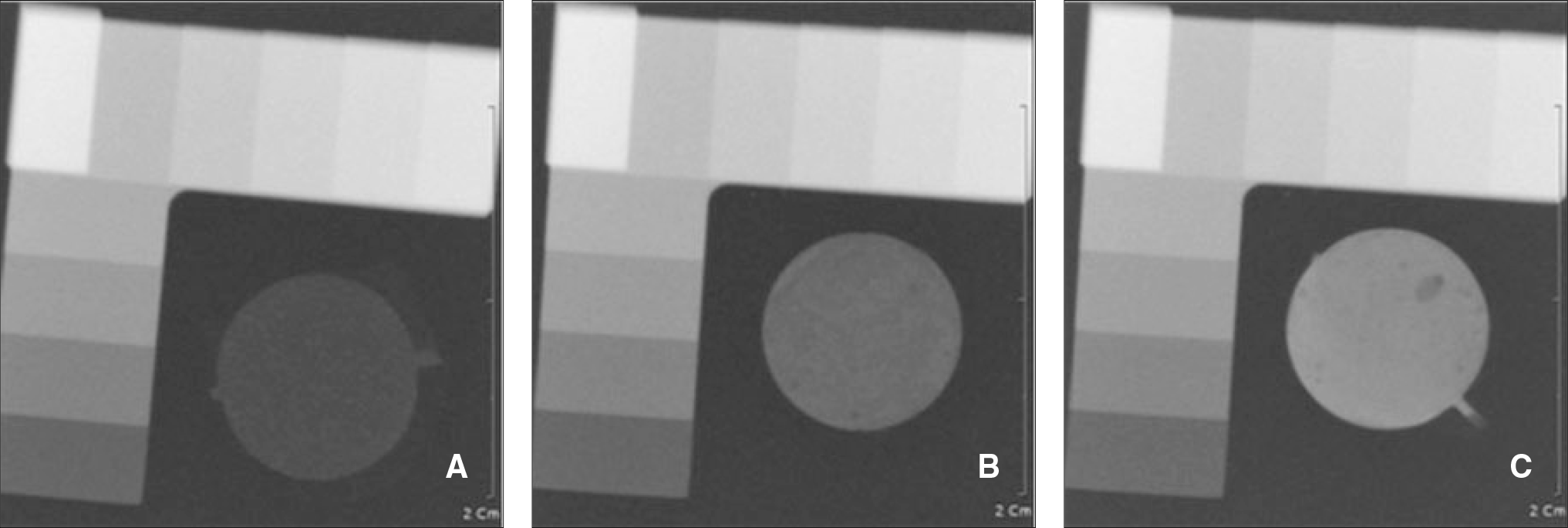
 | Fig. 3.Radiographic image of specimen and Aluminum step wedge. (A) 0.5 mm thickness, (B) 1.0 mm thickness, (C) 2.0 mm thickness. |
 | Fig. 4.Aluminum wedge equivalent thickness values of experimental groups. ∗ : No significant difference. |
Table 1.
Experimental cements used in this study
Table 2.
Aluminum wedge equivalent thickness(mm Al) of cements
| Thickness Cement | 0.5 mm | 1.0 mm | 2.0 mm |
|---|---|---|---|
| Elite Cement 100TM | 4.77 ± 0.641 | 6.45 ± 0.61a | 10.19 ± 0.54∗ |
| Cem-implantTM | 0.52 ± 0.042 | 0.76 ± 0.23bc | 1.48 ± 0.17†‡ |
| Estemp ImplantTM | 0.37 ± 0.073 | 0.97 ± 0.05b | 1.35 ± 0.08† |
| IntercemTM | 0.71 ± 0.074 | 1.14 ± 0.08d | 2.21 ± 0.26# |
| PremierI†implantCementTM | 0.11 ± 0.065 | 0.88 ± 0.05c | 1.50 ± 0.06† |


 PDF
PDF ePub
ePub Citation
 Print
Print


 XML Download