Abstract
They have been recently introduced many aesthetic implant prosthesis using with zirconia and CAD/CAM. However, there are many limitations in their gingival and occlusal region. In this case, submucosal zirconia implant prosthesis were fabricated with CAD/CAM system. The connection of these screw cement retained prosthesis and titanium abutment was designed to 1mm above the fixture. The clinical results were satisfactory on the aesthetics and function.
Go to : 
REFERENCES
1. Ortorp A, Kihl ML, Carlsson GE. A 3-year retrospective and clinical follow-up study of zirconia single crowns performed in a pri-vate practice. J Dent. 2009; 37:731–6.
2. Anusavice KJ. Standardizing failure, success, and survival decisions in clinical studies of ceramic and metal-ceramic fixed dental prostheses. Dent Mater. 2012; 28:102–11.

3. Koenig V, Vanheusden AJ, Le Goff SO, Mainjot AK. Clinical risk factors related to failures with zirconia-based restorations: an up to 9-year retrospective study. J Dent. 2013; 41:1164–74.

4. Marchack BW, Sato S, Marchack CB, White SN. Complete and partial contour zirconia designs for crowns and fixed dental prostheses: a clinical report. J Prosthet Dent. 2011; 106:145–52.

5. Leutert CR, Stawarczyk B, Truninger TC, Ha¨mmerle CH, Sailer I. Bending moments and types of failure of zirconia and titanium abutments with internal implant-abutment connections: a laboratory study. Int J Oral Maxillofac Implants. 2012; 27:505–12.
Go to : 
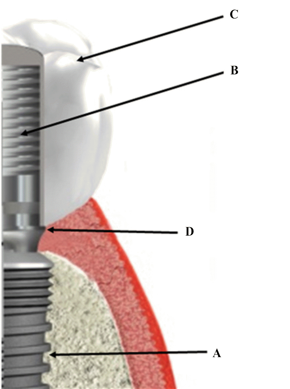
 | Fig. 1.Schematic diagram of submucosal zirconia implant prosthesis. (A) Internal connection fixture, (B) 1 mm cuff titanium abutment, (C) Screw cement retained prosthesis, (D) Prosthesis margin. |
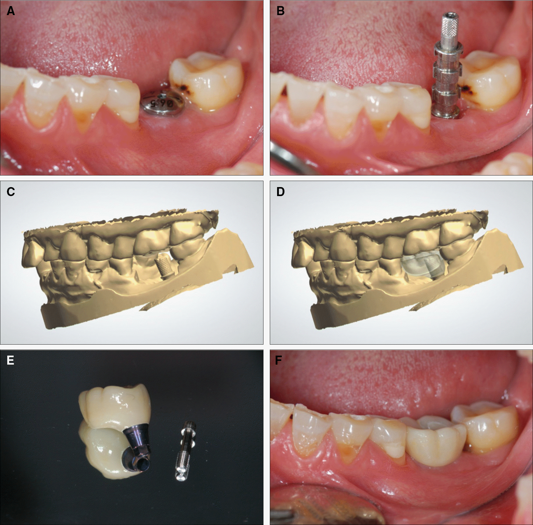
 | Fig. 2.Clinical pictures of case 1. (A) Intraoral view after implant surgery, (B) Pick-up impression coping, (C) Scanning of 1 mm cuff milled titanium abutment, (D) Computer aided design of submucosal prosthesis, (E) Submucosal zirconia implant prosthesis bonded to titanium abutment, (F) Definitive prosthesis. |
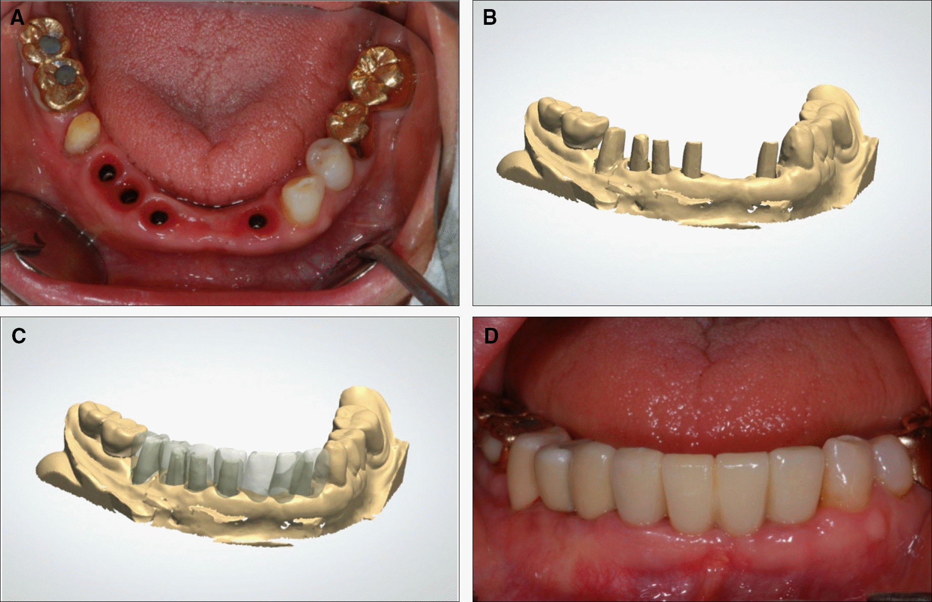
 | Fig. 3.Clinical pictures of case 2. (A) Occlusal view after implant surgery, (B) Scanning of milled titanium abutment, (C) Computer aided design of prosthesis, (D) Definitive prosthesis. |
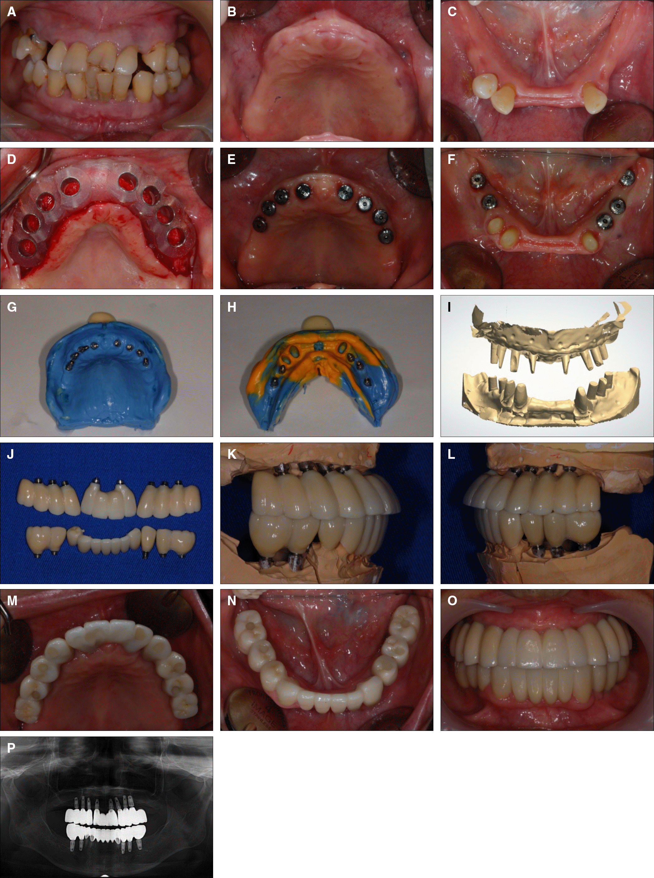
 | Fig. 4.Clinical pictures of case 3. (A) Frontal view of first visit, (B) Occlusal view after maxillary teeth extraction, (C) Occlusal view after mandibular teeth extraction, (D) SurgiGuide with SimPlant program, (E) Occlusal view after maxillary implants surgery, (F) Occlusal view after mandibular implants surgery, (G) Final impression of maxillary implants, (H) Final impression of mandibular implants, (I) Scanning of milled titanium abutment, (J) Submucosal zirconia implant prosthesis bonded to titanium abutment, (K) Right view of occlusal pattern, (L) Left view of occlusal pattern, (M) Occlusal view of maxillary definitive restoration, (N) Occlusal view of mandibular definitive restoration, (O) Frontal view of definitive restoration, (P) Panoramic view of definitive restoration. |


 PDF
PDF ePub
ePub Citation
 Print
Print


 XML Download