Abstract
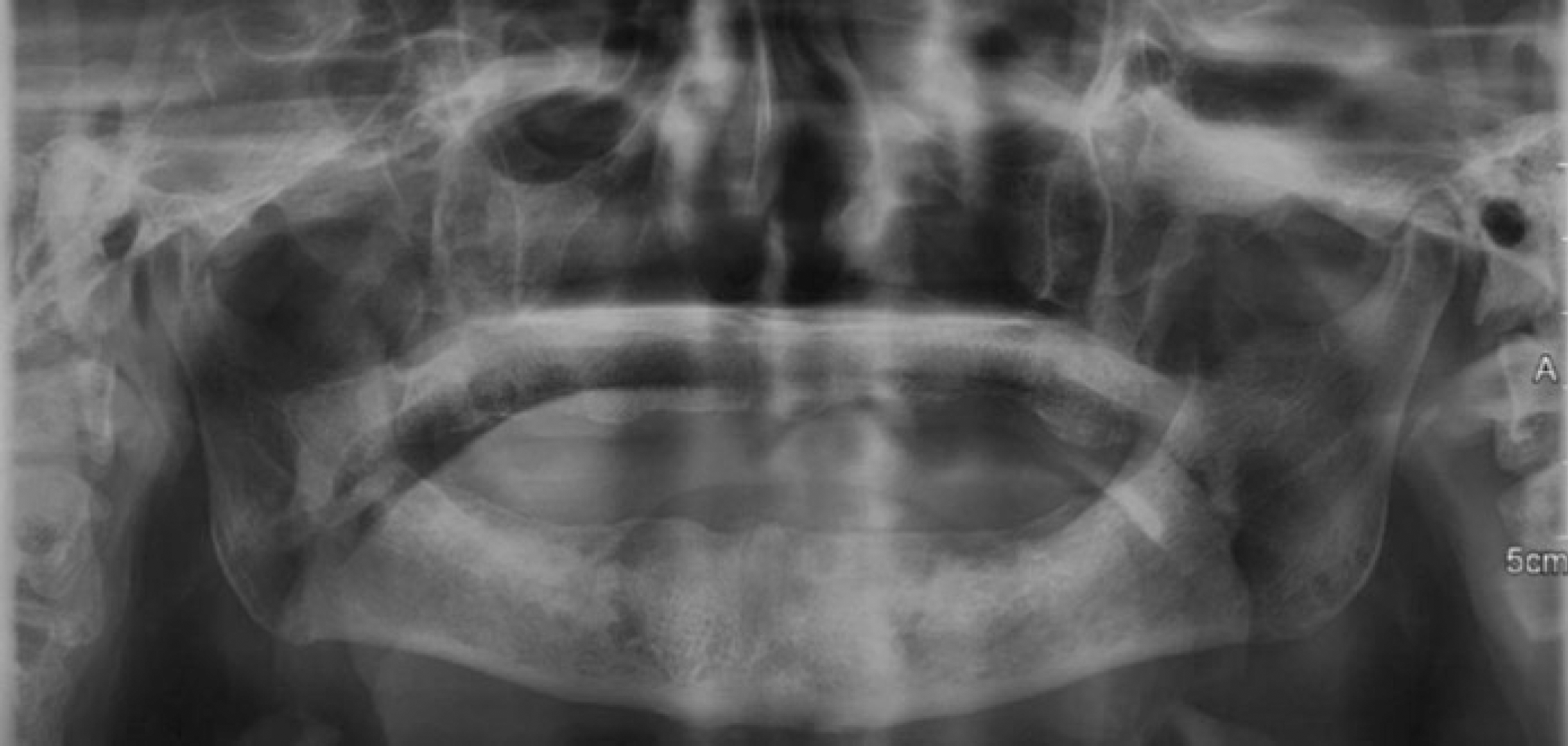
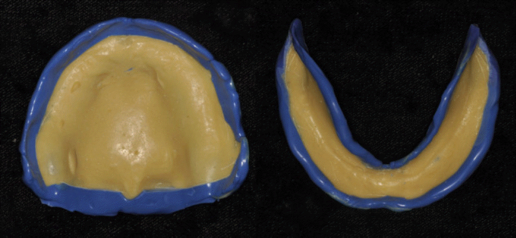
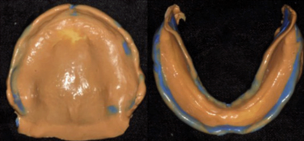
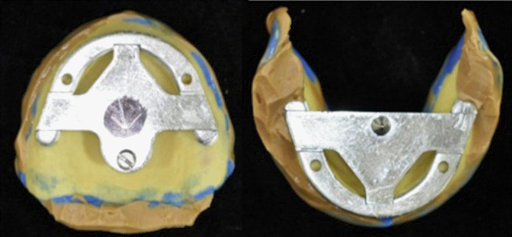
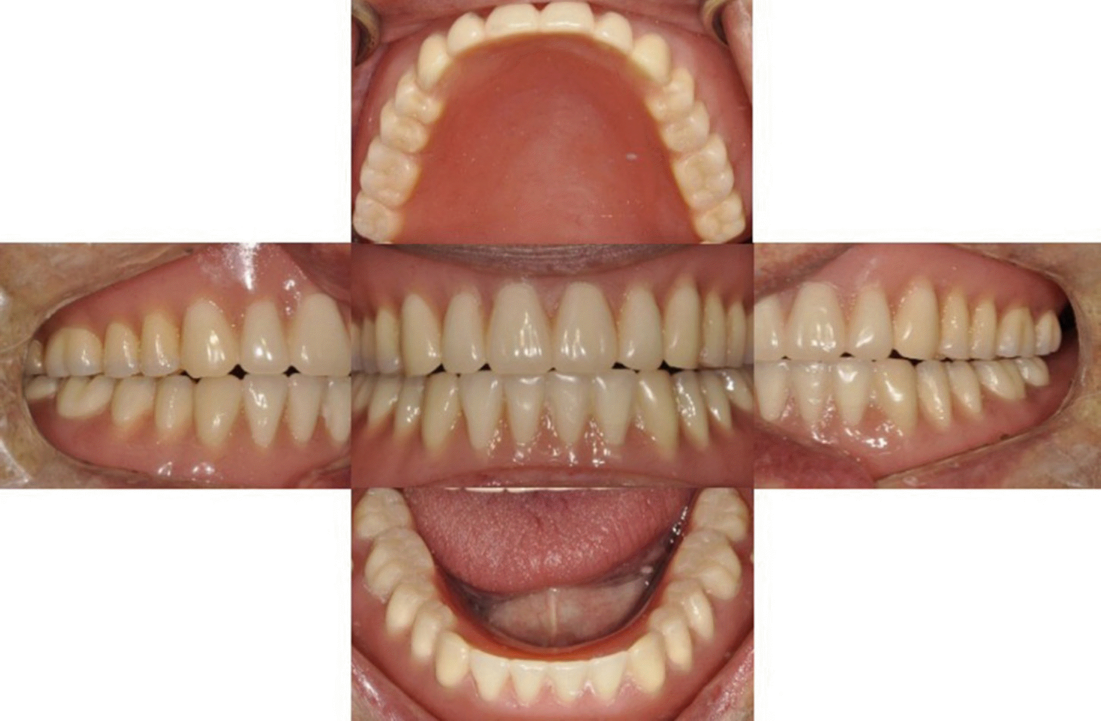
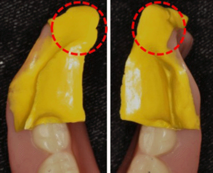
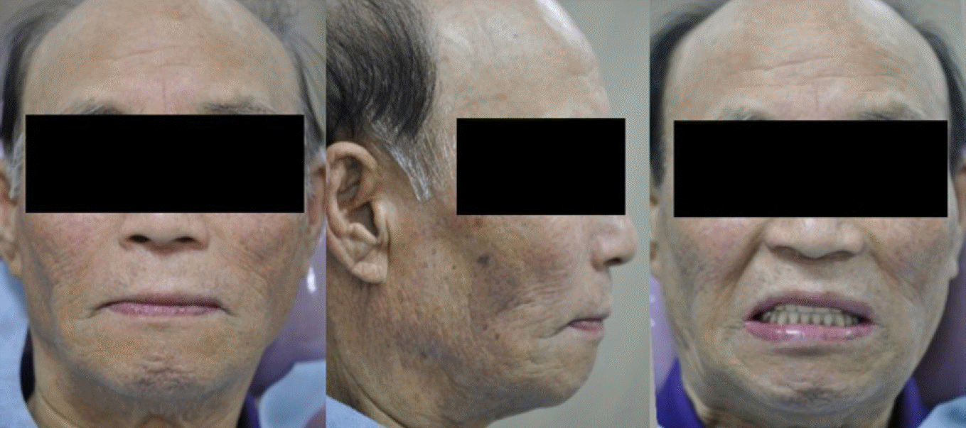
Suction dentures enhance retention and support by forming negative pressure temporarily at the internal surface of denture base at times of swallowing and chewing because the areas surrounding the denture flanges are sealed by mobile mucosa. In this case, an 81-year-old male visited for new dentures. Considering the high expectations for retention and masticatory efficiency of dentures, fabricating complete dentures with suction dentures was planned. Preliminary impression was taken without applying pressure on retromolar pad area and diagnostic cast was fabricated. Afterwards, individual tray was made and final impression was taken, at the same time, gothic arch tracing was done to acquire centric relation and vertical dimension. Then, anatomic teeth were placed on maxilla and non-anatomic teeth were placed on mandible forming lingualized occlusion. Consequently, restoring a complete edentulous patient with complete dentures using mandibular suction denture resulted in recovering satisfying retention and function.
REFERENCES
1. Zarb GA, Bolender CL, Eckert SE, Fenton AH, Jacob RF, Mericske-Stern R. Prosthodontic treatment for edentulous patients: Complete dentures and implant-supported prestheses. 12th ed.Mosby;2004. p. 24–33. p. 73–99.
2. Douglass CW1, Shih A, Ostry L. Will there be a need for complete dentures in the United States in 2020? J Prosthet Dent. 2002; 87:5–8.

3. Ministry of Health & Welfare. The Korean National Oral Health Survey. 2003.
4. Tallgren A. The continuing reduction of the residual alveolar ridges in complete denture wearers: a mixed-longitudinal study covering 25 years. J Prosthet Dent. 2003; 89:427–35.

6. Abe J, Kokubo K, Sato K. Mandibular Suction-effective denture and BPS: A complete guide. Tokyo: Quintessence Publishing Co. Ltd.;2012.
7. Abe J. Difference of preliminary impression takings between conventional mandibular complete denture and the mandibular complete denture intended with effective suction. Pract Prosthodont. 2010; 43:510–24.
8. el-Aramany MA, George WA, Scott RH. Evaluation of the needle point tracing as a method for determining centric relation. J Prosthet Dent. 1965; 15:1043–54.

9. Eun SS, Kweon HS, Chung CH. A study on the physical properties and volumetric stability of sr-ivocap resin system. J Korean Acad Prosthodont. 1998; 36:453–67.


 PDF
PDF ePub
ePub Citation
 Print
Print




 XML Download