Abstract
Fabrication of complete denture by Jiro Abe's method was introduced that enhance the retention and stability of denture by sealing around the denture border with mucous membrane to make negative pressure at the inner surface of denture base when swallowing or occlusion. In this case, taking impression and fabricating complete denture by the Jiro Abe's method for an edentulous patient with severe mandibular alveolar bone resorption allowed us to obtain clinically enhance stability of denture and improve satisfaction of patient.
Go to : 
REFERENCES
1. Misch CE. Contemporary implant dentistry. 3rd ed.Mosby;2008. p. 295–7.
2. Zarb GA, Bolender CL. Prosthodontic treatment for edentulous patients. 12th ed. Mosby;2004. p. 232–51.
3. Nagle RJ, Sears VH. Dental prosthetics; complete dentures. St. Louis: Mosby;1958. p. 155–7.
4. Abe J. Challenge to Lower Complete Denture Suction. J Nippon Dent Rev. 2007; 67:50–89.
5. Saini V, Singla R. Biofunctional prosthetic system: A new era complete denture. J Pharm Bioallied Sci. 2011; 3:170–2.

6. Al-Zubeidi MI, Payne AG. Mandibular overdentures: a review of treatment philosophy and prosthodontic maintenance. N Z Dent J. 2007; 103:88–97.
7. Balch JH, Smith PD, Marin MA, Cagna DR. Reinforcement of a mandibular complete denture with internal metal framework. J Prosthet Dent. 2013; 109:202–5.

8. Wieder M, Faigenblum M, Eder A, Louca C. An investigation of complete denture teaching in the UK: part 2. The DF1 experience. Br Dent J. 2013; 215:229–36.

9. Abe J, Kokubo K, Sato K. Mandibular suction-effective denture and BPS: A complete guide. Quintessence Pub Co.;2012. p. 70–7.
10. Someya S. The anatomical study of the sinew string observed on the buccal mucosa of mandibular second molar and posterior of retromolar pad. J Jpn Acad Gnathol Occlusion. 2008; 28:14–20.
11. Kondo H. Current BPS. J Dent Technol. 2003; 31:518–65.
Go to : 
 | Fig. 2.Initial intraoral photographs. Severe alveolar ridge atropy observed on mandible. (A) Frontal view, (B) Maxillary occlusal view, (C) Mandibular occlusal view. |
 | Fig. 3.Preliminary impression taking with Accu Tray®. (A) Accu Tray®, (B) Impression of maxilla, (C) Impression of mandible. |
 | Fig. 4.Preliminary vertical dimension taking with Centric tray®. (A) Centric tray®, (B) Resting position, (C) Vertical dimension taking at resting position. |
 | Fig. 5.Marking on study cast for individual tray and mounting the study cast. (A) Mark on study cast of maxilla, (B) Mark on study cast of mandible. Mark to do not expand the individual tray over the between the retromolar pad and the mobile buccal mucosa (arrow), (C) Mount the study cast. |
 | Fig. 6.Fabrication individual tray and Gnathometer M® placement with resin. (A) Bite rim mount application on maxillary individual tray, (B) Bite rim mount application on mandibular individual tray, (C) Gnathometer M® placement with resin. |
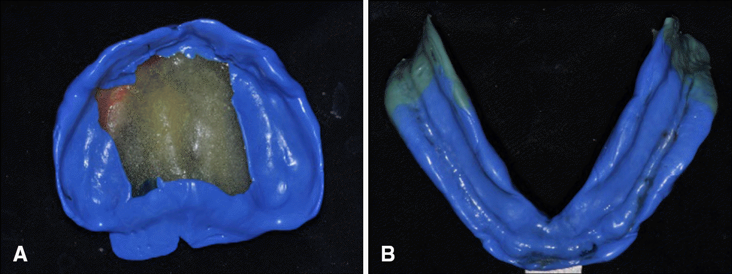
 | Fig. 7.Border molding with functional movement. (A) Border molding of maxilla, (B) Border molding of mandible. |
 | Fig. 8.BTC (Buccal mucosa-Tongue side wall-Contact) point. (A) Both yellow lines indicate BTC point on impression material, (B) Schematic diagram of BTC point (coronal section on retromolar pad area). |
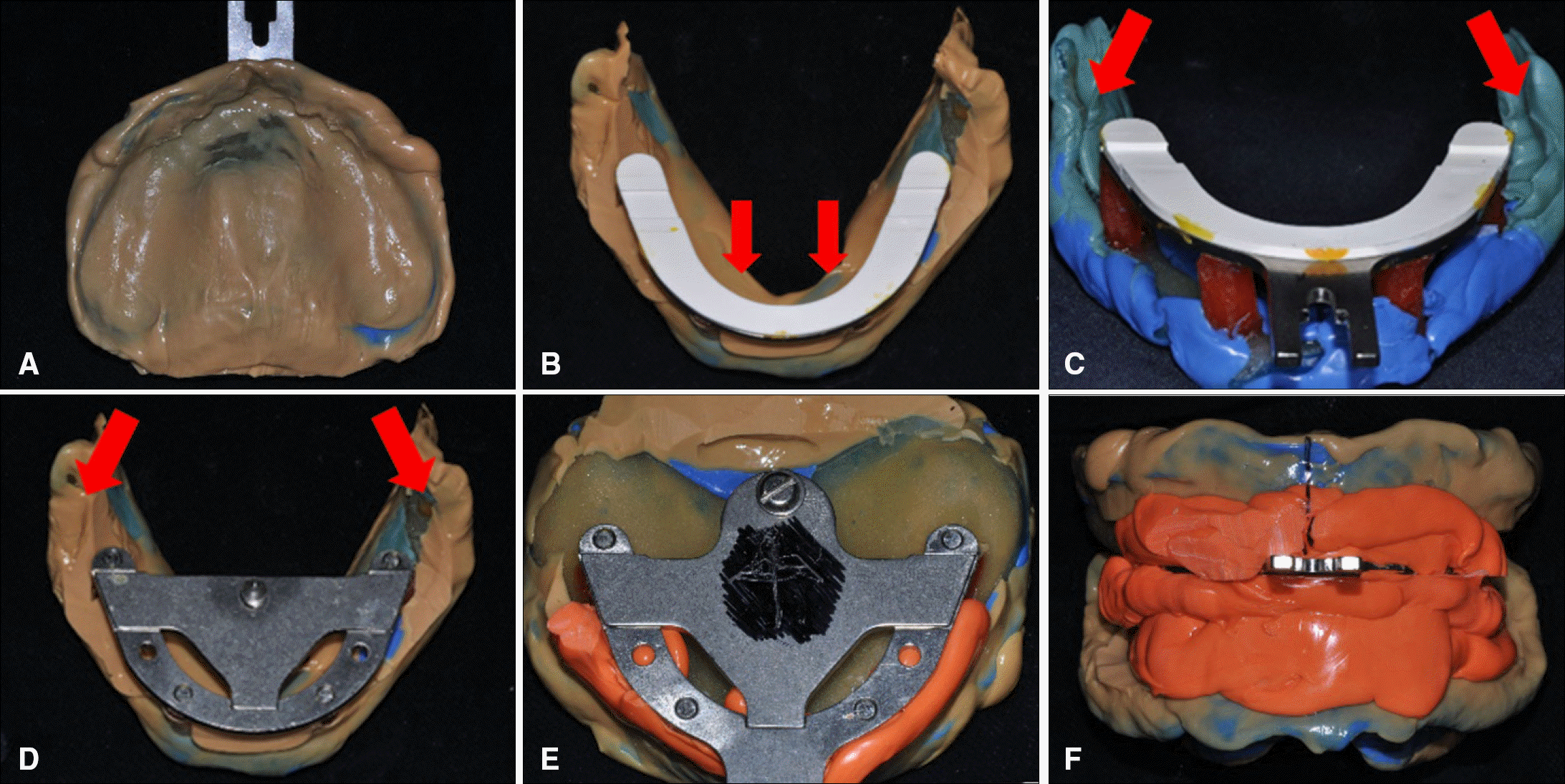
 | Fig. 9.Final impression taking and Gothic arch tracing. (A) Final impression of maxilla, (B) Final impression of mandible. The arrow indicates thick sublingual fold, (C) Before removal of bite rim mount on mandible. Both arrows indicate BTC points, (D) Remove bite rim mount and placement of registration plate with Gothic arch marking pin on mandible. Both arrows indicate BTC points, (E) Remove bite rim mount and placement of registration plate for Gothic arch tracing on maxilla, (F) Final vertical dimension taking by Gothic arch tracing. |
 | Fig. 10.Facebow transfer with UTS® and wax denture delivery. (A) Facebow transfer with UTS®, (B) Wax denture fabrication on master cast, (C) Wax denture delivery. |
 | Fig. 11.Definitive denture fabricate. (A) Frontal view, (B) Maxillary definitive denture, (C) Mandibular definitive denture. The arrow indicates recessus for BTC point. |


 PDF
PDF ePub
ePub Citation
 Print
Print


 XML Download