Abstract
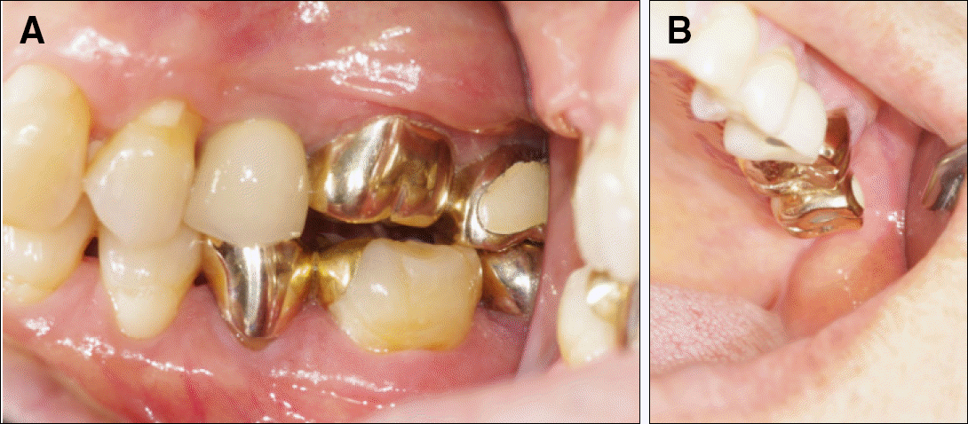
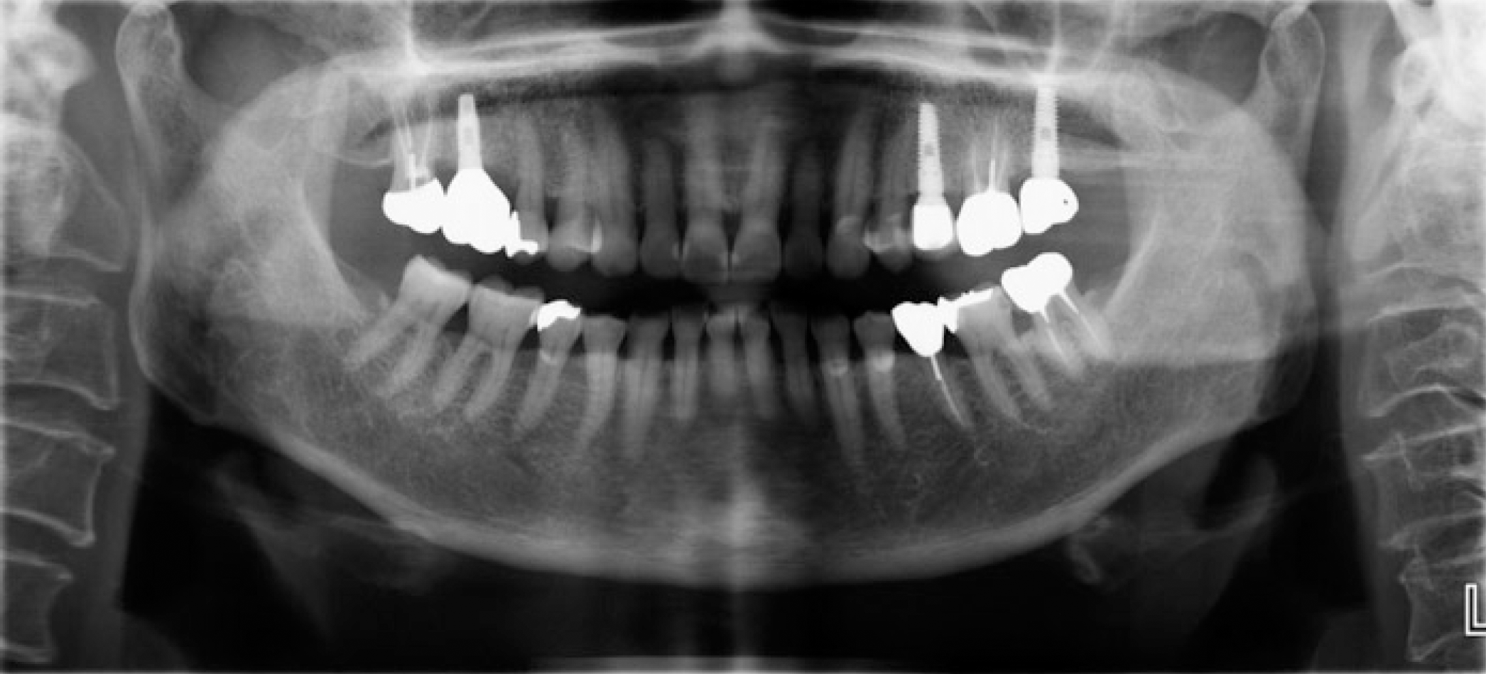
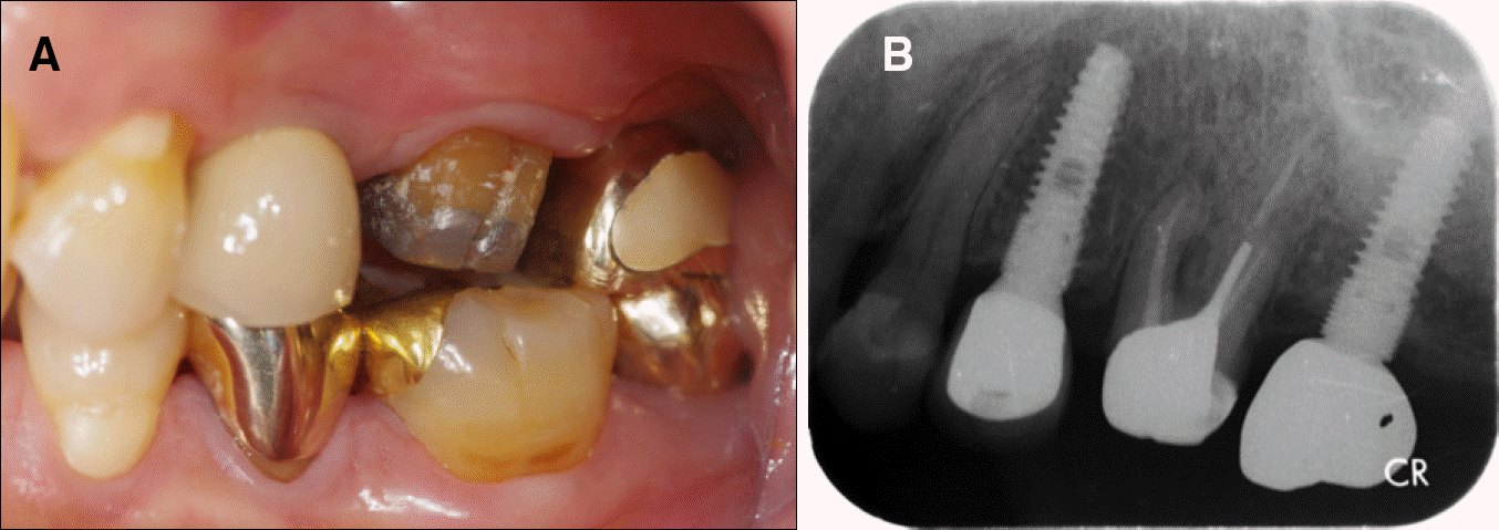
In case of implant-tooth connected prosthesis, a natural tooth tends to intrude. There are several mechanisms that explain an intrusion phenomenon. So it is reco mmended not to connect an implant with a natural tooth. A 68-year-old female had upper left 2nd premolar and 2nd molar extracted and underwent implant surgery on the missing area. We made an implant prosthesis and treated upper left 1st molar with a gold crown. 2.5 year later, the patient complained about loose proximal contact and food impaction between upper left 1st molar and 2nd molar. Mesial side of upper left 2nd molar implant prosthesis was soldered so that proximal contact became tight again. But after 7 months, about 2 mm intrusion of upper left 1 st molar occurred, and the patient felt periodontally originated pain on intruded upper left 1 st molar. After the gold crown on upper left 1 st molar was removed, extrusion occurred and pain was relived.
Go to : 
REFERENCES
1. Schlumberger TL, Bowley JF, Maze GI. Intrusion phenomenon in combination tooth-implant restorations: a review of the literature. J Prosthet Dent. 1998; 80:199–203.

2. Rieder CE, Parel SM. A survey of natural tooth abutment intrusion with implant-connected fixed partial dentures. Int J Periodontics Restorative Dent. 1993; 13:334–47.
3. Lindh T, Dahlgren S, Gunnarsson K, Josefsson T, Nilson H, Wilhelmsson P, Gunne J. Tooth-implant supported fixed prostheses: a retrospective multicenter study. Int J Prosthodont. 2001; 14:321–8.
4. Wang TM, Lee MS, Kok SH, Lin LD. Intrusion and reversal of a free-standing natural tooth bounded by two implant-support-ed prostheses: a clinical report. J Prosthet Dent. 2004; 92:418–22.

5. Sheets CG, Earthman JC. Tooth intrusion in implant-assisted prostheses. J Prosthet Dent. 1997; 77:39–45.

7. Pilcher ES, Gellin RG. Open proximal contact associated with a cast restoration-progressive bone loss: a case report. Gen Dent. 1998; 46:294–7.
8. Miura H. Behavior of the interdental proximal contact relation during function. J Med Dent Sci. 2000; 47:117–22.
Go to : 
 | Fig. 3.Serial periapical image after soldering at mesial side of upper left 2 nd molar implant crown. (A) just after redelivery of 2 nd molar implant crown, (B) after 5 months,(C) after 7 months. |
 | Fig. 4.7 months after redelivery of upper left 2 nd molar crown following soldering at mesial side. About 2 mm intrusion of maxillary left 1 st molar occurred. Buccal gingival swelling and mucosa fold is seen. |
 | Fig. 5.Intraoral photos after removal of crown on upper left 1 st molar. (A) just after removal, (B) 1 month after removal, (C) 1.5 month after removal. |
 | Fig. 6.Periapical radiograph before and after removal of crown on upper left 1 st molar. (A) just before removal, (B) 1 month after removal, (C) 1.5 month after removal. |
 | Fig. 7.Study model 1 month after removal of upper left 1 st molar crown. Planning for preparation of mesial side undercut of upper left 2 nd molar implant crown. |
 | Fig. 8.Temporary crown is made. No occlusal contact, and slight loose proximal contact between posterior tooth. (A) just after setting of temporary crown, (B) 3 weeks after setting of temporary crown. |


 PDF
PDF ePub
ePub Citation
 Print
Print


 XML Download