Abstract
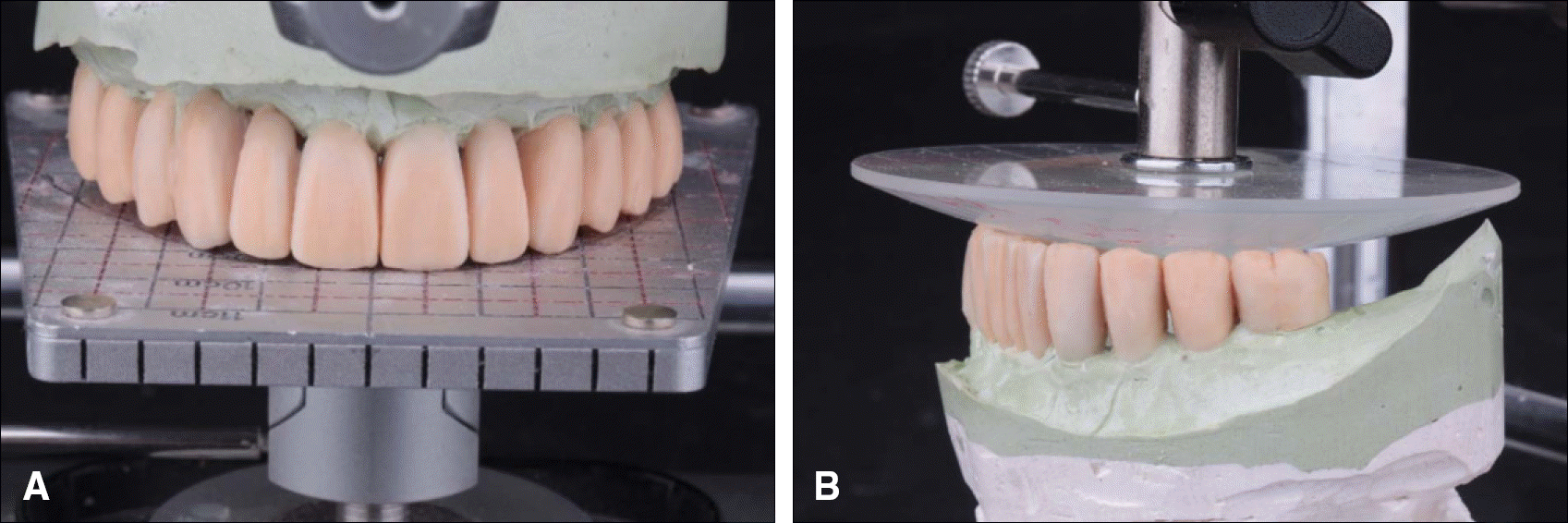
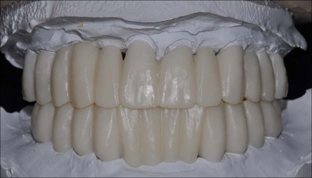
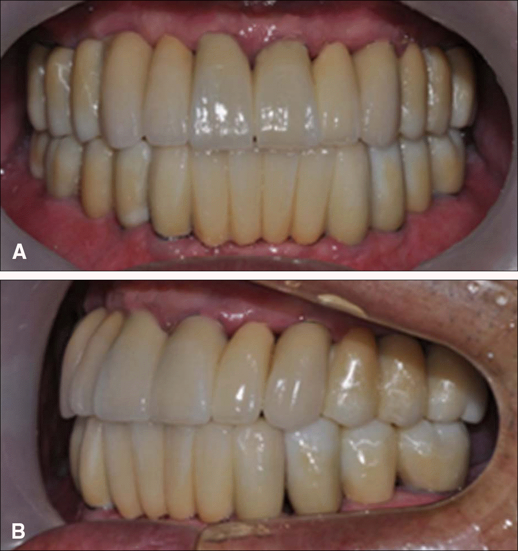
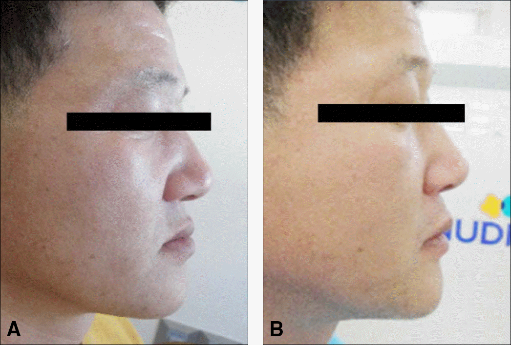
It is important to produce a provisional restoration reflecting the patient's jaw relation, occlusal plane, lip support, shape of teeth, and occlusion type for fully edentulous patients before making a definite prosthesis. The patient introduced in this study showed bad prognosis of remained tooth after severe periodontal diseases. Therefore, remaining teeth were extracted and replaced with dental implants. Provisional restorations were fabricated and the the patient's vertical and horizontal jaw relationship, occlusal plane, amount of overjet and overbite, size of teeth, and length of anterior tooth were recorded. Provisional restorations were scanned and CAD/CAM techniques were used to fabricate a monolithic zirconia bridge, which contour is identical with the provisional restorations. The patient was satisfied with the treatment results on functional, esthetic aspects and the prosthesis retained stable during the four-month clinical observation period.
Go to : 
REFERENCES
1. Cho Y, Raigrodski AJ. The rehabilitation of an edentulous mandible with a CAD/CAM zirconia framework and heat-pressed lithium disilicate ceramic crowns: a clinical report. J Prosthet Dent. 2014; 111:443–7.

2. Karl M, Graef F, Wichmann M, Krafft T. Passivity of fit of CAD/CAM and copy-milled frameworks, veneered frameworks, and anatomically contoured, zirconia ceramic, implant-supported fixed prostheses. J Prosthet Dent. 2012; 107:232–8.

3. Misch CE. Contemporary Implant Dentistry. 3rd ed.St. Louis: CV Mosby;2009. p. 367–88.
4. Rakosi T. Cephalometric Radiography: Significance of Angular and Linear Measurements for Dento-skeletal Analysis. Kuk Jae D.M Publishing Co.;1994. p. 46–77.
5. Kim JC, Jung CW. OP Finder System & Zirconia Prosthesis: Occlusal Plane Finder 2 & Opus 1 Articulator. 2010 DaehanNarae Publishing Inc.;2010. p. 45–9.
6. The council of the faculty of otrhodontics. Textbook of Orthodontics. 2nd ed.DaehanNarae Publishing Inc.;2006. p. 177–87.
Go to : 
 | Fig. 1.First visit views. (A) Intraoral photo, (B) Panoramic radiograph, (C) Extraoral photo. |
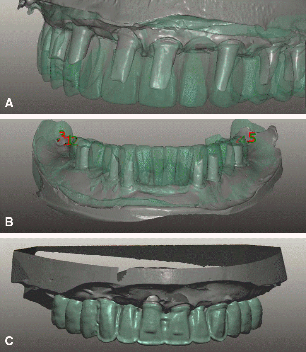 | Fig. 7.Double scanning technique. (A) Maxillary superimposition, (B) Mandibular superimposition, (C) Cutback after maxillary superimposition. |


 PDF
PDF ePub
ePub Citation
 Print
Print


 XML Download