

 PDF
PDF ePub
ePub Citation
 Print
Print


Abstract
Purpose
A method detecting change of jaw or alveolar bone density may be helpful in periodontal care, implant dentistry and evaluation of bone density of whole body.
Materials and methods
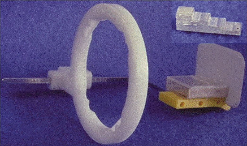
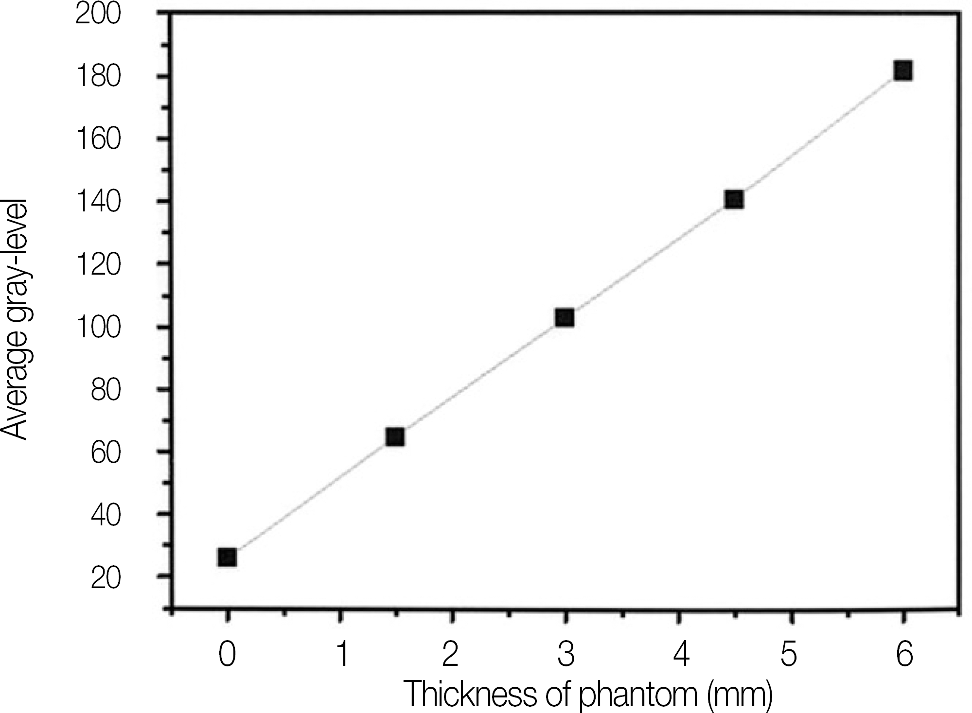
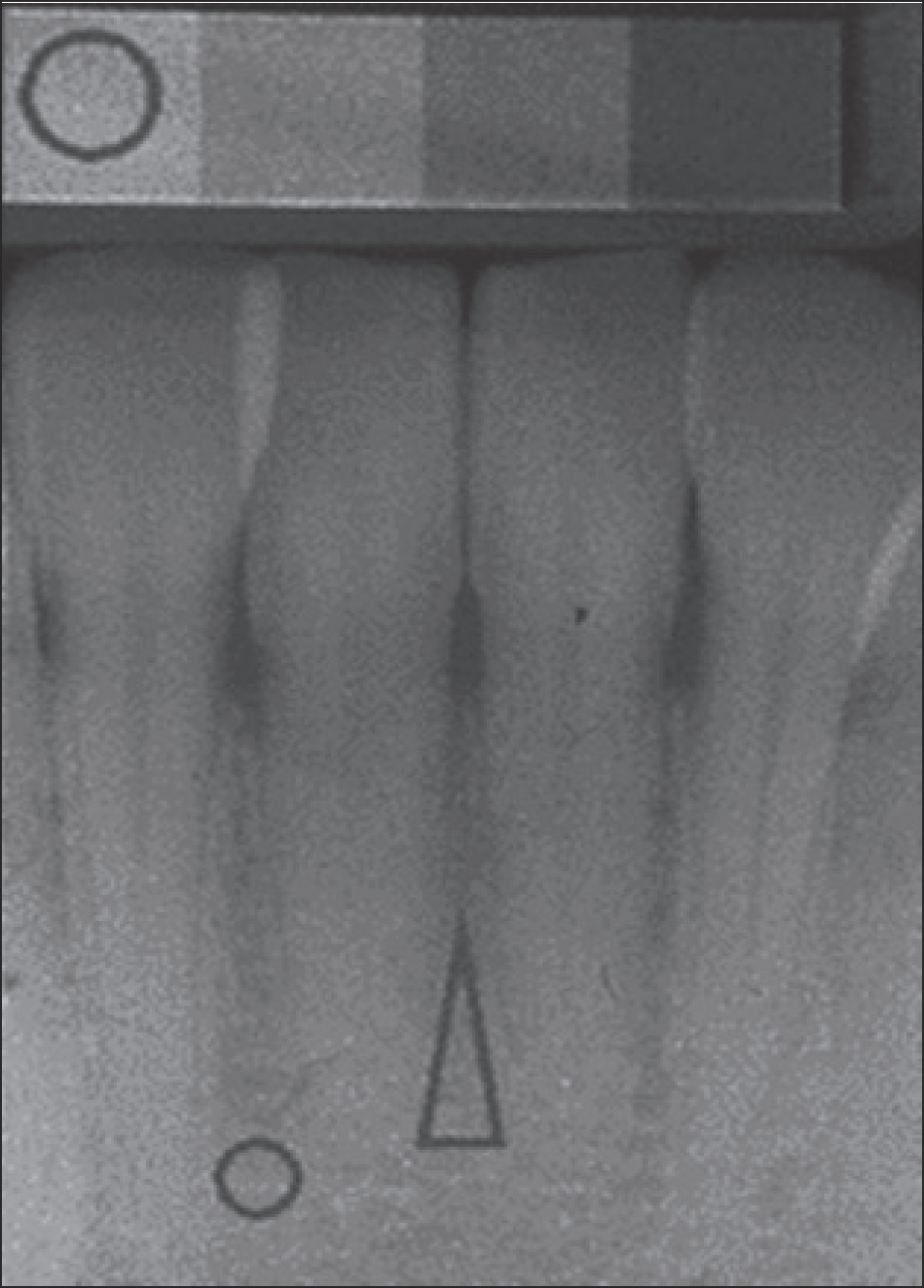
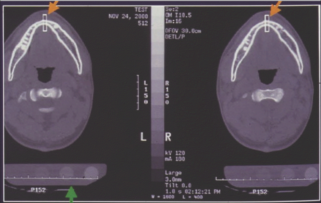
In this study, bone density of intraoral periapical radiography using phantom-integrated XCP is compared with that of quantitative computed tomography (QCT).
Go to : 
REFERENCES
1. Denissen H, Verhey H, de Blieck J, Corten F, Klein C, van Lingen A. Dual X-ray absorptiometry for alveolar bone: precision of peri-implant mineral measurements ex vivo. J Periodontal Res. 1996; 31:265–70.

2. Ellwood R, Horner K, Alexander S, Davies R. A digital subtraction radiography investigation of upper first molar proximal bone density changes in adolescents. J Periodontal Res. 1998; 33:172–7.
3. Bassi F, Procchio M, Fava C, Schierano G, Preti G. Bone density in human dentate and edentulous mandibles using computed tomography. Clin Oral Implants Res. 1999; 10:356–61.
4. Grampp S, Jergas M, Lang P, Steiner E, Fuerst T, Glu¨er CC, Mathur A, Genant HK. Quantitative CT assessment of the lumbar spine and radius in patients with osteoporosis. AJR Am J Roentgenol. 1996; 167:133–40.
5. Kuhl ED, Nummikoski PV. Radiographic absorptiometry method in measurement of localized alveolar bone density changes. Oral Surg Oral Med Oral Pathol Oral Radiol Endod. 2000; 89:375–81.
6. Allen KM, Hausmann E. Analytical methodology in quantitative digital subtraction radiography: analyses of the aluminum reference wedge. J Periodontol. 1996; 67:1317–21.
7. Bra¨gger U, Bu¨rgin W, Fourmousis I, Schmid G, Schild U, Lang NP. Computer-assisted densitometric image analysis of digital subtraction images: in vivo error of the method and effect of thresholding. J Periodontol. 1998; 69:967–74.
8. Ulm C, Kneissel M, Schedle A, Solar P, Matejka M, Schneider B, Donath K. Characteristic features of trabecular bone in edentulous maxillae. Clin Oral Implants Res. 1999; 10:459–67.
9. Southard TE, Wunderle DM, Southard KA, Jakobsen JR. Geometric and densitometric standardization of intraoral radiography through use of a modified XCP system. Oral Surg Oral Med Oral Pathol Oral Radiol Endod. 1999; 87:253–7.
10. Southard KA, Southard TE, Schlechte JA, Meis PA. The relationship between the density of the alveolar processes and that of post-cranial bone. J Dent Res. 2000; 79:964–9.
Go to :
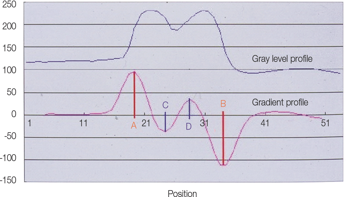 | Fig. 5.Gray level and gradient profile of quantitative computed tomography on mandible. (A) Gradient profile of interface between soft tissue and buccal cortex of mandible,(B) Gradient profile of interface between soft tissue and lingual cortex of mandible, (C) Gradient profile of interface between buccal cortical bone and marrow of mandible,(D) Gradient profile of interface between lingual cortical bone and marrow of mandible. |
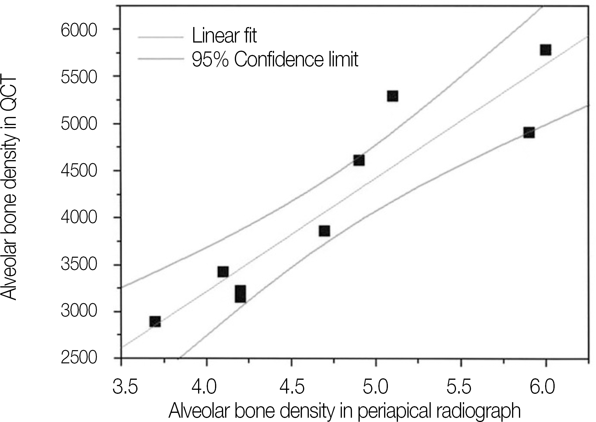 | Fig. 6.A graph of correlation between alveolar bone densities of periapical radiograph and QCT. |
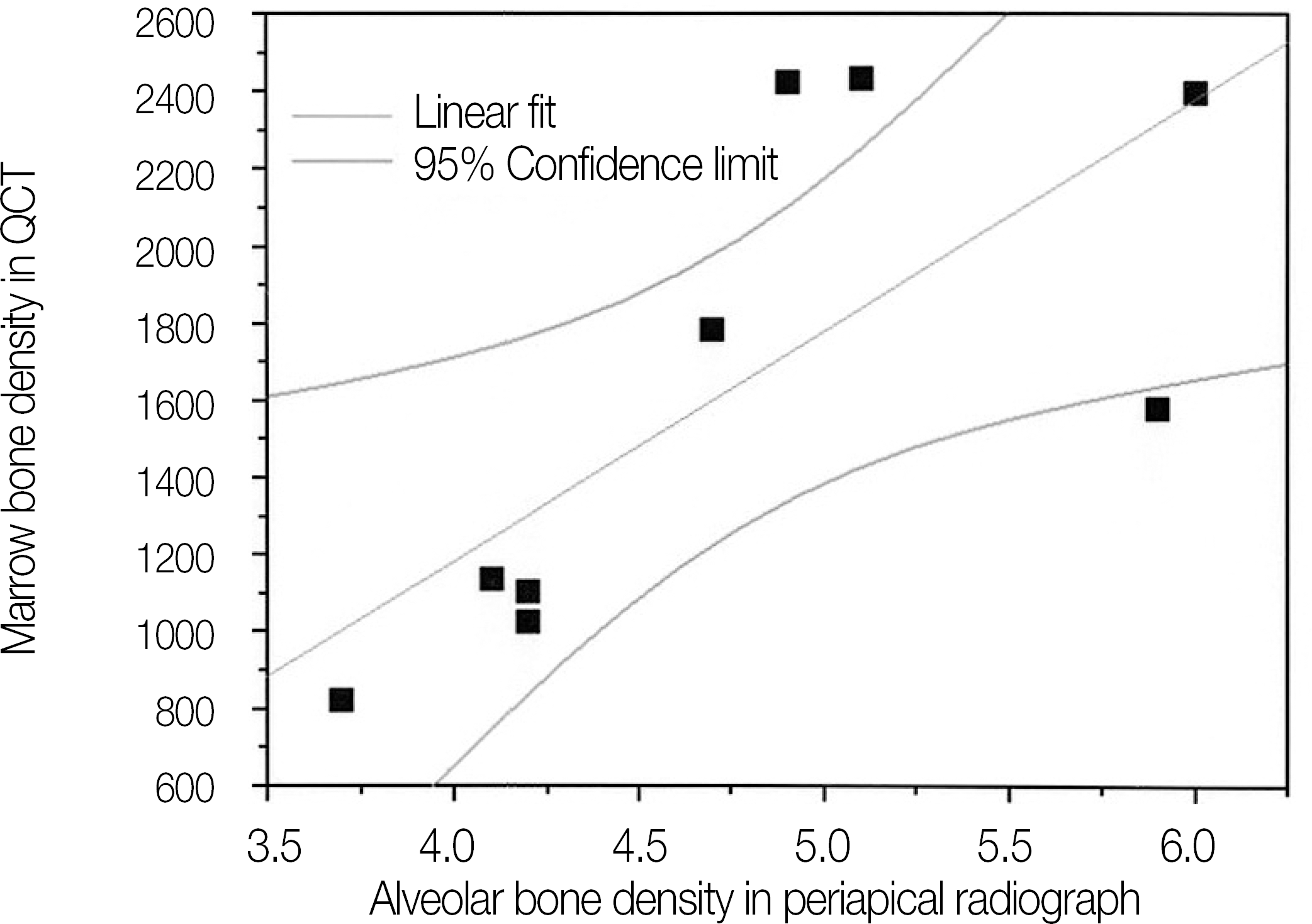 | Fig. 7.A graph of correlation between alveolar bone density of periapical radiograph and marrow density of QCT. |
Table 1.
Bone density of mandibles measured in intraoral radiography (adjusted phantom)
| Subject | 1 | 2 | 3 | 4 | 5 | 6 | 7 | 8 | 9 |
|---|---|---|---|---|---|---|---|---|---|
| Adjusted mean | 6.0 | 4.1 | 4.9 | 4.2 | 5.9 | 4.2 | 5.1 | 3.7 | 4.7 |
 XML Download