Abstract
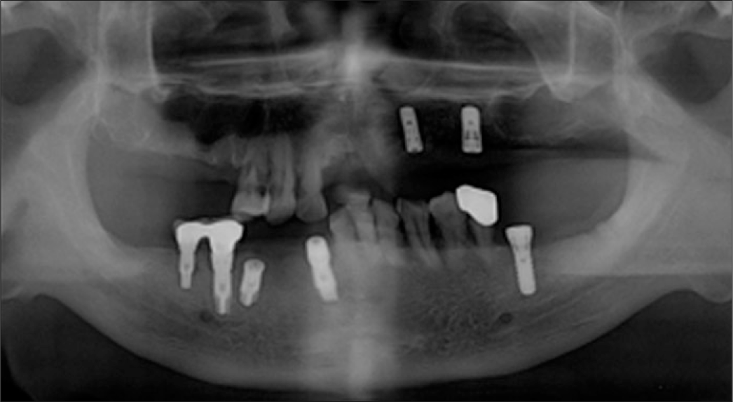
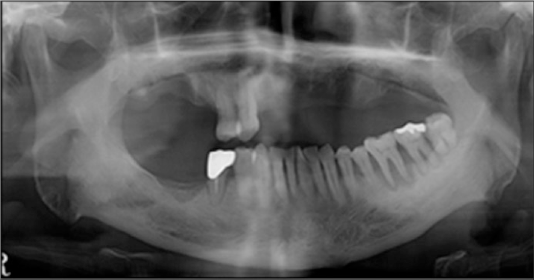
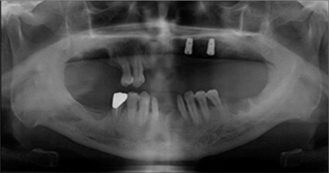
Treatment options for partially edentulous patients are fixed partial denture, removable partial denture and implant supported fixed partial denture. In case of a patient with a few remaining teeth, removable partial denture and implant supported fixed prosthesis are available. For implant fixed prothesis, enough implant fixtures are required and the patient's general condition, local factors and economic status must be considered. When the condition of the abutments and the residual ridge is favorable and the prosthesis is well designed, removable partial denture can be an option. In removable partial denture, the bilateral support is important. If the teeth remain unilateral, harmful stress is put on the abutments by the fulcrum line. In this situation, strategic implantation and implant-retained or assisted removable partial denture is beneficial to the retention and support of the denture. And this can be cost-effective, functional and esthetic choice of treatment. This article describes the prosthodontic rehabilitation of Maxillary Kennedy class I partially edentulous patients. In these two cases, the patients had a small number of teeth and they were restored by the combination of a removable partial denture and dental implants. (J Korean Acad Prosthodont 2014;52:128-35)
Go to : 
REFERENCES
1.Budtz-Jo¨rgensen E. Restoration of the partially edentulous mouth-a comparison of overdentures, removable partial dentures, fixed partial dentures and implant treatment. J Dent. 1996. 24:237–44.
2.Mijiritsky E., Ormianer Z., Klinger A., Mardinger O. Use of dental implants to improve unfavorable removable partial denture design. Compend Contin Educ Dent. 2005. 26:744–6. 748, 750 passim.
3.Chikunov I., Doan P., Vahidi F. Implant-retained partial overdenture with resilient attachments. J Prosthodont. 2008. 17:141–8.

4.Carr AB., Brown DT. McCracken's Removable partial prosthodontics. 12th ed.Mosby: Elsevier;2011. p. 340–1.
5.Chikunov I., Doan P., Vahidi F. Implant-retained partial overdenture with resilient attachments. J Prosthodont. 2008. 17:141–8.

6.Keltjens HM., Kayser AF., Hertel R., Battistuzzi PG. Distal extension removable partial dentures supported by implants and residual teeth: considerations and case reports. Int J Oral Maxillofac Implants. 1993. 8:208–13.
7.Shahmiri RA., Atieh MA. Mandibular Kennedy Class I implant-tooth-borne removable partial denture: a systematic review. J Oral Rehabil. 2010. 37:225–34.

8.Schneider AL., Kurtzman GM. Bar overdentures utilizing the Locator attachment. Gen Dent. 2001. 49:210–4.
Go to : 
 | Fig. 3.Artificial teeth arrangement (A) Frontal view, (B) Occlusal view (Maxilla), (C) Occlusal view (Mandible). |
 | Fig. 4.Kerator® abutments connection and patrix nylons insertion (A) Kerator® abutments connection, (B) Placement of the black processing males with the metal housings on to the Kerator® abutments, (C) Maxillary denture base with enough space for the metal housings, (D) Insertion of the metal housings and patrix nylons. |
 | Fig. 5.Final prosthesis (A) MIC (Maximum intercuspation) frontal view, (B) Upper denture, (C) Lower denture. |
 | Fig. 8.Temporary prosthesis after extraction of the teeth (A) MIC frontal view, (B) Occlusal view (Maxilla), (C) Occlusal view (Mandible). |
 | Fig. 10.Impression taking procedures (A) Die preparation for surveyed FDP fabrication, (B) Pick-up impression of surveyed FDP using Permlastic®, (C) Master cast. |
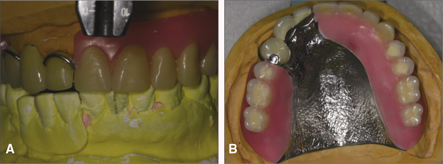 | Fig. 11.Mounted master cast and artificial teeth arrangement (A) Artificial teeth arrangement (frontal view), (B) Artificial teeth arrangement (occlusal view). |


 PDF
PDF ePub
ePub Citation
 Print
Print




 XML Download