Abstract
Comprehensive prosthetic treatment requires considerations from various points of view. The anterior guidance is important factor in prosthodontic treatment of anterior teeth. Lingual surface contour of anterior restoration is so critical that a small mistake of laboratory or clinical process can cause discomfort of patient and disharmony of entire dentition. There are no guidelines for lingual surface contour that fit all patients. Therefore the lingual surface of provisional restoration is most accurately described as a customized one. The dentist transfers the exact information of anterior guidance that has made through long term provisional restoration to the technician. This case introduce that the duplication technique of CAD/CAM system to reproduce the anterior guidance of provisional restoration. This method can improve satisfaction of both patient and dentist.
Go to : 
REFERENCES
1.Dawson PE. Determining the determinants of occlusion. Int J Periodontics Restorative Dent. 1983. 3:8–21.
2.Dawson PE. Evaluation, diagnosis, and treatment of occlusal problems. 1st ed.St Louis: CV Mosby Co.;1974. p. 177–8.
3.Fox CW., Abrams BL., Doukoudakis A. Principles of anterior guidance: development and clinical applications. J Craniomandib Pract 1983-. 1984. 2:23–30.

4.Sabek M., Tre′velo A. Alternative procedure for reconstructing anterior guidance using an autopolymerizing resin pattern. J Prosthet Dent. 1996. 76:550–3.

5.Duret F., Preston JD. CAD/CAM imaging in dentistry. Curr Opin Dent. 1991. 1:150–4.
6.Miyazaki T., Hotta Y., Kunii J., Kuriyama S., Tamaki Y. A review of dental CAD/CAM: current status and future perspectives from 20 years of experience. Dent Mater J. 2009. 28:44–56.

7.Turner KA., Missirlian DM. Restoration of the extremely worn dentition. J Prosthet Dent. 1984. 52:467–74.

8.Mo¨rmann WH., Brandestini M., Lutz F., Barbakow F. Chairside computer-aided direct ceramic inlays. Quintessence Int. 1989. 20:329–39.
9.Beuer F., Schweiger J., Edelhoff D. Digital dentistry: an overview of recent developments for CAD/CAM generated restorations. Br Dent J. 2008. 204:505–11.

10.Smith R. Creating well-fitting restorations with a digital impression system. Compend Contin Educ Dent. 2010. 31:640–4.
11.Ender A., Mehl A. Full arch scans: conventional versus digital impressions—an in-vitro study. Int J Comput Dent. 2011. 14:11–21.
Go to : 
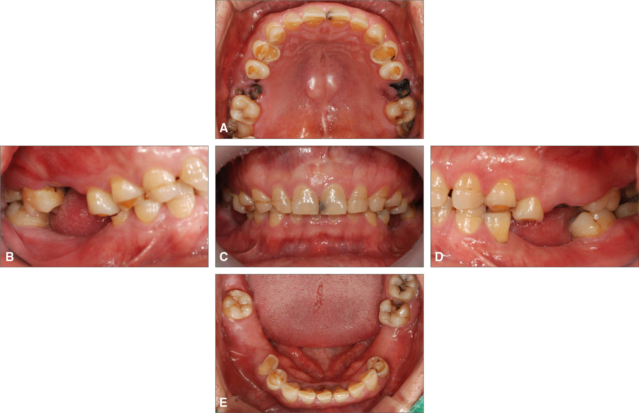
 | Fig. 1.Intraoral view of the patient's preoperative condition showing abrasion-corrosion lesion and loss of vertical dimension. (A, E) Occlusal view of maxilla and mandible, (C) Frontal view, (B, D) Lateral view. |
 | Fig. 3.(A) The new VDO was set by 2.5 mm increase in the anterior teeth (using leaf gage), (B, C, D) Diagnostic wax-up was done on the new VDO. |
 | Fig. 4.Provisional restorations made from a cast of diagnostic wax-up. (A) Frontal view, (B, C) Occlusal view. |
 | Fig. 5.Provisional restorations are reviewed for function prior to making final impressions. (A) Right eccentric position, (B) Left eccentric position, (C) Protrusive position. |
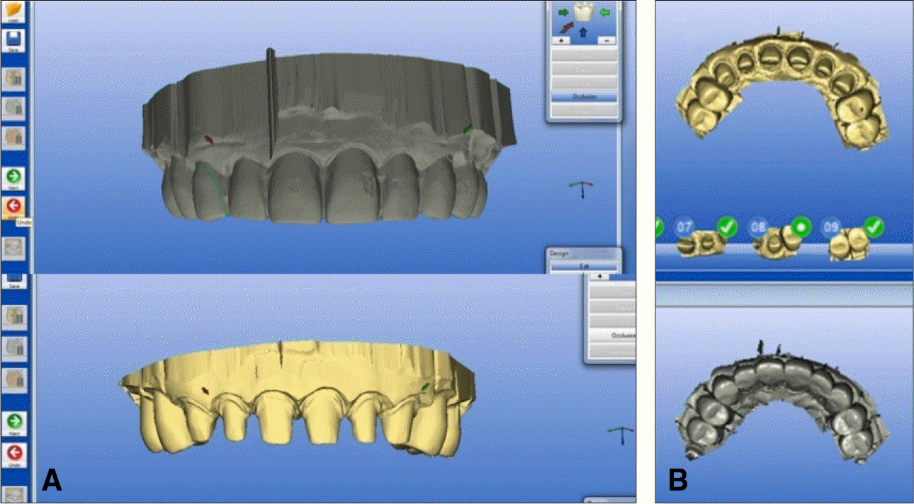
 | Fig. 6.Digitally overlap image of maxillary prepared and temporary teeth. (A) Frontal view, (B) Occlusal view. |
 | Fig. 7.Manufacture anterior teeth one by one by the Correlation method. (A) Right lateral incisor image, (B) Left central incisor image, C: Left canine image. |
 | Fig. 8.(A) Final restorations, (B) Maxillary anterior section and premolars restored with CAD/CAM-designed and -milled monolithic lithium disilicate restorations, molars restored with monolithic zirconia restorations, (C) Mandibular premolars, except for the left second premolar, restored with monolithic lithium disilicate restorations, and remaining molars restored with monolithic zirconia restorations. |
 | Fig. 9.(A) Arrow indicate the depression generated on the provisional restoration from lateral guide of canine during adaptation period of the patient carrying provisional restoration, (B) Duplication of the depression on CAD process, (C) The features of provisional restoration being reproduced on the final prosthesis too. |


 PDF
PDF ePub
ePub Citation
 Print
Print


 XML Download