

 PDF
PDF ePub
ePub Citation
 Print
Print


Abstract
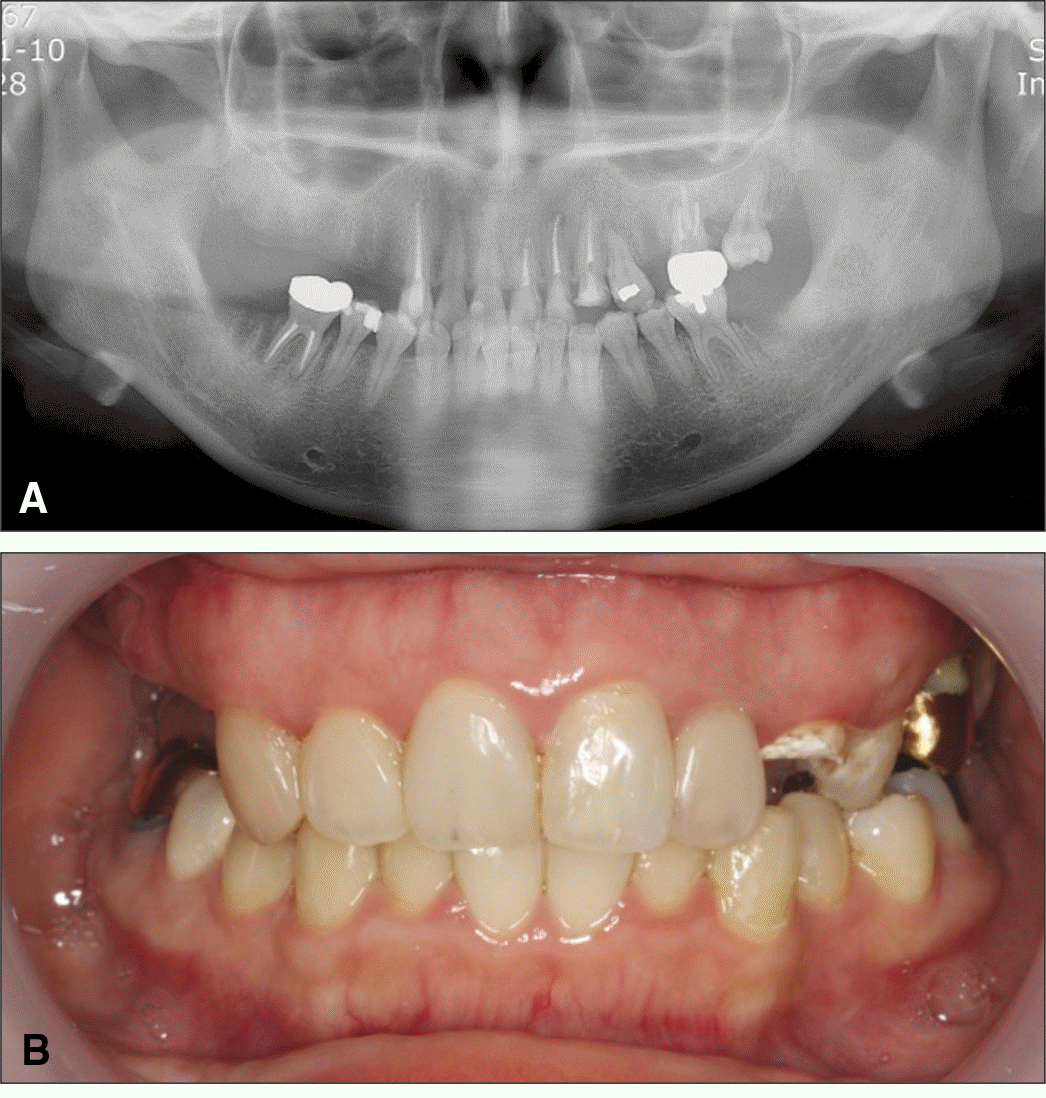
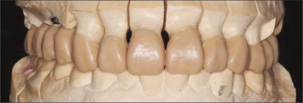
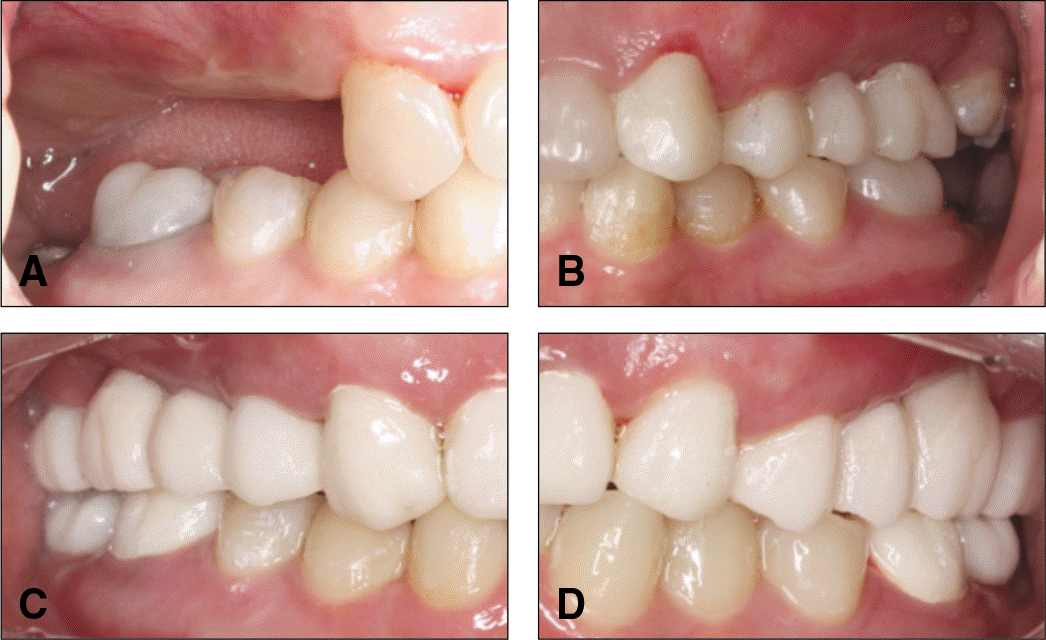
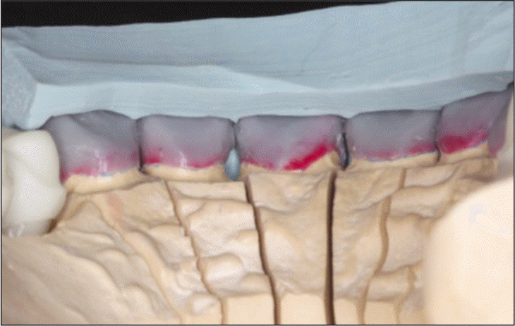
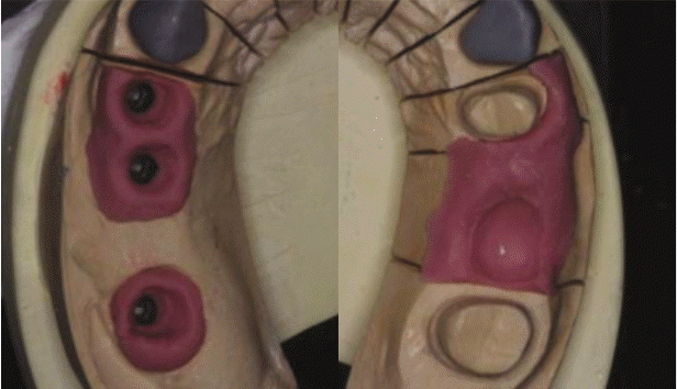
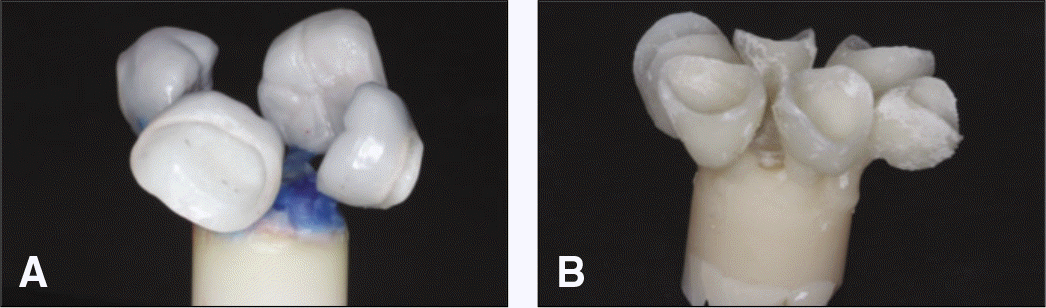
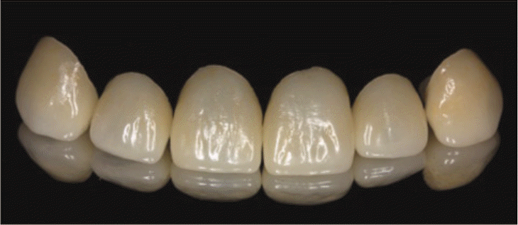
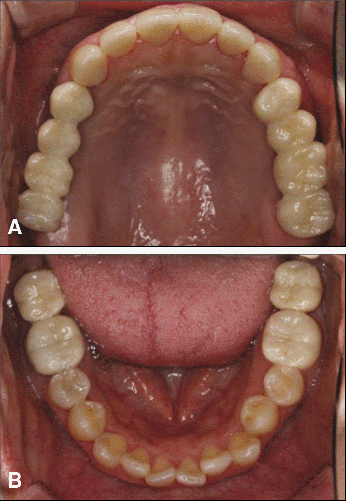
With the introduction of dental implants, restoration of missing teeth with conventional fixed or removable partial dentures is being replaced with implants. Especially, with young patients, not only longevity but also esthetic factors need to be considered. Implant restorations provide long-term success functionally but, esthetic complications such as, marginal exposure due to gingival recession, loss of the papilla and dark color of metal abutments may occur. Recently, zirconia restorations with CAD/CAM technology provide functional, biocompatible and esthetic restorations possible. All-ceramic restorations using the pressed ceramic technique show better fracture toughness values than those of the conventional porcelain veneering technique. Pressed ceramic technique creates the veneer design in wax and the lost wax technique is used to create the restoration. The final contour of the restoration may be controlled during wax-up. A 22-year old female patient was restored with dental implants and zirconia restorations using the pressed ceramic technique presenting short-term but optimistic prognosis. (J Korean Acad Prosthodont 2013;51:47-51)
REFERENCES
1.Conrad HJ., Seong WJ., Pesun IJ. Current ceramic materials and systems with clinical recommendations: a systematic review. J Prosthet Dent. 2007. 98:389–404.

2.Bello A., Jarvis RH. A review of esthetic alternatives for the restoration of anterior teeth. J Prosthet Dent. 1997. 78:437–40.
3.Ozkurt Z., Kazazoğlu E. Clinical success of zirconia in dental applications. J Prosthodont. 2010. 19:64–8.
4.Raigrodski AJ., Hillstead MB., Meng GK., Chung KH. Survival and complications of zirconia-based fixed dental prostheses: a systematic review. J Prosthet Dent. 2012. 107:170–7.
5.Wolf D., Bindl A., Schmidlin PR., Lu¨thy H., Mo¨rmann WH. Strength of CAD/CAM-generated esthetic ceramic molar implant crowns. Int J Oral Maxillofac Implants. 2008. 23:609–17.
6.Beuer F., Edelhoff D., Gernet W., Sorensen JA. Three-year clinical prospective evaluation of zirconia-based posterior fixed dental prostheses (FDPs). Clin Oral Investig. 2009. 13:445–51.
7.Sulaiman F., Chai J., Jameson LM., Wozniak WT. A comparison of the marginal fit of In-Ceram, IPS Empress, and Procera crowns. Int J Prosthodont. 1997. 10:478–84.
8.Stawarczyk B., Ozcan M., Roos M., Trottmann A., Sailer I., Ha¨mmerle CH. Load-bearing capacity and failure types of anterior zirconia crowns veneered with overpressing and layering techniques. Dent Mater. 2011. 27:1045–53.
9.Lin WS., Ercoli C., Feng C., Morton D. The effect of core material, veneering porcelain, and fabrication technique on the biaxial flexural strength and weibull analysis of selected dental ceramics. J Prosthodont. 2012. 21:353–62.
10.Zitzmann NU., Krastl G., Hecker H., Walter C., Waltimo T., Weiger R. Strategic considerations in treatment planning: deciding when to treat, extract, or replace a questionable tooth. J Prosthet Dent. 2010. 104:80–91.
11.Glauser R., Sailer I., Wohlwend A., Studer S., Schibli M., Scha¨rer P. Experimental zirconia abutments for implant-supported single-tooth restorations in esthetically demanding regions: 4-year results of a prospective clinical study. Int J Prosthodont. 2004. 17:285–90.
12.Reshad M., Cascione D., Aalam AA. Fabrication of the mandibular implant-supported fixed restoration using CAD/CAM technology: a clinical report. J Prosthet Dent. 2009. 102:271–8.
13.Zembic A., Sailer I., Jung RE., Ha¨mmerle CH. Randomized-controlled clinical trial of customized zirconia and titanium implant abutments for single-tooth implants in canine and posterior regions: 3-year results. Clin Oral Implants Res. 2009. 20:802–8.
14.Choi JE., Waddell JN., Torr B., Swain MV. Pressed ceramics onto zirconia. Part 1: Comparison of crystalline phases present, adhesion to a zirconia system and flexural strength. Dent Mater. 2011. 27:1204–12.
 XML Download