

 PDF
PDF ePub
ePub Citation
 Print
Print


Abstract
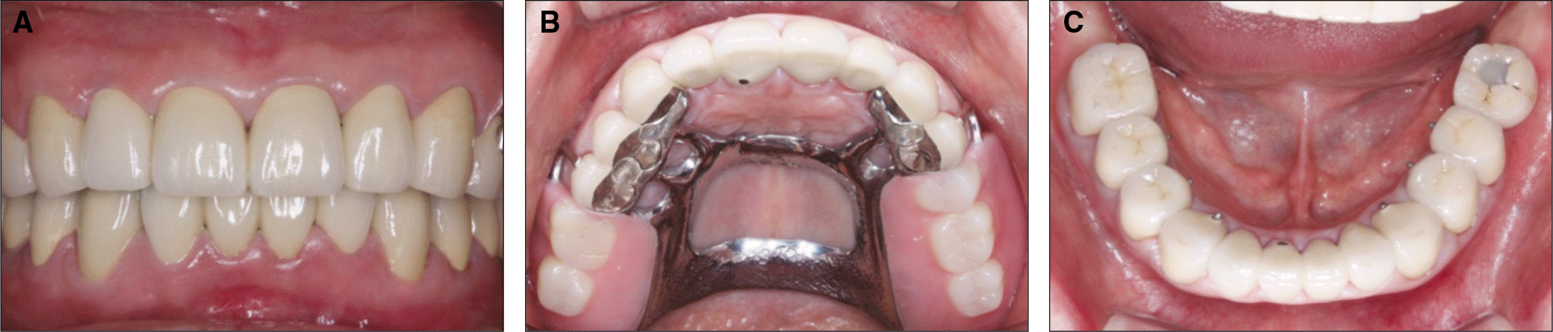
The loss of posterior support may cause attrition of anterior teeth, and loss of occlusal vertical dimension (OVD). The collapse of the posterior support will eventually cause the pathologic change of the TMJ and masticatory muscles, unesthetic facial appearance and decreased masticatory function. Patients with destroyed dentition need extensive prosthetic treatments. Proper diagnosis and treatment planning are necessary for the stability of the neuromuscular system and the TMJ, and esthetic and functional definitive restorations. In this case, 63 year-old male presented with decreased masticatory force and esthetic problems due to pathologic destruction of teeth structure on entire dentition. Based on assessment of OVD including intraoral findings, radiographic examination and diagnostic cast, full-mouth rehabilitation with increase of OVD was planned using fixed partial denture and removable partial denture. Diagnostic wax-up was done after 4 mm increase of OVD determined by assessment of OVD. The OVD was maintained with the overlay type removable interim prostheses for 12 weeks to ascertain his comfort and adaptation to the new OVD. After the adaptation period, second interim prostheses with tooth preparation maintaining the established OVD was delivered. After 4 weeks, final prostheses were fabricated and delivered. After 7 month follow-up period, occlusal stability is maintained. Through this procedure, satisfactory outcomes were achieved both in functional and esthetic aspects. (J Korean Acad Prosthodont 2013;51:39-46)
Go to : 
REFERENCES
1.Briggs P., Bishop K. Fixed prostheses in the treatment of tooth wear. Eur J Prosthodont Restor Dent. 1997. 5:175–80.
2.Hemmings KW., Darbar UR., Vaughan S. Tooth wear treated with direct composite restorations at an increased vertical dimension: results at 30 months. J Prosthet Dent. 2000. 83:287–93.

3.Sato S., Hotta TH., Pedrazzi V. Removable occlusal overlay splint in the management of tooth wear: a clinical report. J Prosthet Dent. 2000. 83:392–5.
4.Berry DC., Poole DF. Attrition: possible mechanisms of compensation. J Oral Rehabil. 1976. 3:201–6.
5.Dahl BL., Krogstad O. The effect of a partial bite-raising splint on the inclination of upper and lower front teeth. Acta Odontol Scand. 1983. 41:311–4.
6.Ramfjord SP., Blankenship JR. Increased occlusal vertical dimension in adult monkeys. J Prosthet Dent. 1981. 45:74–83.
7.Dawson PE. Functional occlusion: from TMJ to smile design. St. Louis; Mo: Mosby;2007. p. 430–52.
8.Willis FM. Features of the face involved in full denture prosthesis. Dent Cosmos. 1935. 77:851–4.
9.Turner KA., Missirlian DM. Restoration of the extremely worn dentition. J Prosthet Dent. 1984. 52:467–74.
10.Nelson SJ. Wheeler's dental anatomy, physiology and occlusion. 9th ed.Saunders. Elsevier Health Sciences;2009. p. 99–139.
11.McGrane HF. Five basic principles of the McGrane full denture procedure. J Florida Dent Soc. 1949. 20:5–8.
12.Fayz F., Eslami A., Graser GN. Use of anterior teeth measurements in determining occlusal vertical dimension. J Prosthet Dent. 1987. 58:317–22.
13.Oh YR., Lee SB., Park NS., Choi DG. A study of intraoral anatomic landmarks of Korean adult upper-jaw. J Korean Acad Prosthodont. 1995. 33:753–68.
14.Park JH., Jeong CM., Jeon YC., Lim JS. A study on the occlusal plane and the vertical dimension in Korean adults with natural dentition. J Korean Acad Prosthodont. 2005. 43:41–51.
15.Lee SJ., Choi DG., Woo YH., Choi BB. A study of the occlusal plane orientation in Korean adults. Kyung Hee Univ Dent J. 1997. 19:753–69.
16.Carlsson GE., Ingervall B., Kocak G. Effect of increasing vertical dimension on the masticatory system in subjects with natural teeth. J Prosthet Dent. 1979. 41:284–9.
17.Abduo J. Safety of increasing vertical dimension of occlusion: a systematic review. Quintessence Int. 2012. 43:369–80.
18.Wessberg GA., O'Ryan FS., Washburn MC., Epker BN. Neuromuscular adaptation to surgical superior repositioning of the maxilla. J Maxillofac Surg. 1981. 9:117–22.
Go to :
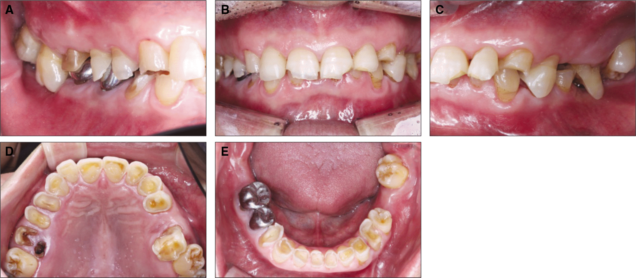 | Fig. 1.Intraoral view before treatment. A: Right lateral view, B: Frontal view, C: Left lateral view, D: Maxillary occlusal view, E: Mandibular occlusal view. |
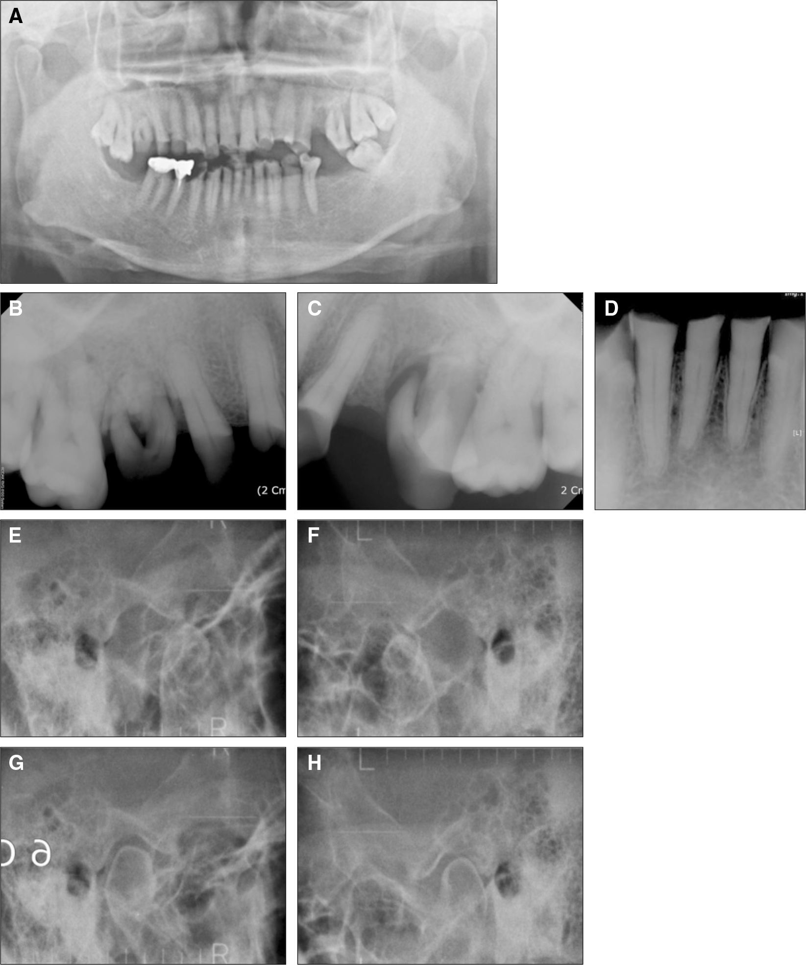 | Fig 2.Radiographic exam before treatment. A: Panoramic view, B: Maxillary right posterior teeth, C: Mandibular left posterior teeth, D: Mandibular anterior teeth, E: TMJ series Right opened, F: Left opened, G: Right closed, H: Left closed. |
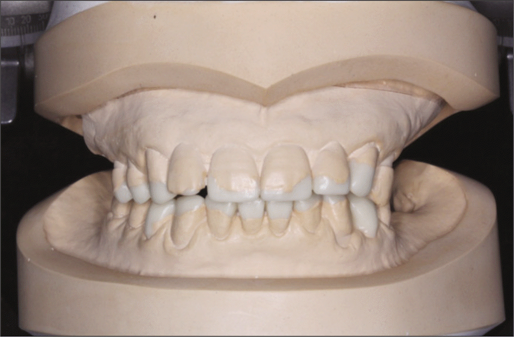
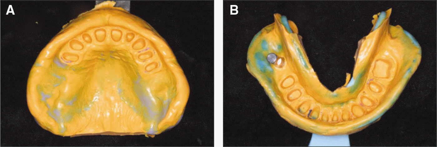
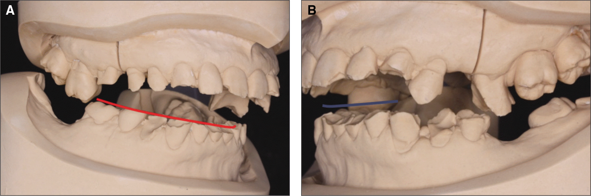 | Fig. 3.Cast analysis. A: Premature contact on maxillary right first molar and mandibular right first molar is observed. There is light curve of Spee, B: Premature contact on maxillary left first molar and mandibular right second premolar is observed. Severe wear on mandibular anterior teeth are observed. |
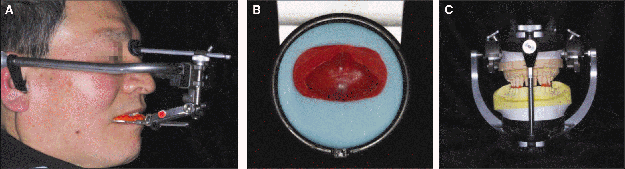
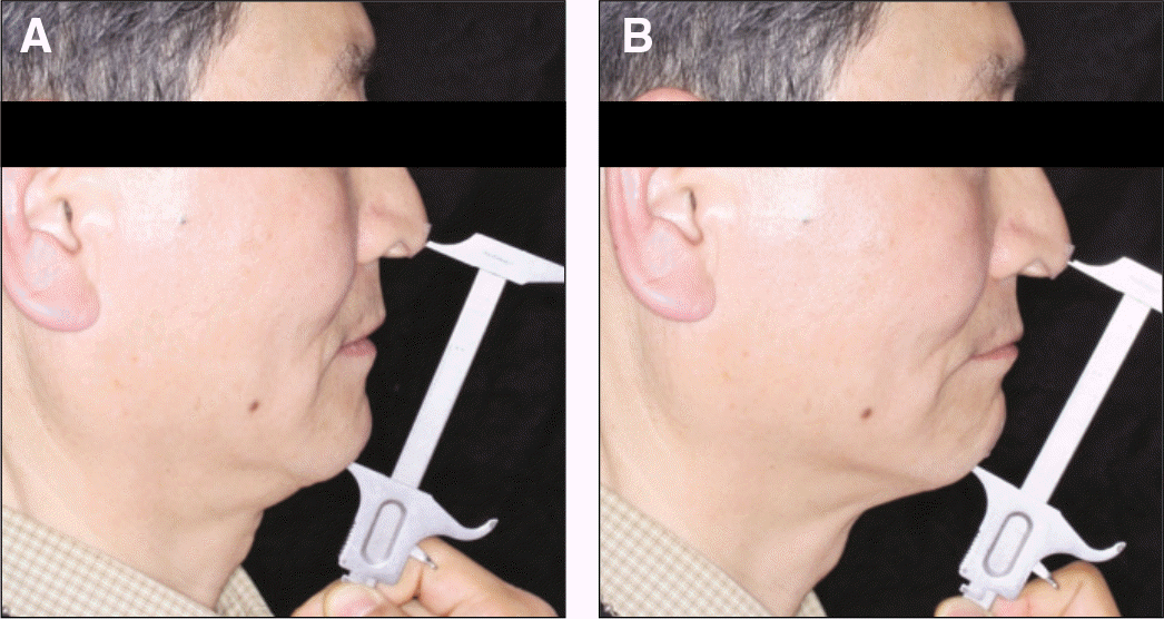 | Fig. 4.Interocclusal distance record. A: Vertical dimension at rest, B: Occlusal vertical dimension. |
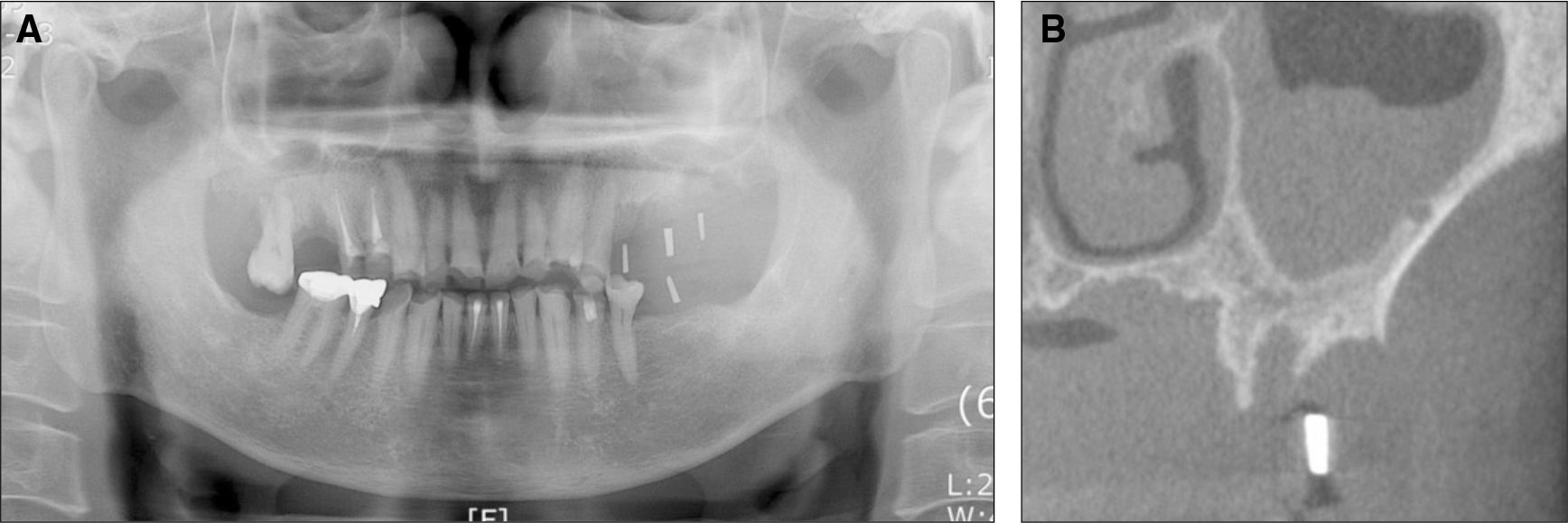 | Fig. 6.Radiograpic exam for implantation. Mucosal thickening on left sinus is observed. A: Panoramic view, B: Sagittal view of left sinus (CBCT). |
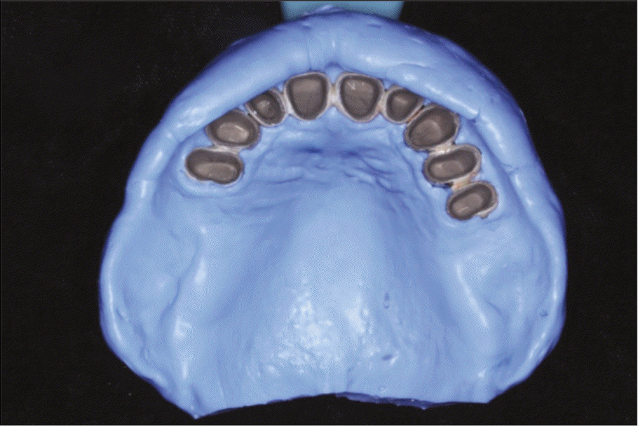
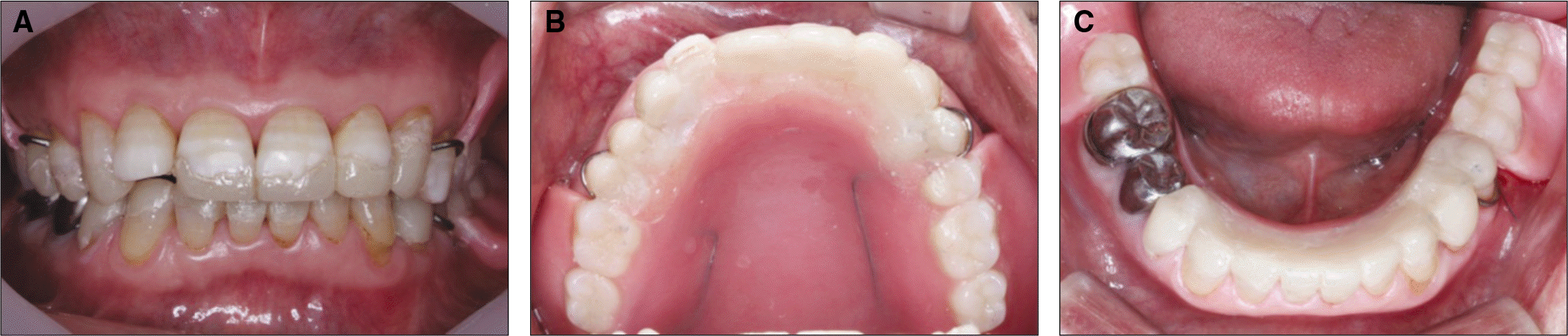 | Fig. 7.Removable interim overlay prosthesis. A: Frontal view, B: Maxillary occlusal view, C: Mandibular occlusal view. |
 XML Download