Abstract
In case of missing of permanent teeth by trauma or innate defect, the decision of treatment modalities and application timing have an important effect on the prognosis of oral rehabilitation. In this case report, interdisciplinary approach between the orthodontic and prosthodontic treatment, the way to re-establish the collapsed occlusal vertical dimension, and implant prosthetic considerations will be discussed. Proper diagnosis on teeth and craniofacial skeleton was made prior to treatment and provisional restorations were used in regard of growth patterns of the patient. Finally, the edentulous areas were restored with fixed implant prostheses. Diagnosis, treatment rationale and prognosis will be discussed thoroughly. (J Korean Acad Prosthodont 2013;51:339-46)
Go to : 
REFERENCES
1.Tuverson DL. Orthodontic treatment using canines in place of missing maxillary lateral incisors. Am J Orthod. 1970. 58:109–27.

2.Nordquist GG., McNeill RW. Orthodontic vs. restorative treatment of the congenitally absent lateral incisor-long term periodontal and occlusal evaluation. J Periodontol. 1975. 46:139–43.

3.Valderhaug J. A 15-year clinical evaluation of fixed prosthodontics. Acta Odontol Scand. 1991. 49:35–40.

4.Hansson O., Moberg LE. Clinical evaluation of resin-bonded prostheses. Int J Prosthodont. 1992. 5:533–41.
5.Ekfeldt A., Carlsson GE., Bo¨rjesson G. Clinical evaluation of single-tooth restorations supported by osseointegrated implants: a retrospective study. Int J Oral Maxillofac Implants. 1994. 9:179–83.
6.Magne P., Magne M., Belser U. Natural and restorative oral esthetics. Part II: Esthetic treatment modalities. J Esthet Dent. 1993. 5:239–46.

7.Dietschi D. Free-hand composite resin restorations: a key to anterior aesthetics. Pract Periodontics Aesthet Dent. 1995. 7:15–25.
8.Rohner F., Cimasoni G., Vuagnat P. Longitudinal radiographical study on the rate of alveolar bone loss in patients of a dental school. J Clin Periodontol. 1983. 10:643–51.

9.Mecall RA., Rosenfeld AL. Influence of residual ridge resorption patterns on implant fixture placement and tooth position. 1. Int J Periodontics Restorative Dent. 1991. 11:8–23.
10.Bergendal B., Bergendal T., Hallonsten AL., Koch G., Kurol J., Kvint S. A multidisciplinary approach to oral rehabilitation with os-seointegrated implants in children and adolescents with multiple aplasia. Eur J Orthod. 1996. 18:119–29.

11.Thilander B., Odman J., Gro¨ndahl K., Lekholm U. Aspects on os-seointegrated implants inserted in growing jaws. A biometric and radiographic study in the young pig. Eur J Orthod. 1992. 14:99–109.

12.Thilander B., Odman J., Gro¨ndahl K., Friberg B. Osseointegrated implants in adolescents. An alternative in replacing missing teeth? Eur J Orthod. 1994. 16:84–95.
13.Kwon KR., Woo YH., Choi DG. The study of relationship between sagittal condylar guide angle and incisal guide angle during mandibular protrusion in normal Korean. J Korean Acad Prosthodont. 1989. 27:11–36.
Go to : 
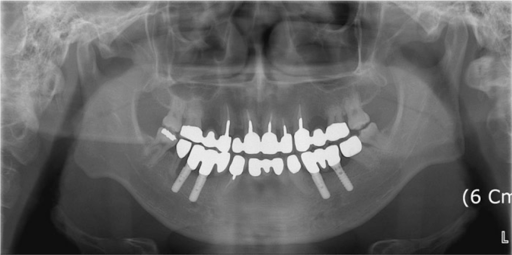
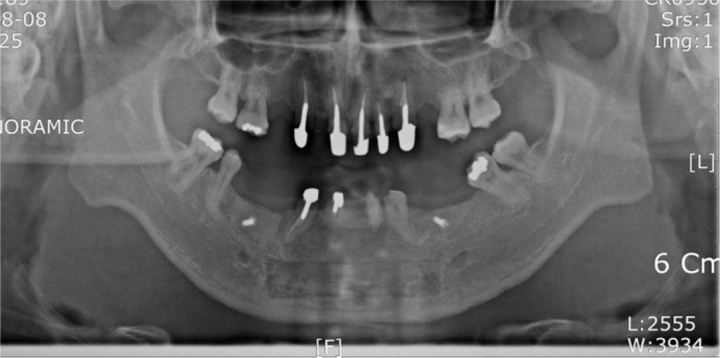 | Fig. 5.Panoramic radiograph after endodontic treatment on maxillary anterior teeth and bone graft on mandibular defect area. |
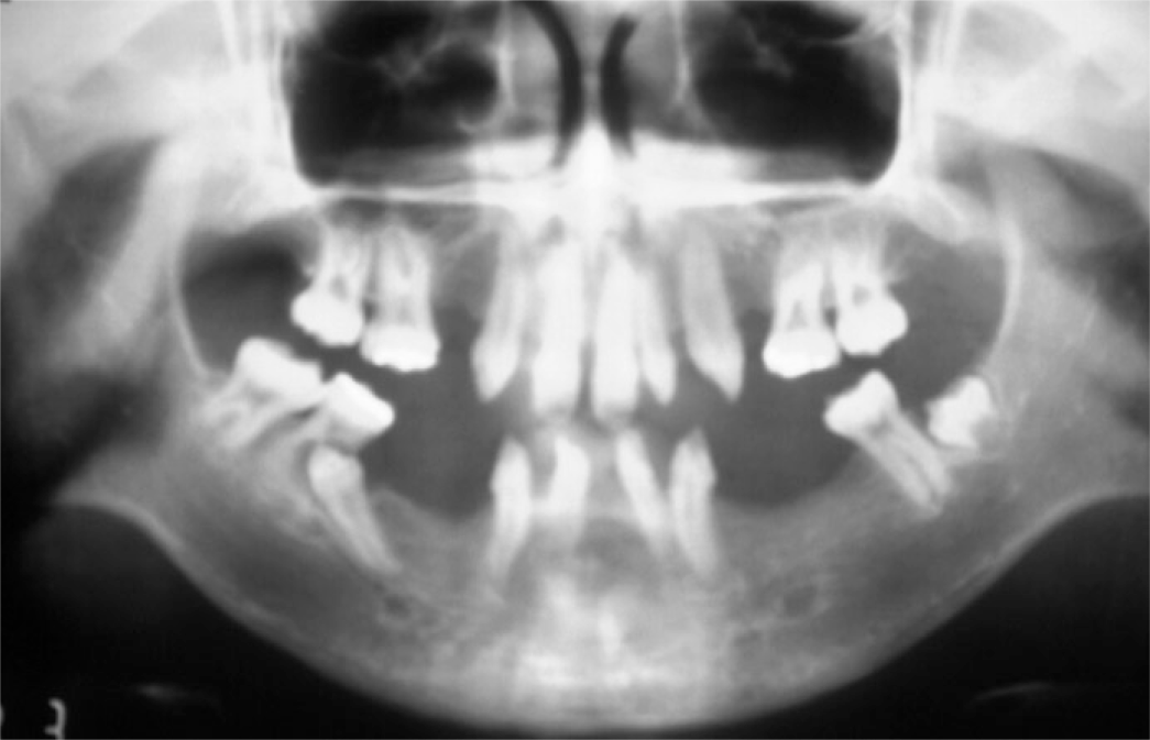
 | Fig. 10.Panoramic radiograph with RSM (January 1995) and root resorption of first molar by erupting first premolar (January 1999). |
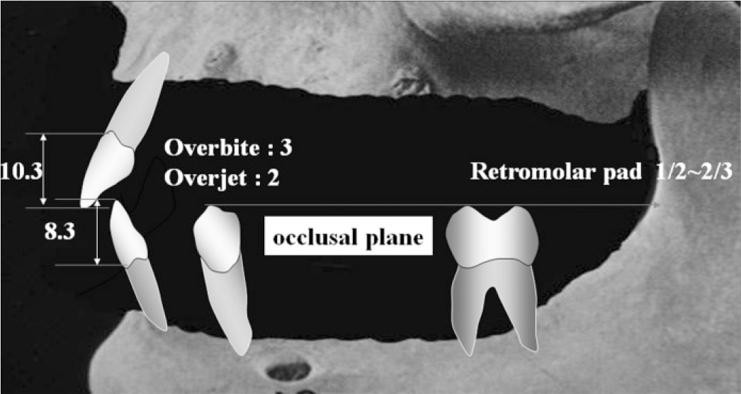 | Fig. 11.Occlusal plane establishment considering clinical crown length of maxillary and mandibular central incisor, vertical and horizontal overlap, height of first molar and height of retromolar pad. |
Table 1.
Diagnosis and treatment objectives


 PDF
PDF ePub
ePub Citation
 Print
Print





 XML Download