Abstract
Purpose
The aim of this retrospective study was to evaluate the influence of implant diameter, length and placement to implant stability.
Materials and methods
Total 90 implants (US II plusTM, Osstem co, Busan, Korea) of 72 patients were determined as experimental samples. The factors of diameters(ø 4 mm, ø 5 mm), lengths (10 mm, 11.5 mm, 13 mm), and implant placement (maxilla, mandible) were analyzed. The stability of the implants was measured by resonance frequency analysis (RFA) at the time of implant placement and impression taking. The difference of ISQ values according to patient's gender was evaluated by Independent t-test. ISQ values were compared between implant diameter, length and placement using one-way ANOVA and Tukey HSD test (α =.05). To compare ISQ values between at the time of surgery and impression taking, paired t-tests were used (α =.05).
Results
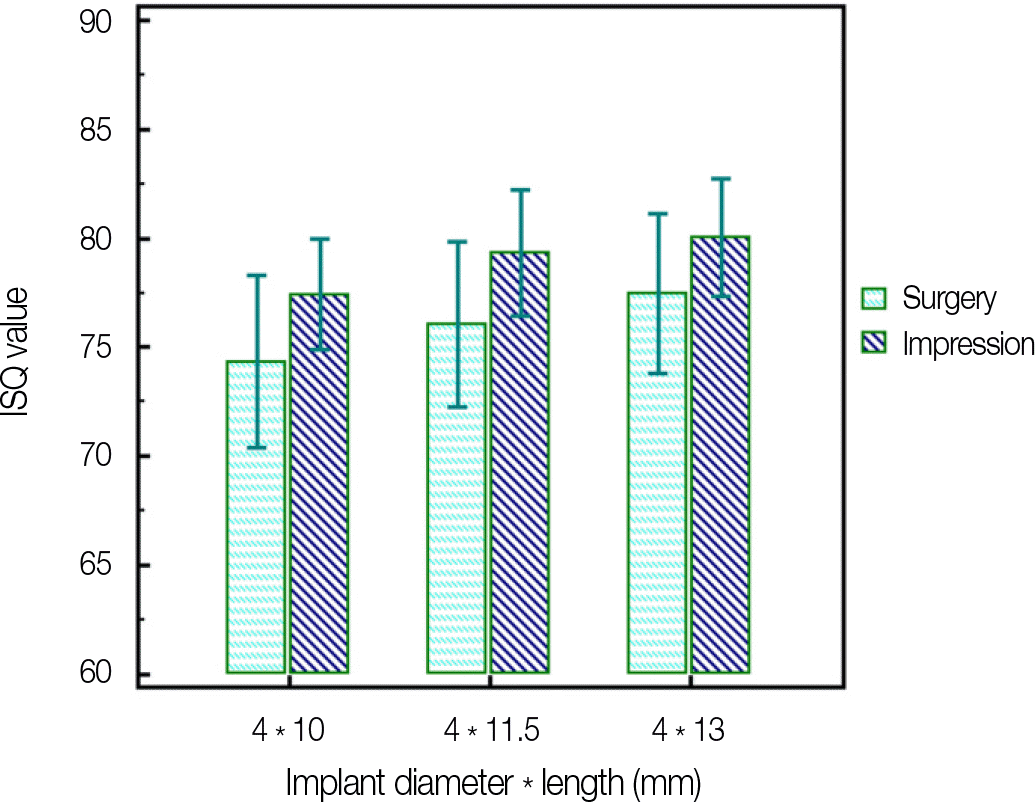
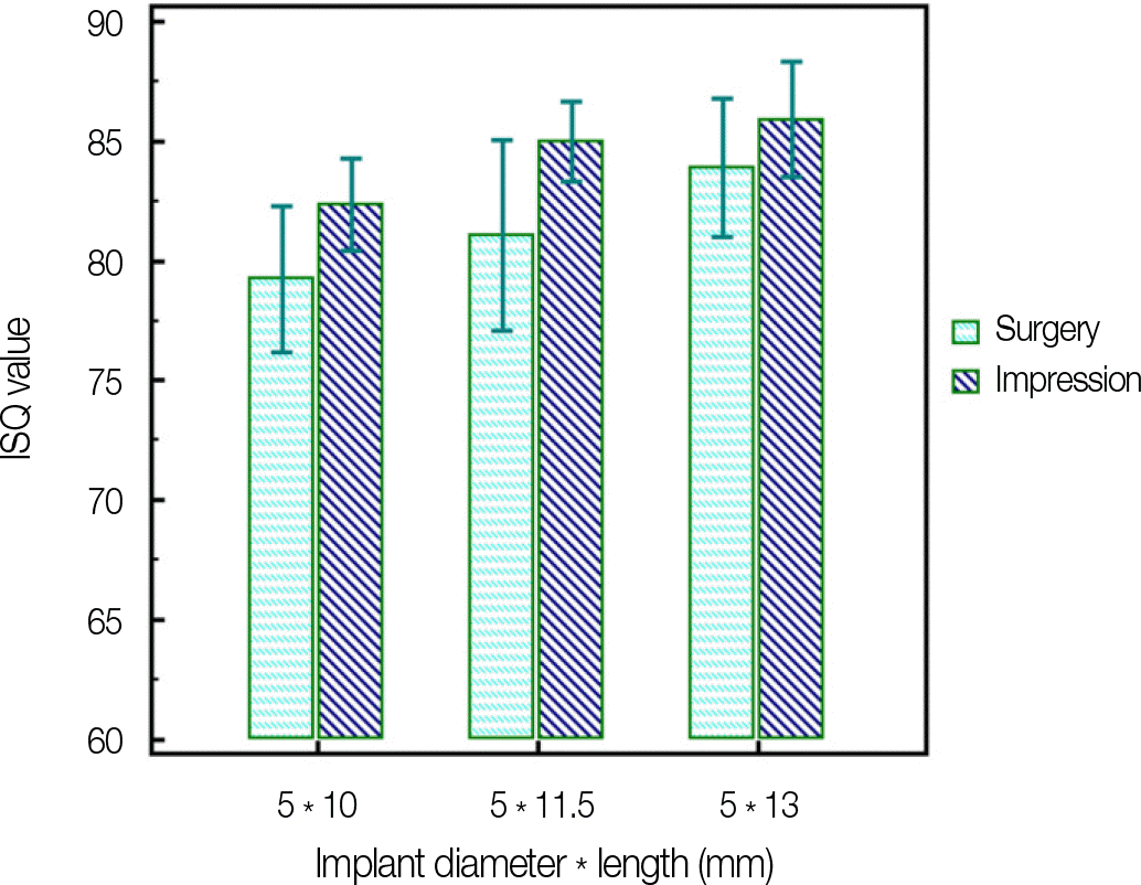
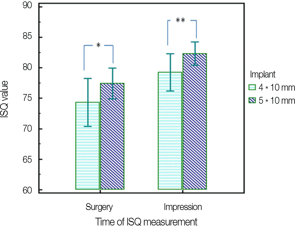
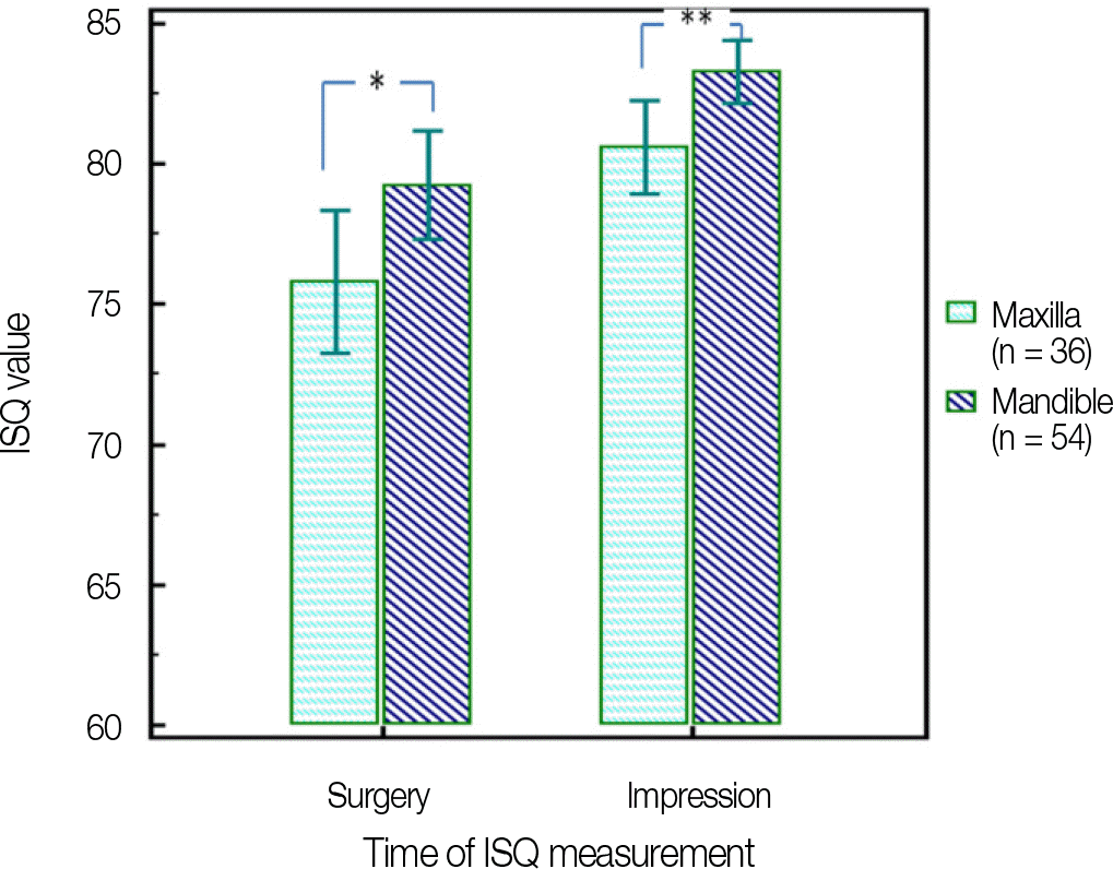
The change of implant length did not show significant different on the ISQ value (P>.05). However, 5 mm diameter implants had higher ISQ values than 4 mm diameter implants (P<.05). Implants placed on the mandible showed significantly higher ISQ values than on the maxilla (P<.05).
Conclusion
In order to increase implant stability, it is better to select the wider implant, and implants placed on mandible are possible to get higher stability than maxilla. ISQ values at impression taking showed higher implant stability than ISQ values at implant placement, it means that RFA is clinically effective method to evaluate the change of implant stability through the osseointegration. The consideration of the factors which may affect to the implant stability will help to determine the time of load applying and increase the implant success rate. (J Korean Acad Prosthodont 2013;51:269-75)
Go to : 
REFERENCES
1.Meredith N. Assessment of implant stability as a prognostic determinant. Int J Prosthodont. 1998. 11:491–501.
2.Cochran DL., Schenk RK., Lussi A., Higginbottom FL., Buser D. Bone response to unloaded and loaded titanium implants with a sandblasted and acid-etched surface: a histometric study in the canine mandible. J. Biomed Master Res. 1998. 40:1–11.
3.Sykaras N., Triplett RG., Nunn ME., Iacopino AM., Opperman LA. Effect of recombinant human bone morphogenetic protein-2 on bone regeneration and osseointegration of dental implants. Clin Oral Implants Res. 2001. 12:339–49.

4.Albreksson T., Jacobsson M. Bone-metal interface in osseointegration. J Prosthet Dent. 1987. 57:597–607.
5.Carlsson L., Ro¨stlund T., Albrektsson B., Albrektsson T. Removal torques for polished and rough titanium implants. Int J Oral Maxillofac Implants. 1988. 3:21–4.
6.Friberg B., Sennerby L., Linden B., Gro¨ndahl K., Lekholm U. Stability measurements of one-stage Bra®nemark implants during healing in mandibles. A clinical resonance frequency analysis study. Int J Oral and Maxillofac Surg. 1999. 28:266–72.
7.Schulte W., Lukas D. Periotest to monitor osseointegration and to check the occlusion in oral implantology. J Oral Implantol. 1993. 19:23–32.
8.Aparicio C. The use of the Periotest value as the initial success criteria of an implant: 8-year report. Int J Periodontics Restorative Dent. 1997. 17:150–61.
9.Meredith N., Alleyne D., Cawley P. Quantitative determination of the stability of the implant - tissue interface using resonance frequency analysis. Clin Oral Implants Res. 1996. 7:261–67.
10.Meredith N., Book K., Friberg B., Jemt T., Sennerby L. Resonance frequency measurements of implant stability in vivo. A cross-sectional and longitudinal study of resonance frequency measurements on implants in the edentulous and partially dentate maxilla. Clin Oral Implants Res. 1997. 8:226–33.
11.Oh JH., Chang MT. Comparison of initial implant stability measured by Resonance frequency analysis between different implant systems. J Korean Acad Periodontol. 2008. 38:529–34.

12.Atsumi M., Park SH., Wang HL. Methods used to assess implant stability: current status. Int J Oral Maxillofac Implants. 2007. 22:743–54.
13.Krennmair G., Waldenberger O. Clinical analysis of wide-diameter frialit-2 implants. Int J Oral Maxillofac Implants. 2004. 19:710–5.
14.Bischof M., Nedir R., Szmukler-Moncler S., Bernard JP., Samson J. Implant stability measurement of delayed and immediately loaded implants during healing. Clin Oral Implants Res. 2004. 15:529–39.
15.Han J., Lulic M., Lang NP. Factors influencing resonance frequency analysis assessed by Osstell mentor during implant tissue integration: II. Implant surface modifications and implant diameter. Clin Oral Implants Res. 2010. 21:605–11.
16.Ohta K., Takechi M., Minami M., Shigeishi H., Hiraoka M., Nishimura M., Kamata N. Influence of factors related to implant stability detected by wireless resonance frequency analysis device. J Oral Rehabil. 2010. 37:131–7.

17.Lee JY., Lee WC., Kim JE., Shin SW. The influence of implant diameter, length and design changes on implant stability quotient (ISQ) value in artificial bone. J Korean Acad Prosthodont. 2012. 50:292–8.

18.Langer B., Langer L., Herrmann I., Jorneus L. The wide fixture: a solution for special bone situations and a rescue for the compromised implant. Part 1. Int J Oral Maxillofac implants. 1993. 8:400–8.
19.Polizzi G., Rangert B., Lekholm U., Gualini F. Lindstrom H. Bra®nemark System Wide Platform implants for single molar replacement: clinical evaluation of prospective and retrospective materials. Clin Implant Dent Relat Res. 2000. 2:61–9.
20.Kim DH., Pang EK., Kim CS., Choi SH., Cho KS. Non-submerged type implant stability analysis during initial healing period by resonance frequency analysis. J Korean Acad Periodontol. 2009. 39:339–48.

21.Scortecci GM., Misch CE., Benner KU. Implants and Restorative dentistry. CRC Press;2001. p. 60.
22.Ivanoff CJ., Sennerby L., Johansson C., Rangert B., Lekholm U. Influence of implant diameters on the integration of screw implants. An experimental study in rabbits. Int J Oral Maxillofac Surg. 1997. 26:141–8.
23.Chung DM., Oh TJ., Lee J., Misch CE., Wang HL. Factors affecting late implant bone loss: a retrospective analysis. Int J Oral Maxillofac Implants. 2007. 22:117–26.
24.Kim JM., Kim SJ., Han I., Shin SW., Ryu JJ. A comparison of the implant stability among various implant systems: clinical study. J Adv Prosthodont. 2009. 1:31–6.

25.Barewal RM., Oates TW., Meredith N., Cochran DL. Resonance frequency measurement of implant stability in vivo on implants with a sandblasted and acid-etched surface. Int J Oral Maxillofac Implants. 2003. 18:641–51.
Go to : 
 | Fig. 1.ISQ values (Mean ± SD) for different implant lengths in ø 4.0 mm implants at the time of surgery and impression taking. There is no statistically significant different according to the implant length. |
 | Fig. 2.ISQ values (Mean ± SD) for different implant lengths in ø 5.0 mm implants at the time of surgery and impression taking. There is no statistically significant different according to the implant length. |
 | Fig. 3.ISQ values (Mean ± SD) for different implant diameter in 10 mm implants at the time of surgery and impression taking. The stars (∗, ∗∗) indicate significant differences between 4 mm and 5 mm diameter implants (P<.05). |
 | Fig. 4.ISQ values (Mean ± SD) depending on implant position (maxilla / mandible) at the time of surgery and impression taking. The stars (∗, ∗∗) indicate significant differences between implants on the maxilla and mandible (P<.05). |
Table 1.
Mean ISQ values according to patient's gender at surgery and impression
| Patient's Gender | n | ISQ values (Mean ± SD) | |
|---|---|---|---|
| Surgery | Impression | ||
| Male | 41 | 77.80 ± 8.27 | 81.12 ± 5.77 |
| Female | 31 | 79.72 ± 5.14 | 82.33 ± 4.35 |
Table 2.
Mean ISQ values for different implant lengths in ø 4.0 mm implants at surgery and impression
| Implant Length | n | ISQ values (Mean ± SD) | |
|---|---|---|---|
| Surgery | Impression | ||
| 10 mm | 15 | 74.33 ± 7.13 | 77.43 ± 4.56 |
| 11.5 mm | 15 | 76.03 ± 6.84 | 79.33 ± 5.21 |
| 13 mm | 15 | 77.47 ± 6.61 | 80.07 ± 4.87 |
Table 3.
Mean ISQ values for different implant lengths in ø 5.0 mm implants at surgery and impression
| Implant Length | n | ISQ values (Mean ± SD) | |
|---|---|---|---|
| Surgery | Impression | ||
| 10 mm | 15 | 79.27 ± 5.51 | 82.33 ± 3.46 |
| 11.5 mm | 15 | 81.07 ± 7.17 | 84.97 ± 3.02 |
| 13 mm | 15 | 83.90 ± 5.27 | 85.90 ± 4.34 |
Table 4.
Mean ISQ values for different implant diameter in 10 mm implants at surgery and impression
| Implant Diameter | n | ISQ values (Mean ± SD) | |
|---|---|---|---|
| Surgery | Impression | ||
| 4 mm | 15 | 74.33 ± 5.51∗ | 77.43 ± 3.46∗∗ |
| 5 mm | 15 | 79.26 ± 7.17∗ | 82.33 ± 3.02∗∗ |


 PDF
PDF ePub
ePub Citation
 Print
Print


 XML Download