Abstract
Purpose
This study was conducted to compare in vivo biofilm formation on titanium surface and zirconia surface.
Materials and methods
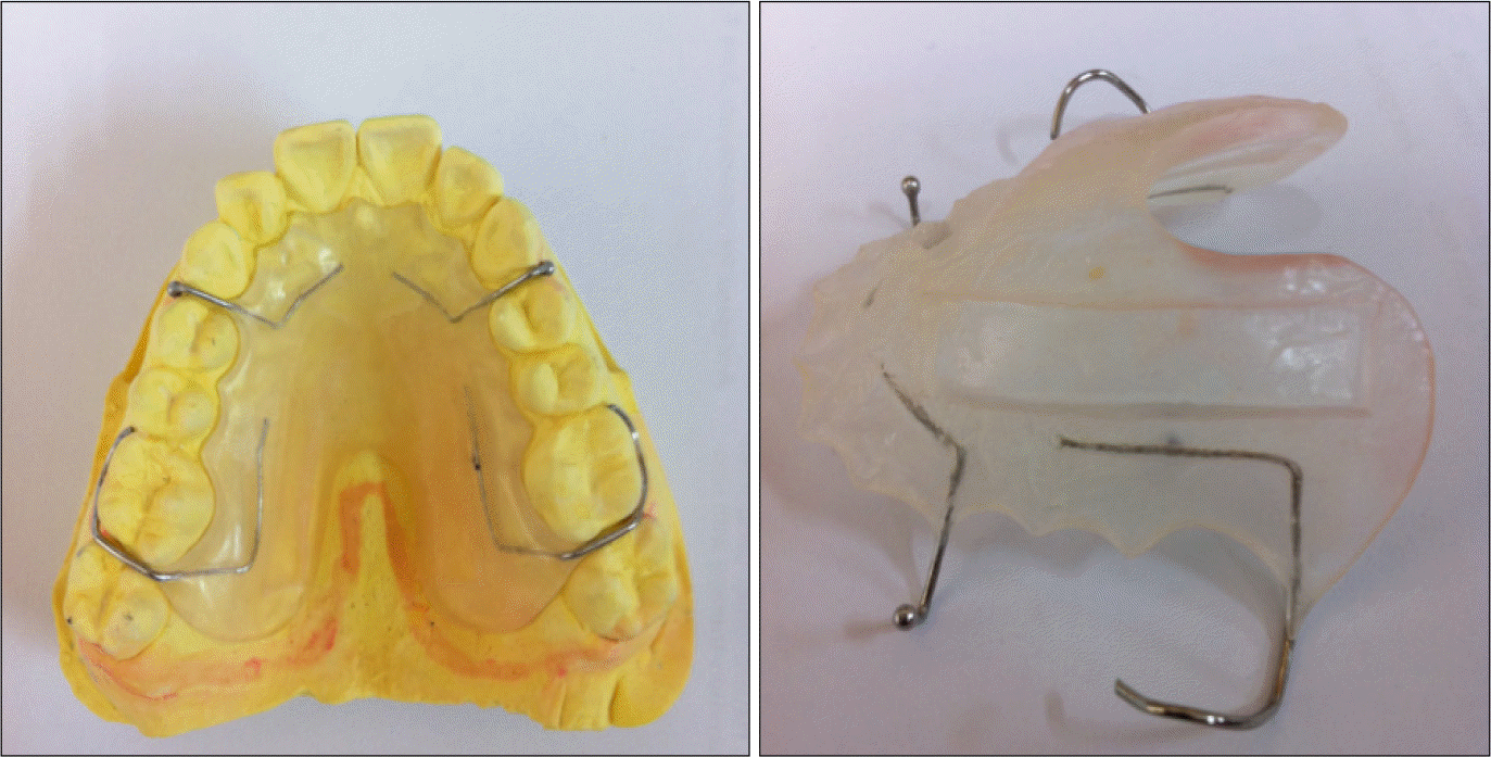
For biofilm formation on titanium and zirconia in oral cavity, after producing oral appliances using acrylic resin and orthodontic wire tailored to 9 subjects, we made titanium and zirconia specimens (6 mm × 6 mm × 2 mm), fixed them on oral appliances and maintained them in oral cavity of test subjects for 24 and 72 hours. Test subjects who have equipped two pairs of specimens maintained oral hygiene not by using toothpaste but only by tooth brushing. After 24 and 72 hours, we removed and observed specimens through scanning electron microscopy (SEM).
Results
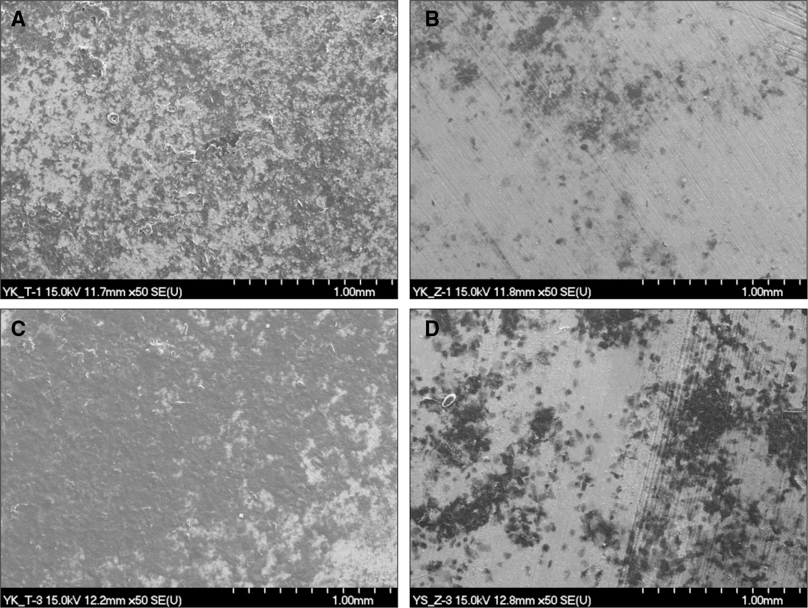
Biofilm formation showed large deviation depending on individuals. For formation comparison between titanium and zirconia for 24 hours, zirconia showed less biofilm formation than titanium. Biofilm formation showed large deviation depending on individuals. As for formation comparison between zirconia and titanium, the degree of biofilm formation in zirconia was less than it was in titanium after a lapse of 24 hours. The result of biofilm formation in 72 hours trial show that zirconia has an inclination to formate less biofilm than it was in titanium.
Go to : 
REFERENCES
1.Jemt T., Laney WR., Harris D., Henry PJ., Krogh PHJ., Pollizi ., Zarb GA., Hermann I. Osseointegrated implants for single tooth replacement: a 1-year report from a multi-center prospective study. Int J Oral Maxillofac Implants. 1991. 6:29–36.
2.Heydecke G., Sierraalta M., Razzong ME. Evolution and use of aluminum oxide single tooth implant abutement: A short review and presentation of two cases. Int J Prosthodont. 2002. 15:448–93.
3.Rasperini G., Magnolione M., Cocconcelli P., Simion M. In vivo early plaque formation on pure titanium and ceramic abutments: a comparative microbiological and SEM analysis. Clin Oral Implants Res. 1998. 9:357–64.

4.Johnson RH., Persson GR. A 3-year prospective study of a single-tooth implant-Prosthodontic complication. Int J Prosthodont. 2001. 14:183–9.
5.Miyazaki T., Hotta Y., Kunii J., Kuriyama S., Tamaki Y. A Review of Dental CAD/CAM: Current Status and Future Perspectives from 20 Years of Experience. Dent Mater J. 2009. 28:44–56.

6.Nakamura K., Kanno T., Milleding P., Ortengren U. Zirconia as a dental implant abutment material: a systematic review. Int J Prosthodont. 2010. 23:299–309.
7.Gomes AL., Montero J. Zirconia implant abutments: a review. Med Oral Patol Oral Cir Bucal. 2011. 16:50–5.

8.Rimondini L., Cerroni L., Carrassi A., Torricelli P. Bacterial colonization of zirconia ceramic surfaces: an in vitro and in vivo study. Int J Oral Maxillofac Implants. 2002. 17:793–8.
9.Scarano A., Piattelli M., Caputi S., Favero GA., Piattelli A. Bacterial adhesion on commercially pure titanium and zirconium oxide disks: an in vivo human study. J Periodontol. 2004. 75:292–6.

10.Steinberg D., Sela MN., Klinger A., Kohavi D. Adhesion of periodontal bacteria to titanium, and titanium alloy powders. Clin Oral Implants Res. 1998. 9:67–72.

11.Quirynen M., Bollen CM., Papaioannou W., Van Eldere J., van Steenberghe D. The influence of titanium abutment surface roughness on plaque accumulation and gingivitis: short-term observations. Int J Oral Maxillofac Implants. 1996. 11:169–78.
Go to : 
 | Fig. 2.Bacterial adhesion on titanium and zirconia Surfaces (magnification ×50). A: 24 hrs titanium surface, B: 24 hrs zirconia surface, C: 72 hrs titanium surface, D: 72 hrs zirconia surface. |
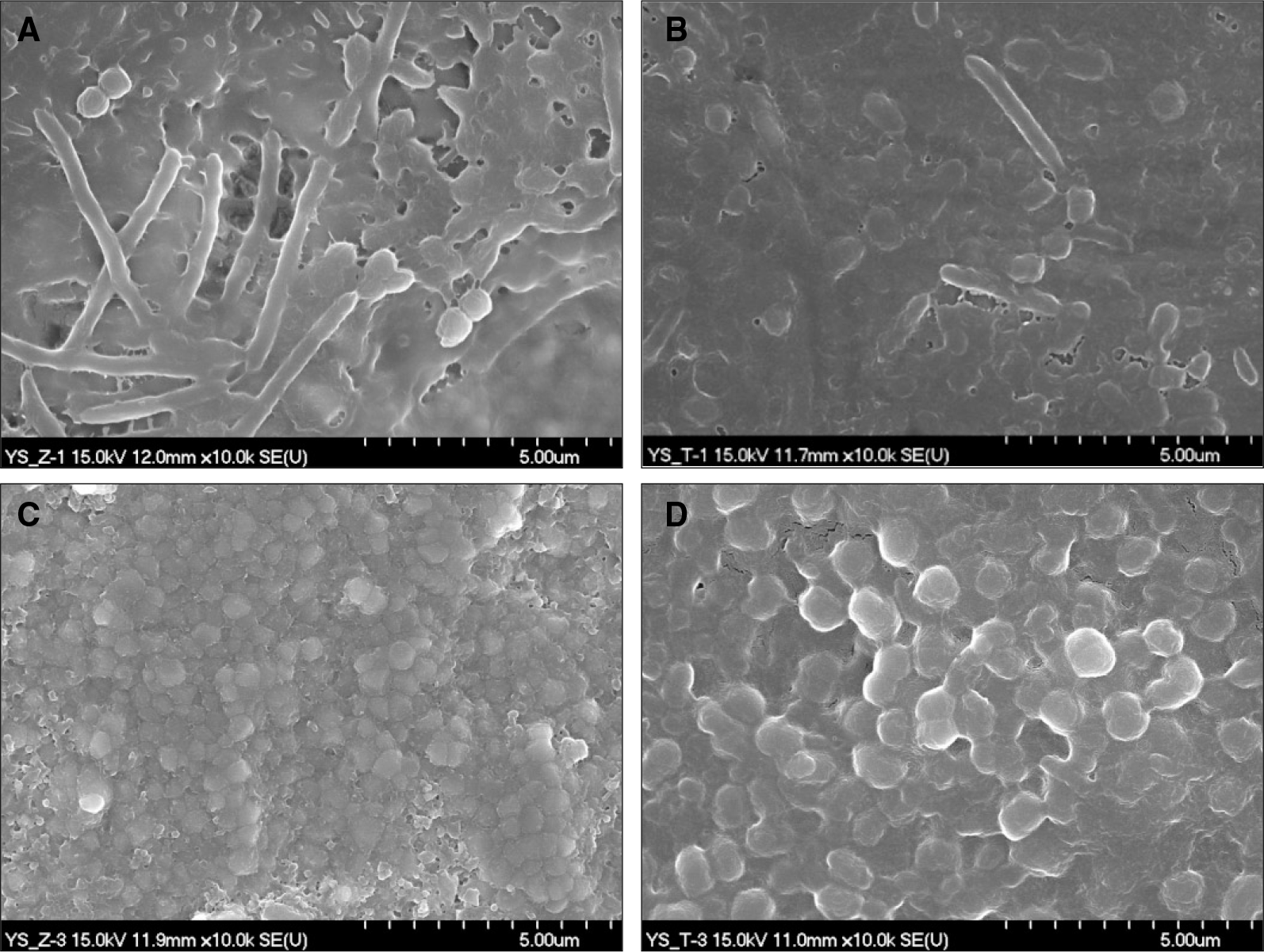
 | Fig. 3.Plaque composition (Rod %) attachment to titanium and zirconia surfaces after 24 hrs and 72 hrs (magnification ×10,000, 12 × 10 ㎛ size specimen). A: 24 hrs zirconia, B: 24 hrs titanium, C: 72 hrs zirconia, D: 72 hrs titanium. |


 PDF
PDF ePub
ePub Citation
 Print
Print


 XML Download