

 PDF
PDF ePub
ePub Citation
 Print
Print


Abstract
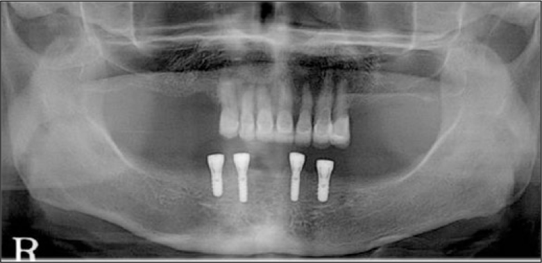
Implant prosthodontics is beneficial for edentulous patients in enhancing the support, retention, stability, phonation and so on. Various types of prosthesis supported by implant, including implant retained- or supported- overdenture for the removable type and ceramo-metal and fixed prostheses with processed acrylic teeth for the fixed type, are frequently used. Treatment planning for the prosthesis with implant must be made after considering individual characteristics such as form of residual ridge, soft tissue, interocclusal relationship, economic status. Fixed prosthesis with processed acrylic teeth (also known as 'implant hybrid prosthesis' or 'bone anchored bridge') has the advantages of both removable and fixed prosthesis such as proper soft tissue profile, esthetic outcome, increased masticatory efficiency and psychological stability. The 73-years-old female patient came to the department of prosthodontics, Dental hospital of Yonsei University. She was diagnosed with Kennedy class I partial edentulism in the maxilla and complete edentulism in the mandible. This article reports a satisfactory clinical and esthetic outcome of full mouth rehabilitation using removable partial denture in the maxilla and implant hybrid prosthesis in the mandible. (J Korean Acad Prosthodont 2013;51:214-20)
Go to : 
REFERENCES
1.Zarb GA., Bolender CL., Eckert SE., Fenton AH., Jacob RF., Mericske-Stern R. Prosthodontic treatment for edentulous patients: Complete dentures and implant-supported prostheses. 12th ed.Mosby;2003.
2.Misch CE. Dental implant prosthetics. 1st ed.Mosby;2004.
3.Chee W., Jivraj S. Treatment planning of the edentulous mandible. Br Dent J. 2006. 201:337–47.

4.Drago C., Gurney L. Maintenance of implant hybrid prostheses: clinical and laboratory procedures. J Prosthodont. 2013. 22:28–35.
5.Adell R., Eriksson B., Lekholm U., Bra�nemark PI., Jemt T. Longterm follow-up study of osseointegrated implants in the treatment of totally edentulous jaws. Int J Oral Maxillofac Implants. 1990. 5:347–59.
6.Ferrigno N., Laureti M., Fanali S., Grippaudo G. A long-term follow-up study of non-submerged ITI implants in the treatment of totally edentulous jaws. Part I: Ten-year life table analysis of a prospective multicenter study with 1286 implants. Clin Oral Implants Res. 2002. 13:260–73.
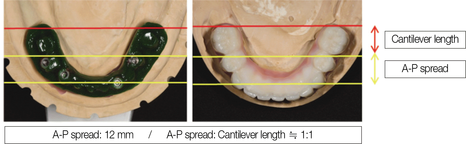
7.McAlarney ME., Stavropoulos DN. Theoretical cantilever lengths versus clinical variables in fifty-five clinical cases. J Prosthet Dent. 2000. 83:332–43.
8.McAlarney ME., Stavropoulos DN. Determination of cantilever length-anterior-posterior spread ratio assuming failure criteria to be the compromise of the prosthesis retaining screw-prosthesis joint. Int J Oral Maxillofac Implants. 1996. 11:331–9.
Go to :
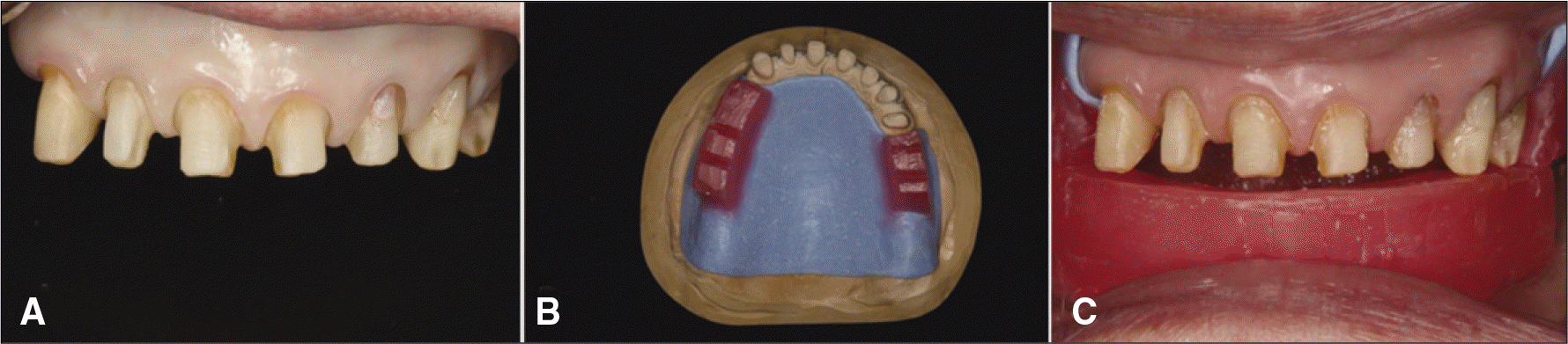 | Fig. 3.Tooth preparation and bite registration for temporary prosthesis (A) Preparation of maxillary teeth (B) Wax rim for temporary denture, (C) Bite registration for temporary prosthesis with wax rim. |
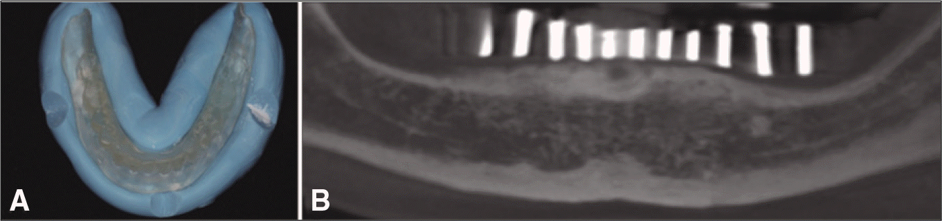 | Fig. 5.Conebeam CT taking with CT stent (A) Temporary denture duplication for CT stent, (B) Conebeam CT image. |
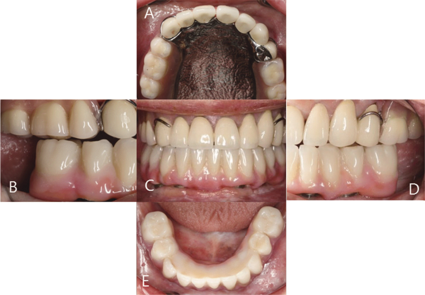
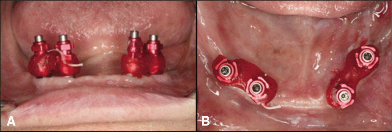 | Fig. 7.(A) Impression taking for definitive restoration (frontal view) (B) (occlusal view). |
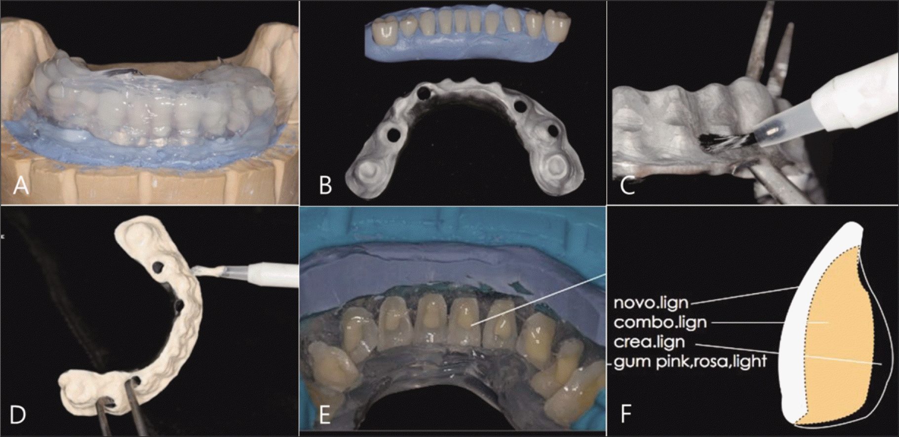 | Fig. 8.Laboratory procedures using resin veneering system. (A) Putty index for veneering teeth alignment duplication (novo.lign) (B) Putty index and metal framework after sandblasting (C) Bonding agent (visio.link) (D) Opaque application (E) Dentine-colored adhesive composite (combo.lign, white arrow) (F) Schematic diagram of visio.lign system. |
 XML Download