Abstract
Purpose
The importance of occlusal contacts of the natural dentition for durability of teeth, mandibular stabilization, and restorative dentistry is well known. The purpose of this study is to analyze the occlusal contact and guidance pattern of Koreans by evaluating the static occlusion on maximal intercuspal position and measuring dynamic occlusion during straight protrusion.
Materials and methods
The occlusal contacts at maximal interincisal position and the occlusal guidance pattern during straight protrusion of 29 subjects were recorded with shimstock foil (Whaledent, Langenau, Germany), T-Scan III (Tekscan Inc., Boston, MA, USA), polyvinylsiloxane registration material (Genie Bite, Sultan Healthcare, Hackensack, NJ, USA) and compared. Occlusal registration procedures were repeated 3 times. The position was fixed to an upright position and the head position was fixed with the Frankfurt horizontal plane paralleling the horizontal plane. Fisher's Exact Test (R-General Public License, ver. 2.14.1) and Pearson's Test were used to assess the significance level of the differences between the experimental groups (α =.05).
Results
When using shimstock foil, T-Scan III system, and polyvinylsiloxane registration material, most of the patients showed contact on anterior, premolar, and molar teeth during maximal intercuspal position. Approximately 51% of maximal intercuspal position showed anterior contact using shimstock foil. When examining the protrusive movement using shimstock foil and T-Scan III system, guidance pattern with the central incisor was the most common.
Conclusion
During maximal intercuspal position, there were cases in which not all of the teeth showed occlusal contact. During mandibular protrusive movements, one or more maxillary central incisors frequently joined in straight protrusion and the posterior teeth were disoccluded. Therefore, the anterior teeth protect the posterior teeth, and vice versa. Thus, mutually protected occlusion should be applied when reconstructing occlusion. (J Korean Acad Prosthodont 2013;51:199-207)
Go to : 
REFERENCES
1.Hochman N., Ehrlich J. Tooth contact location in intercuspal position. Quintessence Int. 1987. 18:193–6.
2.Ehrlich J., Taicher S. Intercuspal contacts of the natural dentition in centric occlusion. J Prosthet Dent. 1981. 45:419–21.

3.Okeson JP. Management of temporomandibular disorders and occlusion. 6th ed. St. Louis, MO: Mosby, Elsevier;2008. p. 101–16.
4.The glossary of prosthodontic terms. J Prosthet Dent. 2005. 94:10–92.
5.Myers GE., Anderson JR Jr. Nature of contacts in centric occlusion in 32 adults. J Dent Res. 1971. 50:7–13.

6.Black GV. Descriptive anatomy of the human teeth. 4th ed.Philadelphia: Wilmington Dental manufacturing Company;1904. p. 1836–915.
7.Ono I. The crushing power and masticating area of the teeth as the foundations of oral hygienics. Dent Cosmos. 1921. 63:1278–83.
8.Hellman M. Variations in occlusion. Dent Cosmos. 1921. 63:608–19.
9.Dawson PE., Arcan M. Attaining harmonic occlusion through visualized strain analysis. J Prosthet Dent. 1981. 46:615–22.

10.Maness WL., Benjamin M., Podoloff R., Bobick A., Golden RF. Computerized occlusal analysis: a new technology. Quintessence Int. 1987. 18:287–92.
11.Chapman RJ., Maness WL., Osorio J. Occlusal contact variation with changes in head position. Int J Prosthodont. 1991. 4:377–81.
12.Saraçoglu A., Ozpinar B. In vivo and in vitro evaluation of occlusal indicator sensitivity. J Prosthet Dent. 2002. 88:522–6.
13.Friel S. Occlusion, observations on its development from infancy to old age. Int J Orthod Oral Surg Radio. 1927. 13:322–43.

14.Millstein PL. An evaluation of occlusal contact marking indicators: a descriptive, qualitative method. Quintessence Int Dent Dig. 1983. 14:813–36.
15.Halperin GC., Halperin AR., Norling BK. Thickness, strength, and plastic deformation of occlusal registration strips. J Prosthet Dent. 1982. 48:575–8.

16.Riise C., Ericsson SG. A clinical study of the distribution of occlusal tooth contacts in the intercuspal position at light and hard pressure in adults. J Oral Rehabil. 1983. 10:473–80.

17.Chapman RJ., Maness WL., Osorio J. Occlusal contact variation with changes in head position. Int J Prosthodont. 1991. 4:377–81.
19.Yun TH., Kim YG. A study on occlusal contact using computerized occlusal analysis system. Korean J Oral Med. 1989. 14:81–8.
20.Beyron H. Optimal occlusion. Dent Clin North Am. 1969. 13:537–54.
21.Pahng WD., Woo YH., Choi BB. The balance of occlusal contats in normal occlusion during intercuspal position on t-scan system. J Korean Acad Prosthodont. 1991. 29:23–38.
22.Choi HC., Lee SB., Choi DG., Park NS. A study of occlusal contact variaton due to change in each head position in normal occlusion. J Korean Acad Prosthodont. 1995. 33:769–79.
23.Berry DC., Singh BP. Effect of electromyographic biofeedback therapy on occlusal contacts. J Prosthet Dent. 1984. 51:397–403.

24.Gazit E., Fitzig S., Lieberman MA. Reproducibility of occlusal marking techniques. J Prosthet Dent. 1986. 55:505–9.

25.Anderson GC., Schulte JK., Aeppli DM. Reliability of the evaluation of occlusal contacts in the intercuspal position. J Prosthet Dent. 1993. 70:320–3.

26.Maness WL., Chapman RJ., Dario LJ. Laboratory evaluation of a direct reading digital occlusal sensor. J Prosthet Dent. 1985. 54:744.

27.Riis D., Giddon DB. Interdental discrimination of small thickness differences. J Prosthet Dent. 1970. 24:324–34.

28.Choi DG., Lee SB., Kwon YH., Choi BB. A study of the normal and abnormal occlusal patterns in adults using the superimposed rubber pattern method. J Korean Acad Prosthodont. 1995. 33:467–91.
29.Chai YA., Park NS., Choi BB. A study of the occlusal contact pattern during mandibular monvements of adult with normal occlusion. J Korean Acad Prosthodont. 1993. 31:565–79.
Go to : 
 | Fig. 2.Number of contact tooth of Mx. in maximal intercuspal position evaluated with shimstock foil (n = 406); CI: central incisor, LI: lateral incisor, C: canine, P1: first premolar, P2: second premolar, M1: first molar, M2: second molar. |
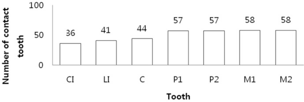 | Fig. 4.Number of contact tooth of Mx. in maximal intercuspal position evaluated with T-Scan III system (n = 406); CI: central incisor, LI: lateral incisor, C: canine, P1: first premolar, P2: second premolar, M1: first molar, M2: second molar. |
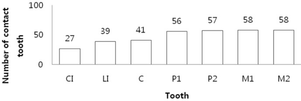 | Fig. 6.Number of contact tooth of Mx. in maximal intercuspal position evaluated with polyvinylsiloxane registration material (n = 406); CI: central incisor, LI: lateral incisor, C: canine, P1: first premolar, P2: second premolar, M1: first molar, M2: second molar. |
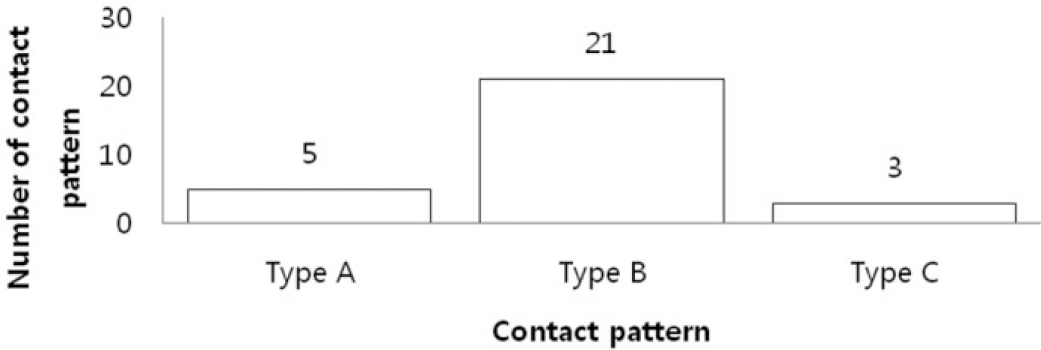
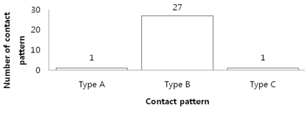
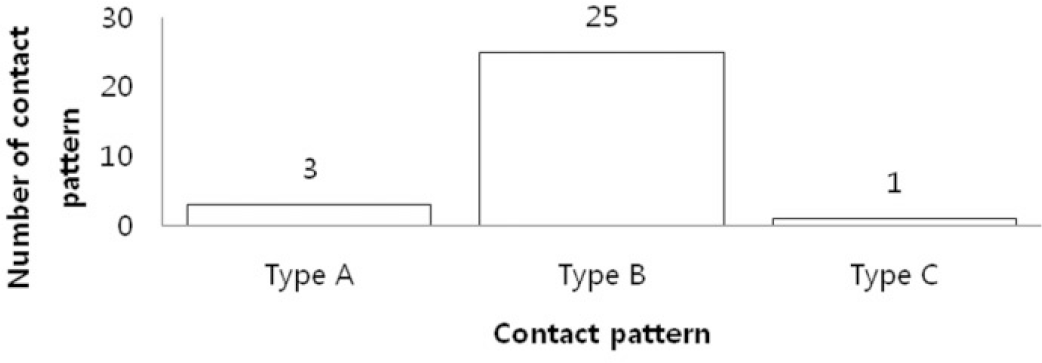 | Fig. 7.Distribution of contact pattern evaluated with polyvinylsiloxane registration material (n = 29). |
 | Fig. 8.Number of contact tooth of Mx. in straight protrusion evaluated with shimstock foil (n = 406); CI: central incisor, LI: lateral incisor, C: canine, P1: first premolar, P2: second premolar, M1: first molar, M2: second molar. |
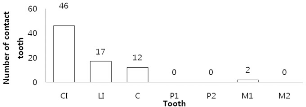 | Fig. 10.Number of contact tooth of Mx. in straight protrusion evaluated with T-Scan III system (n = 406); CI: central incisor, LI: lateral incisor, C: canine, P1: first premolar, P2: second premolar, M1: first molar, M2: second molar. |
Table 1.
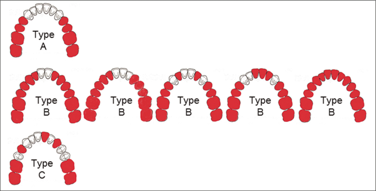
Classification of occlusal contact pattern (maximal intercuspal position)
| Contact pattern | Definition | |
|---|---|---|
| Type A | Non contact on anterior teeth | |
| Type B | Contact on anterior teeth | Contact on premolar & molar teeth |
| Type C | Non contact on premolar teeth | Contact on molar teeth |
Table 2.
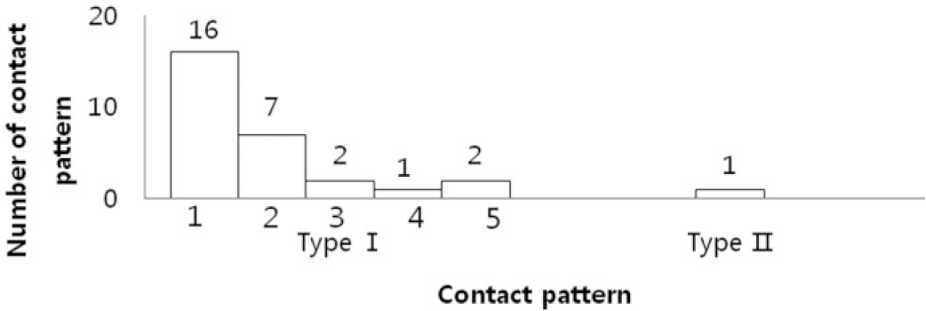
Classification of occlusal guidance pattern (protrusive movement) Guidance pattern Definition


 PDF
PDF ePub
ePub Citation
 Print
Print



 XML Download